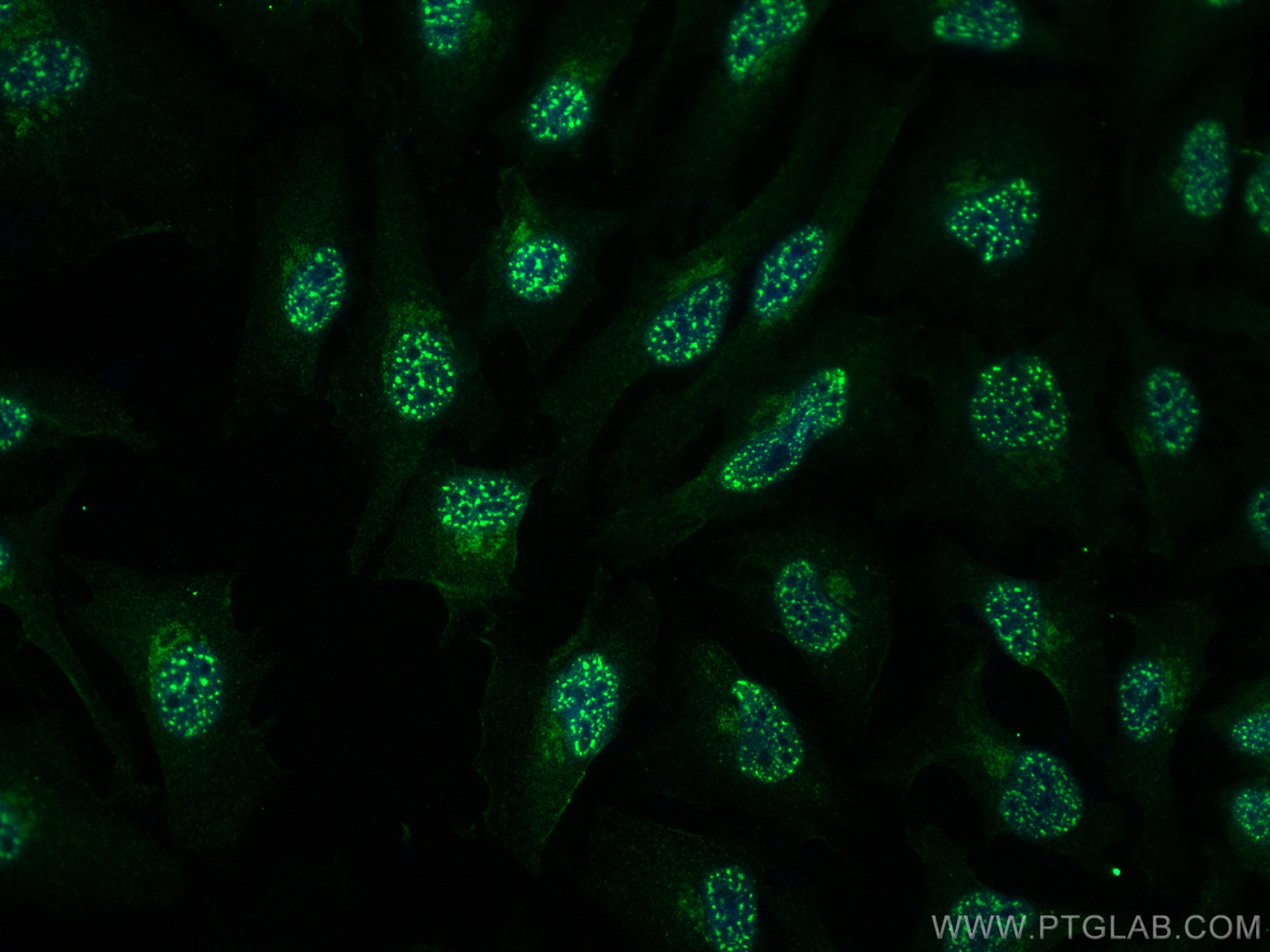
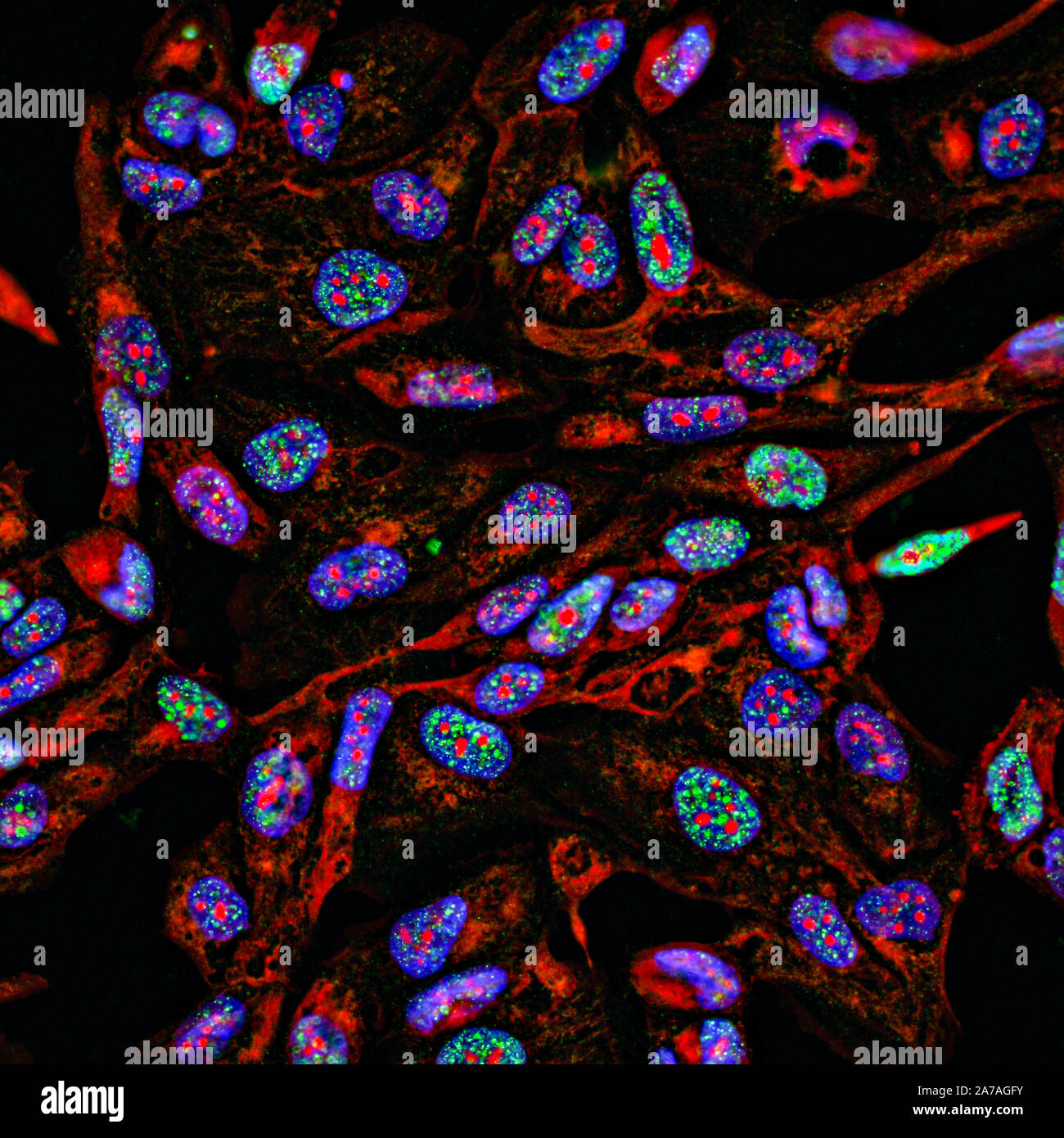
Antibody immunofluorescence is a powerful histological and cell biology technique that utilizes fluorescently labeled antibodies to detect and localize specific proteins or antigens within biological samples. By leveraging the high specificity of antigen-antibody binding, researchers can visualize the spatial distribution of target molecules in cells and tissues under fluorescence microscopy, enabling precise qualitative and quantitative analysis of protein expression and localization.
Fundamental Principles of Immunofluorescence
The core mechanism of immunofluorescence (IF) relies on the selective binding of an antibody to its corresponding antigen. The primary antibody is typically raised against a specific protein of interest. To visualize this binding, the antibody—or a secondary antibody that binds to the primary—is conjugated to a fluorophore, a molecule capable of absorbing light at a specific wavelength and emitting it at a longer wavelength.
When a sample is illuminated with the excitation light, the fluorophore emits light that can be captured by a microscope, revealing the precise location of the target antigen. The success of this process depends on the integrity of the sample, the specificity of the antibody, and the efficiency of the labeling process.
Direct vs. Indirect Immunofluorescence
Researchers must choose between two primary methods for detecting antigens: direct or indirect immunofluorescence. Each approach offers distinct advantages depending on the experimental goal, the availability of antibodies, and the desired signal amplification.
Direct Immunofluorescence
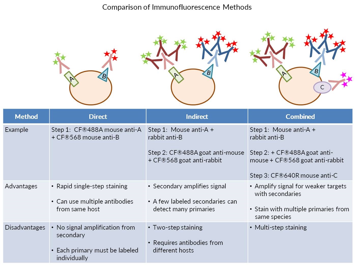
In direct IF, the primary antibody itself is conjugated to a fluorophore. This method is relatively straightforward and faster than indirect labeling, as it requires fewer incubation and washing steps. By reducing the number of steps, researchers can often minimize non-specific background signals. However, direct IF is less sensitive because there is no signal amplification; each antigen is bound by only one fluorophore-conjugated antibody.
Indirect Immunofluorescence
Indirect IF is the more common approach in research settings. In this protocol, an unlabeled primary antibody binds to the target antigen. Subsequently, a labeled secondary antibody, which is directed against the constant (Fc) region of the primary antibody, is applied. This method offers several advantages:
- Signal Amplification: Multiple secondary antibodies can bind to a single primary antibody, significantly increasing the number of fluorophores at the target site and enhancing the signal intensity.
- Versatility: A single labeled secondary antibody can be used with various primary antibodies raised in the same species, making it a cost-effective and flexible option.
- Flexibility: Researchers can easily switch fluorophores by using different secondary antibodies without needing to re-conjugate the primary antibody.
Experimental Workflow and Prerequisites
Executing a successful immunofluorescence experiment requires rigorous preparation and standardized protocols to ensure reproducibility and reliability.
Sample Preparation
The preparation of the sample—whether cell cultures, tissue sections, or clinical biopsies—is critical.
- Fixation: Samples must be fixed to preserve cellular morphology and prevent antigen degradation. Common fixatives include paraformaldehyde (cross-linking) or methanol/acetone (precipitating). The choice depends on the specific antigen and antibody compatibility.
- Permeabilization: If the target antigen is located inside the cell (intracellular), the cell membrane must be permeabilized using detergents such as Triton X-100 or Tween-20 to allow antibodies to access the target.
- Blocking: To prevent non-specific binding of the antibodies to cellular structures, the sample is incubated in a blocking buffer, typically containing bovine serum albumin (BSA) or normal serum from the species in which the secondary antibody was raised.
Antibody Incubation and Washing
Following blocking, the sample is incubated with the primary antibody. After a series of washing steps—essential to remove unbound antibodies and minimize background noise—the secondary antibody is added. These washes typically use phosphate-buffered saline (PBS) with a low concentration of detergent.
Mounting and Imaging
Once the final wash is complete, the sample is mounted on a slide using a mounting medium. This medium serves two purposes: it protects the sample from mechanical damage and includes additives to reduce the rate of photobleaching (fading of the fluorescent signal) during imaging. The sample is then visualized using widefield fluorescence microscopy, confocal microscopy, or super-resolution techniques.
Comparison of Detection Methods
| Feature | Direct Immunofluorescence | Indirect Immunofluorescence |
|---|---|---|
| Sensitivity | Lower | Higher (Signal Amplification) |
| Speed | Faster (Fewer steps) | Slower (More incubations) |
| Cost | Higher (Primary must be labeled) | Lower (Universal secondary) |
| Flexibility | Limited | High |
| Complexity | Low | Moderate |
Troubleshooting Common Issues
Immunofluorescence experiments are sensitive to environmental and chemical variables. Troubleshooting requires a systematic evaluation of each step.
- High Background Noise: This is frequently caused by incomplete washing or insufficient blocking. Ensure the blocking buffer is appropriate for the species of the secondary antibody. Decreasing the concentration of the primary or secondary antibody can also mitigate background signals.
- Weak or No Signal: If the target signal is absent, verify the primary antibody's reactivity with the sample species. Additionally, check the fixation and permeabilization parameters, as harsh fixation can sometimes mask or destroy the antigen. Ensure the fluorophore's excitation/emission spectra are compatible with the microscope's filter sets.
- Photobleaching: If the signal fades rapidly during imaging, try using a mounting medium containing anti-fade reagents. Alternatively, lower the intensity of the excitation light or decrease exposure times.
- Non-Specific Binding: Ensure the secondary antibody does not cross-react with endogenous proteins in the sample. Pre-adsorbing the secondary antibody can help ensure specificity.
Safety and Storage Considerations
Antibodies are biological reagents that require careful handling to maintain their activity. Most antibodies should be stored at -20°C in small aliquots to avoid repeated freeze-thaw cycles, which can lead to denaturation. When handling samples, utilize standard laboratory personal protective equipment (PPE), including gloves and eye protection, particularly when using chemical fixatives like paraformaldehyde, which are toxic and should be handled in a chemical fume hood.
Always verify the manufacturer’s documentation provided with the antibody. As of 2026, many suppliers have shifted toward digital-only documentation. Ensure you maintain access to the original Certificate of Analysis (CoA) for your specific lot number to confirm the recommended dilution factors and validation data for your experimental model.
Frequently Asked Questions
What is the primary difference between immunofluorescence and immunohistochemistry?
Immunofluorescence uses fluorescent tags to label antibodies for detection under a fluorescence microscope. Immunohistochemistry (IHC) typically uses an enzyme-linked secondary antibody (like Horseradish Peroxidase) that produces a colored precipitate visible under a standard brightfield light microscope.
How do I prevent photobleaching during long-term imaging?
Use an anti-fade mounting medium specifically formulated for your imaging conditions. Additionally, minimize exposure to excitation light when you are not actively acquiring images, and consider using more stable, modern synthetic fluorophores.
Can I perform double or triple labeling on the same sample?
Yes, multiplexing is possible. To do this, you must use primary antibodies from different species (e.g., one rabbit, one mouse) and secondary antibodies conjugated to fluorophores that emit at distinct, non-overlapping wavelengths (e.g., green, red, and far-red).
What is the role of the blocking buffer?
The blocking buffer contains proteins (like serum or BSA) that occupy non-specific binding sites on the tissue or cell surfaces. This ensures that the primary and secondary antibodies bind only to the intended target antigens, thereby reducing background noise.
Why does the quality of the primary antibody matter?
The primary antibody is the foundation of the experiment. Its specificity determines whether the fluorescent signal represents the true localization of your target protein or merely an artifact. Always check that the antibody has been validated for your specific application (e.g., IF, IHC, or Western Blot) and sample type.
Recommended Next Steps
To begin or optimize your immunofluorescence work, start by identifying the validated antibodies for your specific target. Visit the databases of reputable biological reagent suppliers or the Protein Atlas to confirm known expression patterns. Before proceeding with expensive experiments, run a pilot study including the necessary controls: a negative control (omitting the primary antibody) and, if possible, a positive control (a sample known to express the protein of interest). Document every step of your protocol, including antibody concentrations and incubation times, to ensure your results are reproducible and publication-ready.