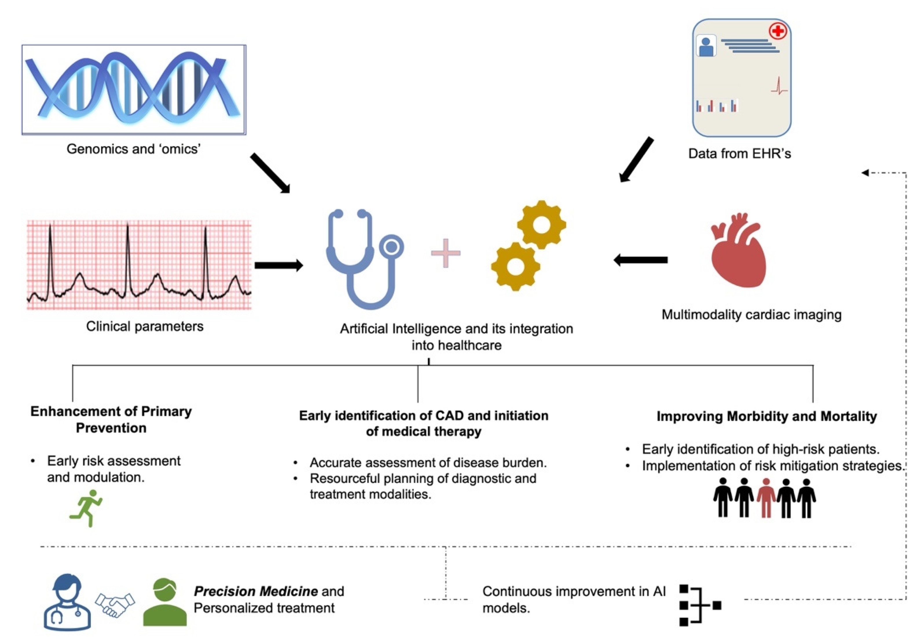
Artificial intelligence (AI) transforms cardiac diagnosis by automating the interpretation of complex datasets, detecting subtle physiological patterns invisible to the human eye, and providing real-time predictive analytics. By 2026, these technologies have shifted from experimental tools to essential clinical components, significantly improving the accuracy of arrhythmia detection, heart failure monitoring, and personalized risk stratification.
The integration of artificial intelligence in cardiology transforms diagnosis through three primary pillars: automated signal processing in electrocardiography (ECG), advanced computer vision for cardiac imaging, and predictive modeling using multi-omic and electronic health record (EHR) data. This evolution addresses a critical bottleneck in modern medicine—the overwhelming volume of diagnostic data—enabling clinicians to focus on high-level decision-making while AI handles the high-velocity screening and quantification tasks.
The Evolution of AI in Electrocardiography (ECG)
Electrocardiography remains the most common diagnostic tool in cardiology due to its low cost and accessibility. However, manual interpretation is subject to inter-observer variability and human fatigue. AI-enhanced ECG interpretation utilizes deep learning, specifically convolutional neural networks (CNNs), to identify arrhythmias such as atrial fibrillation (AFib) with precision that often exceeds that of general practitioners and matches expert rhythmologists.
Detecting Silent Atrial Fibrillation
By 2026, AI algorithms are routinely used to screen for "silent" or paroxysmal AFib. Traditional ECGs only provide a 10-second snapshot of cardiac rhythm, often missing intermittent arrhythmias. AI models trained on millions of hours of telemetry data can now identify subtle "signatures" in a patient’s normal sinus rhythm that indicate a high probability of future AFib episodes.
This predictive capability allows for early intervention, such as the initiation of anticoagulation therapy in high-risk patients, potentially preventing thousands of embolic strokes annually. The transformation lies in moving the ECG from a reactive diagnostic tool to a proactive screening instrument.
Screening for Structural Heart Disease
One of the most significant breakthroughs in AI-augmented cardiology is the ability to detect structural heart disease using a standard 12-lead ECG. Traditionally, conditions like left ventricular dysfunction (low ejection fraction) or hypertrophic cardiomyopathy required expensive and time-consuming echocardiograms for diagnosis.
AI models can now analyze the minute voltage changes and interval durations in an ECG to predict a low ejection fraction with high sensitivity. This serves as an effective "digital biomarker," flagging patients who require urgent follow-up imaging, thereby streamlining the diagnostic pathway and ensuring that specialized resources are directed toward those with the highest clinical need.
Reimagining Cardiac Imaging through Neural Networks
Cardiac imaging, including echocardiography, cardiac magnetic resonance imaging (CMR), and computed tomography (CT), has seen a massive influx of AI integration. The primary challenge in imaging has always been the manual quantification of chamber volumes, wall thickness, and blood flow, which is time-intensive and prone to measurement error.
Automated Quantification in Echocardiography
Echocardiography is the workhorse of cardiac diagnosis. AI software now provides real-time, automated measurement of the Left Ventricular Ejection Fraction (LVEF) and Global Longitudinal Strain (GLS). By 2026, these tools are embedded directly into ultrasound hardware.
When a sonographer places the probe on a patient, the AI automatically selects the best cardiac cycles, segments the endocardial borders, and calculates volumes instantaneously. This reduces the time required for a standard echo study by nearly 30% while ensuring that the measurements are consistent, regardless of which technician performs the scan.
Advanced Analysis in Cardiac MRI and CT
Cardiac MRI provides the most detailed view of myocardial tissue, but its complexity makes it difficult to interpret quickly. AI algorithms in 2026 are capable of automated tissue characterization, identifying areas of fibrosis (scarring) or edema (swelling) with granular detail.
In the realm of Cardiac CT, AI-driven Fractional Flow Reserve (FFR-CT) has transformed the diagnosis of coronary artery disease. Instead of invasive catheterization to determine if a blockage is restricting blood flow, AI analyzes the 3D geometry of the coronary arteries from a non-invasive CT scan to simulate flow dynamics. This "virtual" stress test has significantly reduced the number of unnecessary invasive procedures performed on patients with non-obstructive disease.
Predictive Analytics and Personalized Risk Stratification
The true power of AI in cardiology lies in its ability to synthesize disparate data points to predict future events. Modern cardiology departments now utilize AI-driven risk scores that incorporate data far beyond traditional factors like age, blood pressure, and cholesterol levels.
Integrating Social Determinants and Multi-Omics
By 2026, diagnostic platforms integrate genomic data, proteomic profiles, and social determinants of health (SDOH) into their predictive models. This "precision cardiology" approach allows for the identification of patients who may be genetically predisposed to certain types of heart failure, even if their current clinical presentation is normal.
AI models can process thousands of variables simultaneously to produce a "polygenic risk score." When combined with real-time data from wearable devices, these models can alert clinicians to a patient's worsening status days or weeks before they require hospitalization.
Reducing Heart Failure Readmissions
Heart failure management is one of the most significant burdens on healthcare systems globally. AI-driven remote monitoring platforms now analyze data from implanted devices (like pacemakers and ICDs) or external sensors to detect early signs of fluid overload. By identifying subtle changes in thoracic impedance or heart rate variability, AI can trigger a diagnostic alert, allowing physicians to adjust medications remotely and prevent an acute decompensation.
Technical Infrastructure: Integration into Clinical Workflows
For AI to effectively transform diagnosis, it must be invisible and frictionless. In 2026, the focus has shifted from the algorithms themselves to the infrastructure that supports them.
The primary technical requirements for AI integration in a modern cardiology practice include:
- Interoperability Standards: Robust implementation of HL7 FHIR (Fast Healthcare Interoperability Resources) ensures that AI tools can "read" and "write" directly to the Electronic Health Record (EHR).
- Edge Computing: Many diagnostic devices now perform AI inference locally ("on the edge") to ensure real-time results without the latency associated with cloud processing.
- Data Harmonization: Systems that can standardize data from different manufacturers (e.g., merging ECG data from GE and Philips systems) are essential for consistent AI performance.
Comparison of Diagnostic Methods: Traditional vs. AI-Augmented
| Feature | Traditional Manual Diagnosis | AI-Augmented Diagnosis (2026) |
|---|---|---|
| Speed of Analysis | Minutes to hours (requires human review) | Seconds (automated pre-interpretation) |
| Consistency | Variable (inter-observer differences) | High (standardized algorithmic output) |
| Data Scope | Focused on primary diagnostic signal | Integrates signals, EHR, and genomics |
| Predictive Capability | Reactive (identifies existing disease) | Proactive (identifies future risk) |
| Workflow Impact | High manual burden for clinicians | Automates routine quantification |
| Error Rate | Prone to fatigue and oversight | High sensitivity; requires human verification |
Navigating the Challenges: Bias, Ethics, and the "Black Box"
While the benefits are profound, the transformation of cardiac diagnosis through AI is not without significant hurdles. The "black box" nature of some deep learning models—where it is difficult to understand why an algorithm reached a specific conclusion—remains a point of contention among clinicians.
Algorithmic Bias and Data Diversity
A major concern in 2026 is ensuring that AI models are trained on diverse datasets. If an algorithm is trained primarily on data from one demographic group, its diagnostic accuracy may falter when applied to others. Regulatory bodies like the FDA and EMA now require manufacturers to demonstrate "algorithmic equity," showing that their tools perform consistently across different ethnicities, genders, and socioeconomic backgrounds.
The Role of the Physician in the AI Era
AI is not intended to replace cardiologists; rather, it acts as a "force multiplier." The physician's role is shifting from a data-gatherer to a data-interpreter and counselor. While the AI can identify a 92% probability of a specific cardiomyopathy, the cardiologist must still integrate that finding with the patient's lifestyle, preferences, and overall clinical context to determine the best course of action.
Implementation and Troubleshooting for Clinical Practices
Adopting AI in a clinical setting requires a structured approach to avoid common pitfalls. Practices looking to integrate these tools should consider the following steps:
- Needs Assessment: Identify specific bottlenecks, such as a backlog in ECG readings or high variability in echocardiogram reports.
- Vendor Verification: Ensure the AI tool has cleared relevant regulatory hurdles (e.g., FDA 510(k) clearance) specifically for the intended diagnostic use.
- Pilot Testing: Implement the tool in a "shadow" mode initially, comparing AI outputs with human interpretation to build clinician trust.
- Continuous Monitoring: AI models can suffer from "data drift" as clinical populations change. Regular audits are necessary to ensure the algorithm's accuracy remains stable over time.
Common Mistakes to Avoid
- Over-Reliance: Trusting an AI diagnosis without reviewing the underlying raw data (e.g., the actual ECG tracing).
- Ignoring Integration: Buying "standalone" software that does not connect to the PACS or EHR, leading to fragmented workflows.
- Neglecting Staff Training: Assuming sonographers and nurses do not need to understand how the AI works. Education is vital for effective troubleshooting.
Frequently Asked Questions
Can AI replace a cardiologist for diagnosing heart attacks?
No. While AI can analyze an ECG and suggest the presence of an ST-elevation myocardial infarction (STEMI) with high accuracy, the diagnosis of a heart attack involves clinical symptoms, cardiac biomarkers (like troponin), and physical examination. AI serves as a powerful triage tool to alert the medical team faster, but the final diagnostic and therapeutic decisions remain with the clinician.
Is my heart data safe when processed by AI?
Data privacy is a cornerstone of AI integration in 2026. Most clinical AI tools use de-identified data for processing, and many operate within the hospital’s secure firewall (on-premises or private cloud). Standards such as HIPAA and GDPR strictly govern how this data is handled, ensuring that patient identifiers are stripped before any analysis occurs for research or model training purposes.
Does AI increase the cost of cardiac care?
Initially, the implementation of AI requires an investment in software and infrastructure. However, in the long term, AI is designed to reduce costs by improving diagnostic efficiency, reducing the length of hospital stays through early detection, and preventing unnecessary invasive procedures. Many healthcare systems now view AI as a cost-saving measure due to its ability to prevent expensive complications like stroke and advanced heart failure.
How does AI handle "gray area" or ambiguous diagnostic results?
Advanced AI models in 2026 are designed to provide a confidence score. If an ECG or image is of poor quality or if the findings are borderline, the AI will flag the result as "low confidence" and prompt an immediate review by a human expert. This ensures that the most difficult cases receive the highest level of human scrutiny.
Can wearables like smartwatches provide medical-grade cardiac diagnosis?
While consumer wearables have become highly sophisticated, they are generally classified as "screening" tools rather than "diagnostic" tools. If a smartwatch detects a rhythm abnormality, it provides an alert that must be confirmed by a clinical-grade 12-lead ECG interpreted by a healthcare professional or a medically cleared AI algorithm.
Strategic Recommendation for Healthcare Providers
To leverage the fact that artificial intelligence in cardiology transforms diagnosis, healthcare organizations should move toward a centralized AI-diagnostic hub. This involves consolidating AI tools across different modalities (ECG, Echo, CT) into a single platform that feeds directly into the clinician's workspace. By reducing the number of different interfaces a doctor must use, the clinic can maximize the efficiency gains and accuracy improvements that AI offers.
Clinicians should prioritize the adoption of AI tools that provide "explainable" results—those that highlight the specific areas of an image or segments of an ECG that led to the diagnosis. This transparency is crucial for maintaining clinical standards and ensuring that the final diagnosis is a collaborative effort between human expertise and machine intelligence.