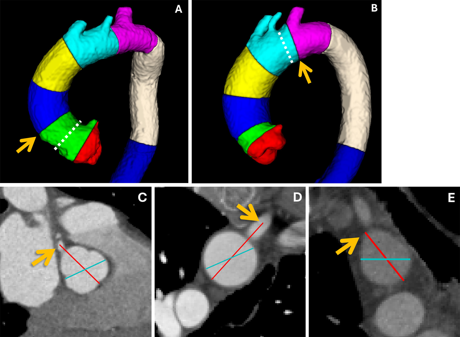
Aortic imaging in clinical practice is the cornerstone of diagnosing, monitoring, and managing life-threatening vascular conditions such as aneurysms, dissections, and intramural hematomas. By utilizing advanced modalities like CT angiography, 4D flow MRI, and echocardiography, clinicians can precisely measure vessel diameter, assess hemodynamic stress, and plan complex endovascular or surgical interventions to prevent catastrophic aortic rupture.
The Essential Role of Aortic Imaging in Modern Medicine
As we navigate the clinical landscape of 2026, aortic imaging has transitioned from a purely diagnostic tool to a comprehensive prognostic framework. The aorta, the body's primary conduit for oxygenated blood, is subject to immense mechanical stress. Structural failures within its walls often remain asymptomatic until a critical event occurs. Consequently, the primary goal of imaging is the early identification of pathology and the meticulous tracking of disease progression.
Clinical practice relies on imaging to answer three fundamental questions: Is the aorta dilated or structurally compromised? What is the immediate risk of rupture or dissection? And, what is the optimal timing and method for intervention? To answer these questions, radiologists and cardiologists employ a multi-modal approach, tailoring the imaging strategy to the patient’s clinical stability, renal function, and the specific segment of the aorta under investigation.
The integration of artificial intelligence (AI) into imaging workflows has further refined this process. In 2026, automated segmentation and volumetric analysis have largely replaced manual "ruler" measurements, reducing inter-observer variability and providing a more accurate assessment of the total aortic burden. This precision is vital, as even a few millimeters of growth can be the difference between conservative management and high-risk surgery.
Comparative Overview of Imaging Modalities
Selecting the appropriate imaging modality requires a balance of speed, accuracy, and patient safety. While some technologies offer superior spatial resolution, others provide functional data regarding blood flow and wall tension.
Computed Tomography Angiography (CTA): The Gold Standard
Computed Tomography Angiography (CTA) remains the first-line imaging modality for nearly all suspected aortic pathologies. Its widespread availability and rapid acquisition times make it indispensable in emergency departments, particularly for diagnosing acute aortic syndromes (AAS).
Modern CTA protocols in 2026 utilize ECG-gating, which synchronizes image acquisition with the cardiac cycle. This is critical for imaging the ascending aorta, as it eliminates motion artifacts caused by the heart’s pulsation. Without gating, a pulsatile aorta can mimic a dissection flap on CT, leading to a false-positive diagnosis. Furthermore, dual-energy CT (DECT) has become more common, allowing for the creation of virtual non-contrast images, which reduces the total radiation dose and helps distinguish between iodine contrast and intramural hemorrhage.
Magnetic Resonance Angiography (MRA): Precision Without Radiation
Magnetic Resonance Angiography (MRA) serves as an excellent alternative for patients requiring long-term surveillance, such as those with Marfan syndrome or bicuspid aortic valve disease. The primary advantage of MRA is the absence of ionizing radiation, which is a significant concern for younger patients who may require dozens of scans over their lifetime.
Technological leaps have made non-contrast MRA more reliable. By using techniques like balanced steady-state free precession (bSSFP), clinicians can visualize the aortic anatomy without the need for gadolinium-based contrast agents, which is beneficial for patients with advanced chronic kidney disease. However, MRA remains limited by its longer acquisition times and incompatibility with certain metallic implants, making it less ideal for unstable patients in the acute phase.
Echocardiography and Transesophageal Echo (TEE)
Echocardiography provides real-time, bedside assessment of the proximal aorta. Transthoracic echocardiography (TTE) is often the initial screening tool used to evaluate the aortic root and the proximal ascending aorta. However, TTE is limited by the "blind spot" created by the air-filled trachea and bronchi, which obscures the distal ascending aorta and the arch.
In contrast, Transesophageal Echocardiography (TEE) offers high-resolution images of the entire thoracic aorta by placing the transducer in the esophagus. In 2026, TEE remains a critical tool in the operating room during aortic repairs and in the emergency assessment of hemodynamically unstable patients who are too sick to be transported to the CT suite. It is particularly effective at identifying small entry tears in aortic dissections and assessing the competence of the aortic valve.
Clinical Indications and Diagnostic Pathways
The choice of imaging is dictated by the clinical presentation. Understanding the specific requirements for different aortic diseases ensures that patients receive the most accurate diagnosis with minimal delay.
Acute Aortic Syndromes (AAS)
Acute aortic syndromes, including Aortic Dissection (AD), Intramural Hematoma (IMH), and Penetrating Atherosclerotic Ulcer (PAU), represent surgical emergencies. The diagnostic pathway is focused on rapid identification and classification using the Stanford system:
- Stanford Type A: Involves the ascending aorta; usually requires immediate surgical intervention.
- Stanford Type B: Involves only the descending aorta; often managed medically or with endovascular stents.
For AAS, CTA is the preferred modality due to its speed. A high-quality scan must extend from the neck to the mid-thighs to assess for malperfusion of the carotid, celiac, mesenteric, and iliac arteries.
Chronic Management of Abdominal Aortic Aneurysms (AAA)
For asymptomatic Abdominal Aortic Aneurysms (AAA), imaging is used for screening and surveillance. Ultrasound is the primary tool for initial screening in high-risk populations (men over 65 with a history of smoking). Once an aneurysm reaches a diameter of 4.0–4.5 cm, the frequency of imaging increases.
When the aneurysm approaches the intervention threshold—typically 5.5 cm in men and 5.0 cm in women—a thin-slice CTA is performed to assess the anatomy for Endovascular Aneurysm Repair (EVAR). Clinicians must evaluate the "landing zones" (the healthy segments of the aorta above and below the aneurysm) and the tortuosity of the iliac arteries to ensure a stent-graft can be safely deployed.
Post-Surgical Surveillance and Endoleaks
Following EVAR or open repair, life-long surveillance is mandatory to detect complications such as endoleaks, where blood continues to flow into the aneurysm sac despite the presence of a stent. In 2026, Contrast-Enhanced Ultrasound (CEUS) has gained popularity for this purpose. CEUS uses microbubble contrast agents to detect low-flow endoleaks that might be missed on standard CT, offering a radiation-free alternative for routine follow-ups.
Technical Specifications and Measurement Protocols
Consistency in measurement is the most critical factor in aortic imaging. Discrepancies in how the aorta is measured can lead to inappropriate clinical decisions. The current standard in 2026 emphasizes the "Double-Oblique" technique. Since the aorta is often tortuous, a standard axial slice may cut the vessel at an angle, resulting in an oval shape and an overestimation of the diameter. The double-oblique method uses multiplanar reconstruction to ensure the measurement is taken exactly perpendicular to the long axis of the blood flow.
Measurement Standards
| Feature | Preferred Method | Clinical Significance |
|---|---|---|
| Dimension | Outer-to-Outer wall | Standardizes measurement across different modalities (CT and MRI). |
| Plane | Perpendicular to center-line | Prevents overestimation due to vessel tortuosity. |
| Timing | Diastolic phase (in gated CT) | Provides the most stable and reproducible diameter. |
| Growth Rate | Volumetric analysis | AI-assisted volume tracking is more sensitive than diameter changes alone. |
Clinicians must also document the presence of mural thrombus (clotting along the wall) and calcification, as these factors influence both the risk of embolization and the success of endovascular procedures.
The Impact of Artificial Intelligence in 2026 Imaging
The role of Artificial Intelligence in aortic imaging has evolved from a research curiosity to a clinical necessity. AI algorithms are now routinely used for automated triage. When a CTA is performed in a remote hospital, AI can scan the raw data for signs of a dissection flap or a ruptured aneurysm and immediately alert the vascular surgery team at a regional center, potentially saving hours of waiting for a radiologist's formal report.
Moreover, "Radiomics"—the extraction of quantitative data from medical images—allows AI to analyze the texture and density of the aortic wall. In 2026, these tools help identify "vulnerable" aneurysms—those that are likely to rupture even if they haven't met the traditional size criteria for surgery. This shift toward biological rather than purely anatomical markers is the new frontier of personalized vascular medicine.
Emerging Roles: PET-CT and 4D Flow MRI
While CTA and MRA provide structural details, newer modalities offer insights into the inflammatory and hemodynamic state of the aorta.
- PET-CT (Positron Emission Tomography): Using FDG (fluorodeoxyglucose) tracers, PET-CT can identify active inflammation within the aortic wall. This is particularly useful in diagnosing Aortitis (inflammation of the aorta) and identifying infected aneurysms or vascular grafts.
- 4D Flow MRI: This advanced technique captures blood flow patterns in three dimensions over the cardiac cycle (the fourth dimension is time). It allows clinicians to visualize turbulence and calculate Wall Shear Stress (WSS). High WSS is increasingly recognized as a predictor of where a dissection might expand or where a new aneurysm might form, providing a map for preventative treatment.
Comparative Table: Choosing the Right Modality
| Modality | Speed | Spatial Resolution | Radiation | Best For |
|---|---|---|---|---|
| CTA (Gated) | Very High | Excellent (<1mm) | Yes | Acute dissection, surgical planning, EVAR follow-up. |
| MRA | Moderate | Good | No | Long-term surveillance, bicuspid valve, renal sparing. |
| TTE | High | Limited | No | Bedside screening, aortic root assessment. |
| TEE | High | Excellent | No | Intraoperative monitoring, unstable patients. |
| Ultrasound | High | Fair | No | AAA screening, monitoring small aneurysms. |
Cost-Effectiveness and Resource Allocation
In the healthcare environment of 2026, the cost-effectiveness of aortic imaging is measured not just by the price of the scan, but by the "downstream" savings of preventing a rupture. An emergency aortic repair is significantly more expensive and has a higher mortality rate than an elective, planned procedure.
Standardized imaging protocols reduce costs by eliminating redundant tests. For example, if a patient receives a high-quality, gated CTA at a community hospital, that data can be digitally transferred to a tertiary center, preventing the need for a repeat scan. Furthermore, the use of ultrasound for routine AAA monitoring remains the most cost-effective strategy for population health, provided the sonographers follow standardized measurement protocols to ensure data reliability.
Challenges and Future Directions
Despite technological advancements, several challenges persist in aortic imaging. The management of "borderline" aneurysms (e.g., a 5.2 cm aneurysm in a healthy man) remains a point of clinical debate. Imaging can provide the size, but it cannot yet perfectly predict the precise moment of wall failure.
Future developments are focusing on Molecular Imaging, where contrast agents are designed to bind to specific proteins involved in wall degradation, such as matrix metalloproteinases (MMPs). Visualizing these proteins would allow doctors to see the disease process at a molecular level before any structural change occurs. Additionally, the move toward "Virtual Reality" (VR) surgical planning, where surgeons can "walk through" a patient’s 3D-rendered aorta before making the first incision, is becoming standard in high-volume vascular centers.
FAQ
What is the most accurate way to measure an aortic aneurysm?
In 2026, the gold standard is the double-oblique perpendicular measurement using ECG-gated CTA. This method ensures that the diameter is measured across the true cross-section of the vessel, preventing the overestimation that occurs when the aorta is measured on a standard axial slice. Automated AI-based volumetric analysis is also increasingly used to provide a more comprehensive view of aneurysm growth.
Why is ECG-gating important for thoracic aortic imaging?
The heart’s contraction causes the ascending aorta to move significantly. In a standard CT scan, this motion creates "blurring" or double-images that can look exactly like a dissection flap. ECG-gating times the X-ray pulses to specific moments in the cardiac cycle (usually mid-diastole) when the aorta is most still, ensuring a crystal-clear image of the vessel wall.
Can I have an aortic scan if I have poor kidney function?
Yes, though it requires specific precautions. For CT, clinicians may use low-volume contrast protocols or pre-scan hydration. Alternatively, non-contrast MRA techniques (like bSSFP) can provide excellent images of the aorta without the need for iodine or gadolinium, making it the preferred choice for patients with severe renal impairment.
Is ultrasound enough to monitor an abdominal aortic aneurysm?
For small aneurysms (under 4.0 cm), ultrasound is usually sufficient for monitoring. However, as the aneurysm grows or if the anatomy is complex, a CTA is required. Ultrasound can sometimes be limited by bowel gas or patient obesity, and it does not provide enough detail for surgical or endovascular planning.
What is 4D Flow MRI, and do I need it?
4D Flow MRI is an advanced scan that visualizes the direction and velocity of blood flow within the aorta. While not necessary for every patient, it is highly valuable for those with complex conditions like bicuspid aortic valves or chronic dissections. It helps doctors understand how the blood flow is stressing the aortic wall, which can help predict if an aneurysm will grow more quickly.
Practical Recommendation
For clinicians, the priority should be the implementation of standardized, ECG-gated protocols for all thoracic aortic imaging to ensure diagnostic accuracy. For patients, particularly those with a family history of aortic disease or a history of smoking, proactive screening via ultrasound is the most effective way to detect silent aneurysms. If a diagnosis is made, ensure that follow-up imaging is performed at the same facility using the same modality when possible, as this allows for the most accurate "apples-to-apples" comparison of aortic growth over time. In 2026, the shift toward AI-assisted volumetric tracking offers the most sensitive way to monitor these changes and should be utilized where available.