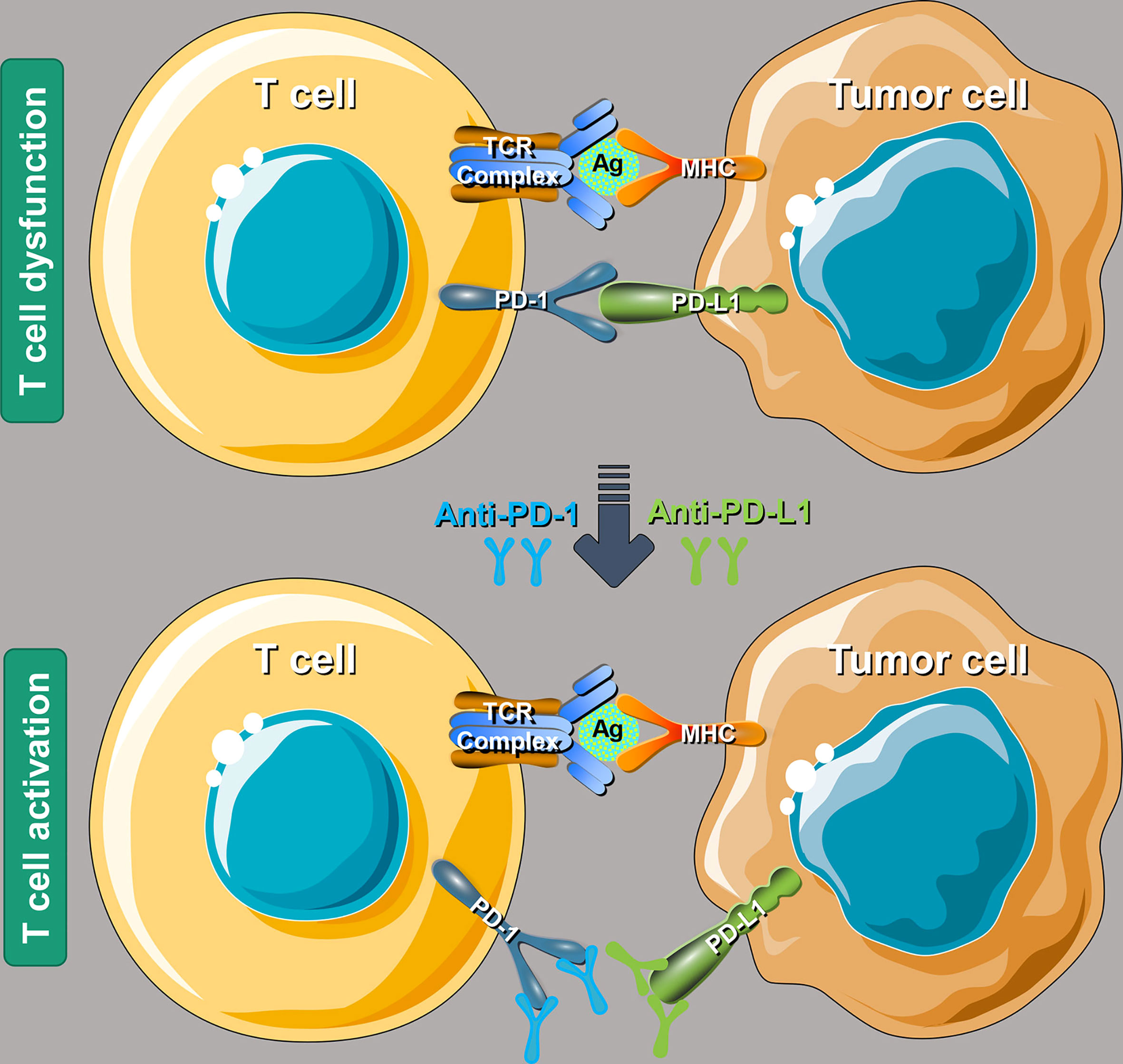
Anti-PD-1 antibodies are a class of immune checkpoint inhibitors that block the interaction between the PD-1 receptor on T cells and its ligands (PD-L1/PD-L2) on tumor cells. By preventing this immune-evasion mechanism, these therapies reactivate the body’s cytotoxic T cells to recognize and destroy cancer cells across diverse malignancies.
The Molecular Basis of PD-1 Inhibition
Programmed Cell Death Protein 1 (PD-1) is a type I transmembrane protein originally identified in 1992. It belongs to the CD28/CTLA-4 family of T-cell regulators. In a healthy physiological state, the PD-1 pathway acts as a critical "brake" on the immune system, preventing overactivation and maintaining self-tolerance to avoid autoimmune destruction of healthy tissues. The receptor is primarily expressed on the surface of activated T cells, B cells, and natural killer (NK) cells.
When PD-1 binds to its ligands—Programmed Death-Ligand 1 (PD-L1) or Programmed Death-Ligand 2 (PD-L2)—it triggers an inhibitory signal through its intracellular immunoreceptor tyrosine-based inhibitory motif (ITIM) and immunoreceptor tyrosine-based switch motif (ITSM). This signaling cascade recruits phosphatases like SHP-2, which dephosphorylate key elements of the T-cell receptor (TCR) signaling pathway. The result is a reduction in T-cell proliferation, cytokine production (such as IFN-gamma and IL-2), and survival.
Cancer cells exploit this regulatory mechanism. By upregulating PD-L1 on their surface—often in response to inflammatory signals in the tumor microenvironment—malignant cells can "shut off" infiltrating T cells, leading to a state known as T-cell exhaustion. Anti-PD-1 antibodies are monoclonal antibodies engineered to bind with high affinity to the PD-1 receptor, physically blocking the ligand interaction and allowing the T cell to regain its cytotoxic functionality.
Pharmacological Landscape of PD-1 Inhibitors
As of 2026, the pharmacological landscape has matured significantly since the initial approvals of nivolumab and pembrolizumab in 2014. These agents are typically designed as humanized or fully human IgG4 monoclonal antibodies. The choice of the IgG4 isotype is intentional; unlike IgG1, IgG4 has a low affinity for C1q and Fc-gamma receptors, which minimizes antibody-dependent cellular cytotoxicity (ADCC) or complement-dependent cytotoxicity (CDC) against the very T cells the drug is intended to activate.
Key Therapeutic Agents in 2026
The following table provides a comparison of the most widely utilized anti-PD-1 antibodies in clinical practice.
| Drug Name | Generic Name | Manufacturer | Primary Indications (Examples) |
|---|---|---|---|
| Keytruda | Pembrolizumab | Merck & Co. | Melanoma, NSCLC, HNSCC, MSI-H/dMMR, RCC |
| Opdivo | Nivolumab | Bristol Myers Squibb | Melanoma, NSCLC, RCC, Hodgkin Lymphoma |
| Libtayo | Cemiplimab | Regeneron/Sanofi | CSCC, BCC, NSCLC |
| Jemperli | Dostarlimab | GSK | dMMR Endometrial Cancer, Rectal Cancer |
| Zynyz | Retifanlimab | Incyte | Merkel Cell Carcinoma |
| Loqtorzi | Toripalimab | Coherus/Junshi | Nasopharyngeal Carcinoma |
While these drugs share the same fundamental mechanism, they differ in their binding epitopes on the PD-1 molecule, their binding affinities, and their half-lives. For instance, pembrolizumab and nivolumab bind to distinct regions of the PD-1 N-terminal loop. By 2026, the introduction of subcutaneous formulations for many of these agents has revolutionized administration, reducing clinic time from hours to minutes and improving patient quality of life.
Clinical Indications and Therapeutic Expansion
The reach of anti-PD-1 therapy has expanded from palliative, late-line treatments for metastatic disease to first-line standards of care and, increasingly, adjuvant and neoadjuvant settings.
Solid Tumors
In non-small cell lung cancer (NSCLC), anti-PD-1 therapy is a cornerstone of treatment. Patients with high PD-L1 expression (TPS ≥ 50%) often receive pembrolizumab monotherapy, while those with lower expression typically receive a combination of anti-PD-1 and platinum-doublet chemotherapy. In melanoma, these agents have transformed a previously terminal diagnosis into a manageable chronic condition for many, with five-year survival rates exceeding 50% when used in combination with anti-CTLA-4 agents.
Hematological Malignancies
While checkpoint inhibitors are predominantly associated with solid tumors, they have shown remarkable efficacy in classical Hodgkin lymphoma (cHL). Patients with cHL often harbor genetic alterations at chromosome 9p24.1, leading to constitutive over-expression of PD-L1 and PD-L2. Anti-PD-1 antibodies provide high response rates in patients who have failed autologous stem cell transplantation and brentuximab vedotin.
The Shift to Earlier Intervention
A major trend in 2026 is the use of anti-PD-1 antibodies in the neoadjuvant (pre-surgical) setting. Data from trials in lung, breast, and colon cancers indicate that administering immunotherapy while the primary tumor is still intact allows for a more robust systemic immune response, potentially eliminating micro-metastases and improving pathological complete response (pCR) rates.
Biomarkers for Patient Selection
Not all patients respond to anti-PD-1 therapy. Identifying those most likely to benefit is a primary goal of precision oncology. Several biomarkers are currently used in clinical decision-making.
- PD-L1 Expression: Measured via immunohistochemistry (IHC), this remains the most common biomarker. It is often reported as a Tumor Proportion Score (TPS) or a Combined Positive Score (CPS), which includes tumor cells, lymphocytes, and macrophages.
- Microsatellite Instability (MSI-H) and dMMR: Tumors with DNA mismatch repair deficiencies accumulate thousands of mutations, creating "neoantigens" that the immune system can easily recognize once the PD-1 brake is removed. In 2017, the FDA granted the first tissue-agnostic approval for pembrolizumab in MSI-H/dMMR solid tumors.
- Tumor Mutational Burden (TMB): A high number of mutations per megabase (mut/Mb) of DNA is often correlated with better response rates to checkpoint inhibition, regardless of PD-L1 status.
- Gene Expression Profiling (GEP): Emerging in 2026, "inflamed" gene signatures are being used to predict response more accurately than IHC alone by evaluating the presence of interferon-gamma-related mRNA.
Managing Immune-Related Adverse Events (irAEs)
Because anti-PD-1 antibodies nonspecifically activate the immune system, they can induce a unique spectrum of side effects known as immune-related adverse events (irAEs). These are essentially "autoimmune-like" reactions where the immune system attacks healthy organs.
Common irAEs include:
- Dermatologic: Rash, pruritus, and vitiligo (often a positive prognostic sign in melanoma).
- Gastrointestinal: Immune-mediated colitis, characterized by diarrhea and abdominal pain.
- Endocrine: Hypophysitis, thyroiditis, and Type 1 diabetes mellitus. Unlike many other irAEs, endocrine toxicities are often permanent and require lifelong hormone replacement.
- Pulmonary: Pneumonitis, which can be life-threatening if not identified early.
Management of irAEs follows a standardized grading system (CTCAE). Grade 1 events often allow for continued treatment with close monitoring. Grade 2 events usually require withholding the drug and initiating low-dose corticosteroids. Grade 3 and 4 events typically require permanent discontinuation and high-dose systemic immunosuppression with agents like methylprednisolone, infliximab, or mycophenolate mofetil.
Resistance Mechanisms and the 2026 Outlook
Despite the success of anti-PD-1 therapy, primary resistance (no response) and acquired resistance (relapse after initial response) remain significant challenges. Primary resistance is often linked to an "immune-cold" tumor microenvironment, characterized by a lack of T-cell infiltration or the presence of immunosuppressive cells like myeloid-derived suppressor cells (MDSCs) and regulatory T cells (Tregs).
Acquired resistance may occur through several pathways:
- Loss of HLA Expression: The tumor "hides" by downregulating the machinery needed to present antigens to T cells.
- Alternative Checkpoints: Tumors may upregulate other inhibitory receptors such as LAG-3, TIGIT, or TIM-3.
- IFN-gamma Signaling Mutations: Defects in the JAK/STAT pathway can render tumor cells insensitive to the killing effects of the immune system.
To combat resistance, 2026 has seen the rise of "next-generation" combinations. This includes dual-checkpoint inhibition (e.g., Nivolumab + Relatlimab for LAG-3) and the pairing of anti-PD-1 agents with Antibody-Drug Conjugates (ADCs). The goal is to use the ADC to kill a portion of tumor cells and release antigens, which then "primes" the immune system for the anti-PD-1 agent to work more effectively.
Frequently Asked Questions
What is the difference between PD-1 and PD-L1 inhibitors?
Anti-PD-1 antibodies (like pembrolizumab) bind to the receptor on the T cell itself. Anti-PD-L1 antibodies (like atezolizumab) bind to the ligand on the tumor cell or surrounding immune cells. While both disrupt the same pathway, anti-PD-1 agents block interactions with both PD-L1 and PD-L2, whereas anti-PD-L1 agents only block the PD-L1 interaction but allow the PD-1/PD-L2 interaction to remain intact, which some theorize may reduce certain side effects like pneumonitis.
How long does a patient stay on anti-PD-1 therapy?
In the metastatic setting, the standard of care has historically been two years of treatment, provided the disease does not progress and side effects remain manageable. However, by 2026, "treatment holidays" and monitoring via circulating tumor DNA (ctDNA) are increasingly used to determine if a patient with a complete response can safely stop therapy earlier.
Can anti-PD-1 antibodies be used for all types of cancer?
No. While many cancers respond, some "cold" tumors like pancreatic cancer and certain types of prostate cancer have shown very limited response to PD-1 monotherapy. These require combination strategies to recruit immune cells into the tumor before the checkpoint inhibitor can be effective.
Are the side effects of immunotherapy worse than chemotherapy?
Generally, anti-PD-1 therapy is better tolerated than traditional cytotoxic chemotherapy. Patients do not typically experience hair loss, severe nausea, or significant myelosuppression. However, the nature of the side effects is different; irAEs can occur at any time (even months after stopping treatment) and can be more unpredictable than chemotherapy side effects.
What should I do if I experience a side effect during treatment?
It is vital to report any new symptoms—especially cough, diarrhea, or extreme fatigue—to your oncology team immediately. Early intervention with corticosteroids can often reverse an irAE quickly, whereas delaying treatment can lead to severe organ damage or the need to stop immunotherapy permanently.
Practical Recommendations for 2026
For clinicians and patients navigating the use of anti-PD-1 antibodies in 2026, the priority remains balanced between maximizing efficacy and minimizing long-term toxicity.
- Prioritize Biomarker Testing: Always ensure comprehensive genomic profiling and PD-L1 IHC are performed before initiating first-line therapy to determine if monotherapy, combination therapy, or a different modality is most appropriate.
- Early irAE Recognition: Maintain a high index of suspicion for immune-mediated toxicities. Patient education regarding the specific symptoms of pneumonitis and colitis is essential for safety.
- Consider Clinical Trials for Non-Responders: For patients who do not respond to initial PD-1 blockade, 2026 offers numerous trials focusing on triplet therapies and personalized neoantigen vaccines that may overcome resistance.
- Logistical Optimization: Where available, utilize subcutaneous administration to reduce the burden on both infusion centers and patients' daily lives.
For more information on current guidelines and approved indications, consult the National Cancer Institute or the American Society of Clinical Oncology (ASCO).