The leg raise test for appendicitis, known clinically as the psoas sign or Cope's psoas test, is a physical examination maneuver used to evaluate lower right abdominal pain. Pain triggered by raising or extending the right leg suggests that an inflamed appendix is contacting the underlying psoas muscle, causing peritoneal irritation.
Understanding the Leg Raise Test for Appendicitis (The Psoas Sign)
When individuals experience acute pain in the lower right quadrant of the abdomen, healthcare providers perform targeted physical examination maneuvers to identify the anatomical source. Among these, the leg raise test—medically referred to as the psoas sign—is one of the most recognized bedside clinical tests for suspected acute appendicitis.
The appendix is a small, finger-shaped pouch attached to the cecum (the beginning of the large intestine) located in the right lower abdomen. When obstruction of the appendiceal lumen occurs due to fecal material, lymphoid hyperplasia, or foreign bodies, inflammation develops rapidly. As swelling progresses, the inflamed tissue rubs against surrounding structures within the abdominal cavity.
The leg raise test relies on the anatomical proximity of the appendix to the psoas major muscle, a large muscle extending from the lower spine across the pelvis to the upper thighbone (femur). By contracting or stretching this muscle through specific leg movements, clinicians can determine whether the movement provokes irritation in the right lower abdominal quadrant.
It is critical to recognize that while physical exam maneuvers like the psoas sign provide valuable clinical clues, they are diagnostic components rather than definitive standalone tests. A positive leg raise test strongly points toward peritoneal irritation, but a negative test never rules out appendicitis.
Anatomical Basis: Why Leg Movement Triggers Abdominal Pain
To understand why a simple movement of the leg can elicit abdominal pain, one must examine the spatial relationship between the abdominal wall, the appendix, and the posterior musculoskeletal structures.
The Anatomy of the Psoas Major Muscle
The psoas major muscle originates from the transverse processes and vertebral bodies of the twelfth thoracic (T12) through the fifth lumbar (L5) vertebrae. It travels downward along the back wall of the abdominal cavity (the retroperitoneum), passes under the inguinal ligament, and inserts into the lesser trochanter of the femur. The primary functions of the psoas major are:
- Hip flexion: Lifting the thigh upward toward the torso.
- Postural stabilization: Maintaining trunk alignment while standing, sitting, and walking.
Anatomical Variations of the Appendix
The anatomical position of the appendix varies significantly among individuals:
- Anterior / Pelvic Appendix: Positioned freely over the anterior pelvic brim or hanging into the pelvis.
- Retrocecal Appendix: Positioned behind the cecum along the retroperitoneum, lying directly over or immediately adjacent to the right psoas major muscle. Studies published in anatomical literature show that approximately 65% of individuals have a retrocecal appendix placement.
- Posterior Abdominal Wall:
- Lumbar Vertebrae L1-L5
- Psoas Major Muscle (runs behind abdominal organs)
- Inflamed Retrocecal Appendix (direct contact when inflamed; irritates overlaying peritoneum during muscle stretch or contraction)
The Mechanism of Friction and Peritoneal Irritation
The abdominal cavity is lined by a thin, sensitive membrane called the parietal peritoneum, which contains a high density of somatic pain receptors. When an appendix becomes acutely inflamed (acute appendicitis), the inflammation spreads to its outer coating and the adjacent parietal peritoneum covering the psoas muscle.
When a patient performs a leg raise or undergoes passive hip extension:
- The right psoas major muscle contracts (during active flexion) or stretches (during passive extension).
- The muscle belly shifts in size and orientation, directly compressing or rubbing against the inflamed appendix and parietal peritoneum.
- This friction triggers somatic pain signals transmitted via spinal nerves, manifesting as sharp, localized pain in the right lower quadrant (RLQ).
How the Psoas Sign Test Is Performed
In a clinical setting, healthcare providers perform the leg raise test using two distinct variations: the passive extension method and the active straight-leg raise method. Both aim to alter the tension of the psoas muscle, but they approach the movement differently.
- Passive Psoas Sign Test (Patient on Left Side, Hip Extended):
- Torso
- Hip Joint
- Clinician gently pulls leg back
- Active Psoas Sign Test (Patient Supine, Leg Raised Against Resistance):
- Head / Torso
- Hip Joint
- Patient lifts right leg against clinician's hand
1. Passive Extension (Standard Clinical Technique)
The passive technique is the classical method described in medical literature:
- Positioning: The patient lies on their left side (left lateral decubitus position) with their legs slightly bent at the knees.
- Execution: The clinician stabilizes the patient's hip with one hand and gently takes hold of the patient's right leg with the other. The clinician then slowly extends the right hip by pulling the straight leg backward behind the plane of the torso.
- Mechanism: This action stretches the psoas major muscle across the retroperitoneum.
- Positive Result: Increased or sharp pain felt in the right lower quadrant of the abdomen during backward extension.
2. Active Straight-Leg Raise (Resistance Technique)
The active technique relies on muscle contraction rather than muscle stretching:
- Positioning: The patient lies flat on their back (supine position) on the examination table.
- Execution: The patient is instructed to keep their right leg straight and lift it off the table. The clinician places a hand on the patient's right thigh just above the knee and applies downward resistance while the patient attempts to flex the hip upward.
- Mechanism: Active contraction of the psoas major against resistance exerts direct mechanical force against the retrocecal tissue.
- Positive Result: Reproduction or aggravation of sharp pain in the right lower abdomen.
Interpreting Pain Responses: True Positive vs. Musculoskeletal Pain
It is vital to distinguish between abdominal pain caused by peritoneal inflammation and hip or lower back discomfort caused by joint or muscle strain:
- True Positive Psoas Sign: Sharp, focal pain deep in the right lower abdomen (McBurney's point region) triggered specifically by psoas movement.
- False Positive / Non-Appendiceal Response: Dull aching in the lower back, discomfort inside the hip joint, or pain in the groin muscle, which typically indicates muscular tightness, lumbar radiculopathy, or hip osteoarthritis rather than appendicitis.
Diagnostic Accuracy: Sensitivity and Specificity of Physical Exams
No physical exam maneuver for appendicitis is completely accurate when used in isolation. Clinical accuracy in diagnostic medicine is measured by two key parameters: sensitivity (the ability of a test to correctly identify those with the disease) and specificity (the ability of a test to correctly identify those without the disease).
Medical research published via the National Center for Biotechnology Information (NCBI) evaluates the psoas sign as follows:
- Sensitivity (14% – 42%): The psoas sign has low sensitivity. This means that out of 100 patients with confirmed appendicitis, only about 14 to 42 will test positive for the psoas sign. The remaining 58 to 86 patients will test negative despite having an inflamed appendix. This low sensitivity occurs because the psoas sign is positive primarily when the appendix is retrocecal in location; an inflamed appendix located anteriorly or in the pelvis will not irritate the psoas muscle.
- Specificity (85% – 97%): The psoas sign demonstrates high specificity. If a patient presents with acute abdominal pain and exhibits a clear, positive psoas sign, there is an 85% to 97% probability that retroperitoneal or peritoneal inflammation—most commonly acute appendicitis—is present.
Clinical Takeaway on Accuracy
A negative leg raise test does not rule out appendicitis. A patient with a completely negative psoas sign can still suffer from an inflamed appendix that requires urgent surgical intervention. Conversely, a positive psoas sign strongly increases clinical suspicion and warrants immediate diagnostic imaging and emergency surgical consultation.
Comparing Diagnostic Physical Exam Maneuvers
Clinicians rely on a battery of physical signs to assess right lower quadrant pain. Understanding how the psoas sign compares to other classical signs provides a broader perspective on appendicitis diagnostics.
| Physical Exam Maneuver | Clinical Method | Anatomical Target | Primary Sensitivity Range | Primary Specificity Range | Clinical Meaning |
|---|---|---|---|---|---|
| Psoas Sign (Leg Raise) | Passive extension or active flexion of right hip against resistance | Retrocecal appendix / Psoas major muscle | Low (14% – 42%) | High (85% – 97%) | Pain indicates retrocecal appendiceal inflammation touching psoas. |
| McBurney’s Point Tenderness | Deep palpation 1/3 distance from anterior superior iliac spine to umbilicus | Base of appendix / Parietal peritoneum | High (50% – 94%) | Moderate (75% – 86%) | Focal tenderness at the anatomical position of the appendiceal base. |
| Rovsing Sign | Palpation of left lower quadrant causes pain in right lower quadrant | Right lower quadrant parietal peritoneum | Moderate (22% – 68%) | Moderate to High (58% – 96%) | Gas shift or peritoneal movement irritates inflamed right side. |
| Obturator Sign | Passive internal rotation of flexed right hip | Pelvic appendix / Obturator internus muscle | Low (8% – 28%) | High (80% – 95%) | Pain indicates pelvic appendiceal inflammation touching obturator muscle. |
| Dunphy Sign (Cough Test) | Patient coughs forcefully | Peritoneal cavity lining | Moderate to High (60% – 80%) | Moderate (50% – 70%) | Increased intra-abdominal pressure causes localized peritoneal pain. |
| Rebound Tenderness (Blumberg Sign) | Deep abdominal press followed by sudden release of pressure | Parietal peritoneum | High (63% – 95%) | Moderate (70% – 80%) | Sudden release causes movement of parietal peritoneum, signaling peritonitis. |
Alternative Conditions That Cause a Positive Psoas Sign
While acute appendicitis is the primary concern when a psoas sign is positive, any inflammatory condition adjacent to the psoas major muscle can produce a positive result. Clinicians consider several differential diagnoses:
- Psoas Abscess: A collection of pus within the psoas muscle sheath, often caused by bacterial infections spreading from the spine, kidneys, or intestines.
- Iliopsoas Bursitis: Inflammation of the bursa located between the iliopsoas muscle and the hip joint capsule.
- Renal Pathologies (Pyelonephritis / Kidney Stones): Severe infections of the right kidney or stones descending the right ureter, which lies anterior to the psoas muscle.
- Gynecological Emergencies: Conditions such as a ruptured right ovarian cyst, ectopic pregnancy, or pelvic inflammatory disease (PID) can cause local inflammation affecting surrounding retroperitoneal tissues.
- Inflammatory Bowel Disease (Crohn's Disease): Severe terminal ileitis or retroperitoneal fistulas secondary to Crohn's disease can irritate the psoas muscle.
- Retroperitoneal Hematoma: Bleeding into the retroperitoneal space, often seen in trauma patients or individuals taking anticoagulant medications.
Red Flag Symptoms Accompanying Abdominal Pain
Acute appendicitis is a progressive condition. If left untreated, an inflamed appendix can undergo ischemia, necrosis, and eventual perforation (rupture) within 24 to 72 hours of symptom onset. Perforation leaks infectious material into the peritoneal cavity, leading to peritonitis, abdominal abscess formation, and life-threatening sepsis.
- Progression of Acute Appendicitis:
- Early Obstruction (Dull Periumbilical)
- Transmural Inflammation (Sharp RLQ Pain / Positive Psoas Sign)
- Perforation (Generalized Peritonitis)
Anyone assessing abdominal pain must monitor for emergency red flags. Immediate emergency medical evaluation is required if abdominal pain is accompanied by any of the following symptoms:
- High Fever or Chills: Temperature exceeding 101°F (38.3°C), indicating systemic infection.
- Severe, Inconsolable Abdominal Pain: Pain that rapidly worsens, makes walking impossible, or causes the patient to curl into a fetal position.
- Persistent Vomiting: Inability to keep any fluids or liquids down for more than several hours.
- Abdominal Rigidity: The abdominal wall feels hard, board-like, and involuntarily tense to the touch.
- Diffused Pain Spreading Across the Entire Abdomen: Transition from localized right lower quadrant pain to generalized, severe pain throughout the whole belly (suggesting appendix rupture).
- Signs of Shock: Dizziness, lightheadedness, rapid heart rate, confusion, pale cold clammy skin, or reduced urination.
Modern Clinical Evaluation and Diagnostic Workup
In emergency departments and surgical clinics, a physical maneuver like the leg raise test is only the initial step in a systematic diagnostic workup.
1. Clinical Risk Stratification Scores
Clinicians aggregate physical exam findings, vital signs, and laboratory markers into validated scoring systems to determine appendicitis risk:
- The Alvarado Score (MANTRELS): Evaluates Migration of pain, Anorexia, Nausea/vomiting, Tenderness in RLQ, Rebound tenderness, Elevated temperature, Leukocytosis (high white blood cell count), and Shift to the left of neutrophils.
- Appendicitis Inflammatory Response (AIR) Score: Incorporates quantitative laboratory values like C-reactive protein (CRP) and polymorphonuclear leukocytes to categorize patients into low, intermediate, or high risk.
2. Laboratory Testing
Standard blood and urine tests provide supportive objective data:
- Complete Blood Count (CBC): Elevated white blood cell count (leukocytosis, >10,000 cells/mcL) indicates an active immune response to infection or inflammation.
- C-Reactive Protein (CRP): Elevated CRP levels reflect systemic inflammation.
- Urinalysis: Performed to rule out urinary tract infections (UTIs) or kidney stones. Small numbers of red or white blood cells can appear in urine if an inflamed appendix lies directly against the ureter or bladder.
- Pregnancy Test (hCG): Essential for all females of childbearing potential to rule out ectopic pregnancy.
3. Diagnostic Imaging Standards
definitive diagnosis relies heavily on diagnostic imaging. Guidelines established by organizations such as the American College of Radiology (ACR) define preferred imaging modalities based on patient demographics:
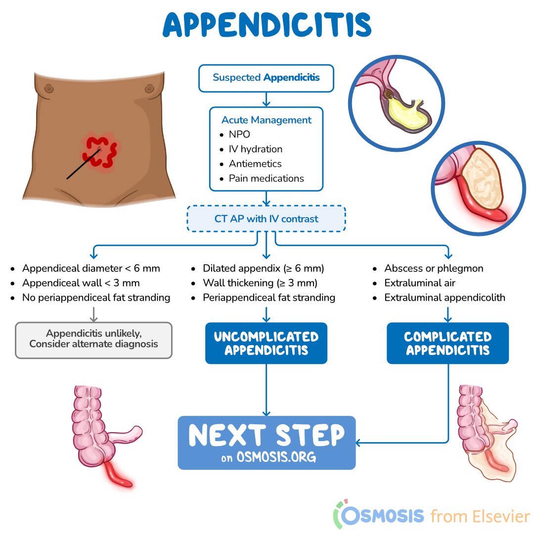
- Abdominal Ultrasound: The primary initial imaging modality for children, adolescents, and pregnant patients to avoid ionizing radiation. An inflamed appendix appears on ultrasound as a non-compressible, blind-ending tubular structure with a diameter greater than 6 millimeters.
- Computed Tomography (CT) Scan of the Abdomen and Pelvis: The gold standard diagnostic tool for adults and elderly patients, offering diagnostic sensitivity and specificity exceeding 95%. CT imaging clearly shows appendiceal enlargement, wall thickening, fat stranding, appendicoliths (calcified fecal stones), or fluid collections.
- Magnetic Resonance Imaging (MRI): Utilized primarily in pregnant patients when ultrasound findings are non-diagnostic or inconclusive, providing detailed soft-tissue visualization without radiation exposure.
- Patient with Suspected Appendicitis:
- Physical Exam & Lab Diagnostics (Psoas Sign, WBC, CRP, Alvarado Score)
- Pediatric / Pregnant Patient:
- Abdominal Ultrasound (Non-ionizing radiation)
- Diagnostic: Surgical Consult
- Inconclusive: Pelvic MRI / CT Scan
- Abdominal Ultrasound (Non-ionizing radiation)
- Adult Patient:
- CT Scan Abdomen/Pelvis (High Sensitivity >95%)
- Positive: Surgical Consult
- Negative: Evaluate Other Causes of Pain
- CT Scan Abdomen/Pelvis (High Sensitivity >95%)
- Pediatric / Pregnant Patient:
- Physical Exam & Lab Diagnostics (Psoas Sign, WBC, CRP, Alvarado Score)
Frequently Asked Questions
Can I perform the leg raise test on myself at home?
While you can physically raise your right leg at home to see if it causes abdominal pain, self-testing is unreliable and should not replace a professional medical examination. A positive test indicates significant intra-abdominal irritation requiring immediate medical assessment, while a negative test does not rule out serious medical conditions, including appendicitis.
What is the difference between the psoas sign and the obturator sign?
Both are physical examination signs for appendicitis, but they test different anatomical positions of the appendix. The psoas sign (leg raise or hip extension) tests for friction against the psoas muscle, indicating a retrocecal appendix. The obturator sign (internal rotation of the flexed right hip) tests for friction against the obturator internus muscle, indicating a pelvic appendix.
Why does my leg hurt when I have appendicitis?
Appendicitis primary pain is abdominal, but nerve pathways sharing spinal segments (such as L1-L3) and direct contact between an inflamed retrocecal appendix and the psoas muscle can cause pain to radiate into the right hip, upper thigh, or groin during movement.
If my leg raise test is negative, does that mean I don't have appendicitis?
No. The leg raise test (psoas sign) has a low sensitivity (roughly 14% to 42%). This means that more than half of all people with appendicitis test negative on the psoas sign, particularly if their appendix is not positioned directly against the psoas muscle. If you have severe abdominal pain, seek emergency care regardless of leg raise test results.
Is appendicitis always treated with surgery?
Appendectomy (surgical removal of the appendix) remains the definitive standard treatment for acute appendicitis. In select uncomplicated cases, intravenous antibiotic therapy may be evaluated by surgical teams, but surgery is the primary treatment to prevent rupture and recurrence.
What to Do Next
If you or someone you are caring for is experiencing right lower abdominal pain and suspects appendicitis:
- Do Not Delay Medical Evaluation: Appendicitis can progress to rupture within 24 to 72 hours. Seek immediate care at an emergency department or urgent care facility equipped with diagnostic imaging.
- Avoid Pain Relievers, Laxatives, or Heating Pads Before Assessment: Do not take anti-inflammatory medications or apply heating pads to the abdomen, as heat can increase localized inflammation and increase the risk of appendiceal rupture. Do not take laxatives or use enemas, as increased intestinal motility can trigger perforation.
- Refrain from Eating or Drinking: If surgical intervention is necessary, having an empty stomach minimizes anesthesia risks. Avoid consuming food, water, or beverages until evaluated by a physician.
- Prepare Relevant Information for the Clinical Team: Be ready to inform the medical staff about when the pain started, whether it migrated from near the belly button to the right lower side, any associated nausea, vomiting, or fever, and your full medical history.
Disclaimer: This article is for informational and educational purposes only and does not constitute medical advice, diagnosis, or treatment. Always consult a qualified healthcare provider or emergency medical services immediately regarding any acute abdominal pain or medical condition.