The assessment of the twelve cranial nerves is a systematic neurological examination used to evaluate the function of nerves emerging directly from the brain. Clinicians test each nerve individually through specialized sensory, motor, or mixed maneuvers—such as checking visual acuity for Cranial Nerve II or observing facial symmetry for Cranial Nerve VII—to localize brainstem lesions, diagnose neuropathies, and monitor central nervous system disorders.
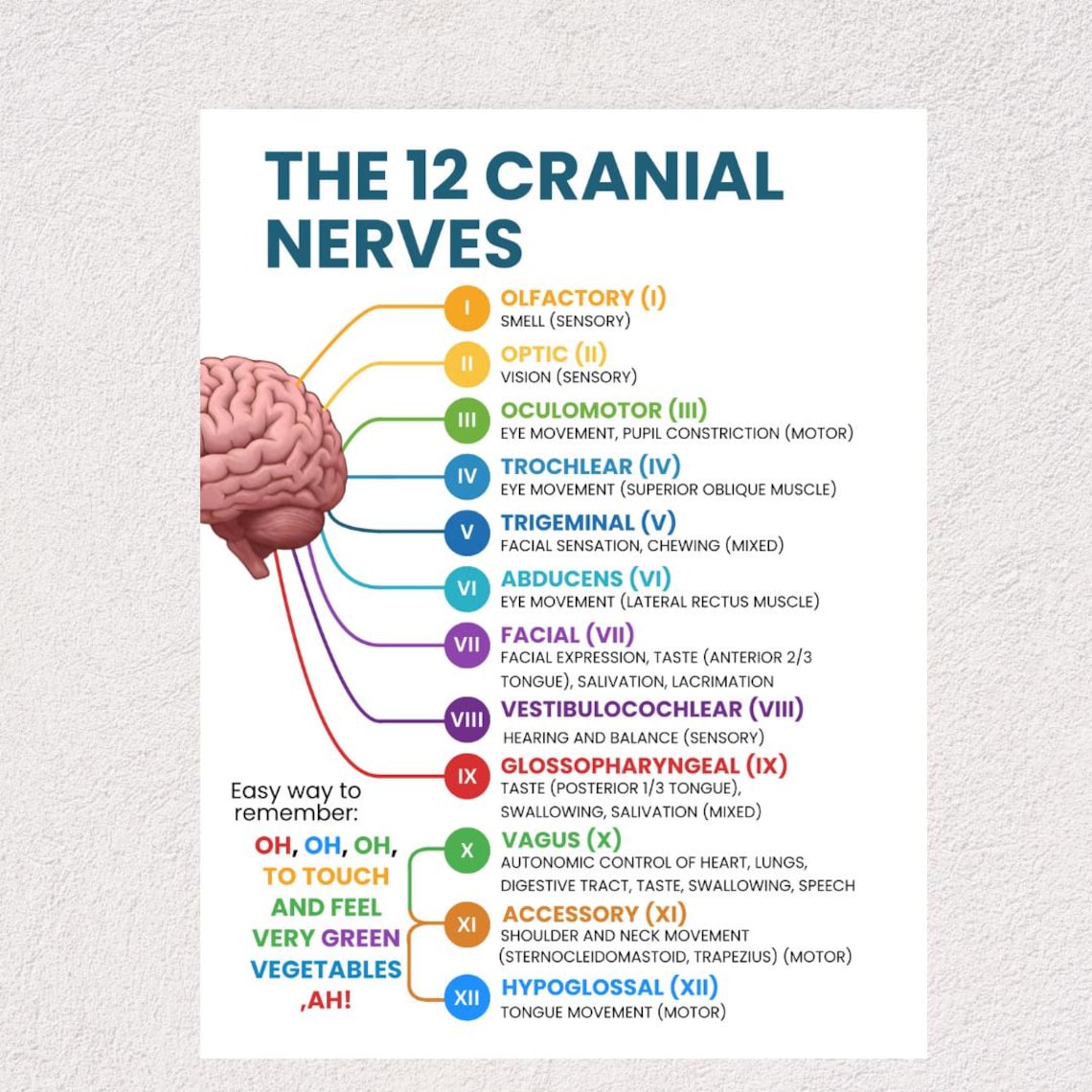
Evaluating the peripheral nervous system requires a structured, anatomical approach. The human nervous system features twelve pairs of cranial nerves that supply motor, sensory, and parasympathetic innervation primarily to the head, neck, and specialized organs, with the exception of the vagus nerve, which extends deep into the thoracic and abdominal cavities.
Performing a rigorous cranial nerve examination is a fundamental competency in neurology, emergency medicine, primary care, and physical therapy. This guide outlines the anatomical pathways, specific clinical testing methods, expected normal findings, pathological deviations, and common examination pitfalls associated with all twelve cranial nerves.
Anatomical Framework and Examination Principles
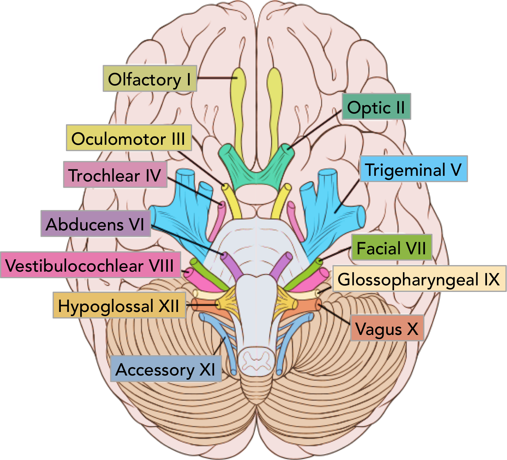
Before touching a patient, a clinician must understand the craniocaudal organization of the cranial nerves. The first two nerves (Olfactory and Optic) originate from the cerebrum and diencephalon, respectively, whereas the remaining ten pairs emerge from nuclei within the brainstem (midbrain, pons, and medulla).
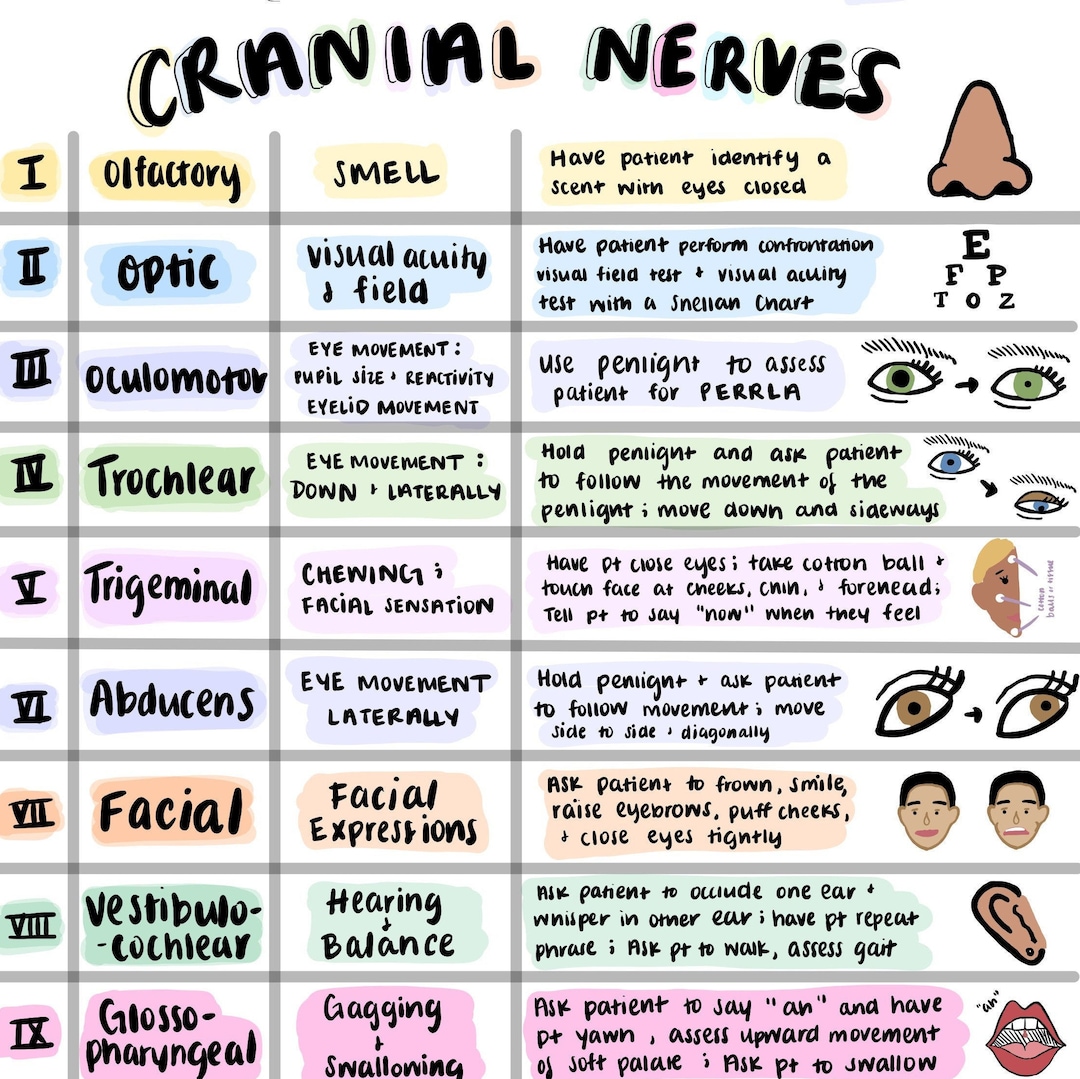
An organized examination prevents omissions and ensures symmetry, which is the cornerstone of neurological assessment. Every test must compare one side of the body to the contralateral side. Furthermore, clinicians must account for patient positioning, lighting, and patient comfort. Standardized equipment should be readily available, including a Snellen chart, ophthalmoscope, penlight, tongue depressors, cotton wisps, a sharp object (such as a sterile safety pin or broken cotton tip), aromatic substances (like coffee or mint), and tuning forks (typically 512 Hz).
Clinical Disclaimer: This guide is intended for educational and training purposes for healthcare professionals and students. It does not replace formal clinical training, institutional protocols, or direct medical judgment.
Detailed Assessment of Cranial Nerves I through XII
Cranial Nerve I: Olfactory Nerve (Sensory)
The olfactory nerve transmits sensory information regarding smells from the nasal mucosa through the cribriform plate of the ethmoid bone to the olfactory bulb.
- Prerequisites and Preparation: Ensure the nasal passages are clear of congestion, polyps, or acute obstruction. Test each nostril separately while occluding the opposite nostril.
- Testing Procedure: Use non-irritating, familiar scents such as coffee, vanilla, or cinnamon. Avoid harsh chemicals like ammonia, which stimulate free nerve endings of the trigeminal nerve (Cranial Nerve V) rather than the olfactory receptors. Instruct the patient to close their eyes, occlude one nostril, sniff the substance presented to the open nostril, and identify it.
- Normal Findings: The patient accurately identifies and differentiates common scents bilaterally.
- Abnormal Findings and Pathology: Anosmia (complete loss of smell) or hyposmia (reduced sense of smell) can result from head trauma (shearing of the cribriform plate), viral upper respiratory infections, neurodegenerative disorders like Parkinson's disease, or frontal lobe tumors (e.g., olfactory groove meningioma). Parosmia refers to a distorted sense of smell.
Cranial Nerve II: Optic Nerve (Sensory)
The optic nerve conveys visual impulses from the retina to the visual cortex. Its assessment encompasses visual acuity, visual fields, color vision, and a direct fundoscopic examination.
- Testing Procedure:
- Visual Acuity: Use a Snellen chart positioned 20 feet away for distance vision, or a Rosenbaum pocket card at 14 inches for near vision, testing each eye independently with corrective lenses worn if normally required.
- Visual Fields: Perform confrontation testing. Sit or stand directly opposite the patient at arm's length. Instruct the patient to cover one eye while you cover your opposite eye, fixing gazes on each other's noses. Move your fingers or a target from the periphery into each of the four visual quadrants.
- Fundoscopy: Use an ophthalmoscope to inspect the optic disc for sharp margins, color, and signs of papilledema or optic atrophy.
- Normal Findings: Intact visual acuity matching baseline or standard norms, full visual fields matching the examiner's peripheral vision, and a sharp, pink optic disc.
- Abnormal Findings and Pathology: Decreased visual acuity suggests refractive errors, cataracts, or optic neuritis. Visual field defects such as bitemporal hemianopsia indicate pituitary adenoma compression at the optic chiasm, whereas homonymous hemianopsia points to a post-chiasmatic stroke or lesion. Papilledema (swelling of the optic disc) indicates elevated intracranial pressure.
Cranial Nerves III, IV, and VI: Oculomotor, Trochlear, and Abducens Nerves (Motor)
These three nerves control extraocular movements (EOMs) and, in the case of Cranial Nerve III, pupillary constriction and eyelid elevation. They are evaluated together through coordinated testing.
| Cranial Nerve | Primary Muscle(s) Innervated | Main Movement / Function | Common Deficit Sign |
|---|---|---|---|
| CN III (Oculomotor) | Superior, inferior, medial rectus; inferior oblique; levator palpebrae superioris; sphincter pupillae | Upward, downward, and medial gaze; eyelid elevation; pupillary constriction | Ptosis, "down and out" gaze, fixed dilated pupil |
| CN IV (Trochlear) | Superior oblique muscle | Intorsion and depression of the eye (looking down and in) | Inability to look down and in; head tilt to compensate for diplopia |
| CN VI (Abducens) | Lateral rectus muscle | Abduction of the eye (lateral gaze) | Failure of lateral gaze; horizontal diplopia |
- Testing Procedure:
- EOMs: Instruct the patient to follow your finger or a target through the "H" pattern (six cardinal fields of gaze) without moving their head. Observe for smooth tracking, nystagmus, and gaze-evoked weakness.
- Convergence: Move the target toward the bridge of the patient's nose to test medial rectus function.
- Pupillary Light Reflex: Shine a light obliquely into one eye, observing direct constriction of that pupil (direct response) and consensual constriction of the opposite pupil (indirect response). Test accommodation by shifting gaze from a distant object to a near target.
- Normal Findings: Full, smooth, and symmetrical extraocular movements without nystagmus or diplopia. Pupils are equal, round, reactive to light, and accommodate appropriately (PERRLA).
- Abnormal Findings and Pathology: A CN III palsy presents with a "down and out" eye, ptosis, and a fixed, dilated pupil (which can be a medical emergency if caused by an expanding posterior communicating artery aneurysm). A CN IV palsy causes vertical or torsional diplopia, mitigated by head tilting away from the affected side. A CN VI palsy results in esotropia and an inability to abduct the affected eye.
Cranial Nerve V: Trigeminal Nerve (Mixed)
The trigeminal nerve provides sensory innervation to the face and motor innervation to the muscles of mastication. It features three major divisions: ophthalmic ($V_1$), maxillary ($V_2$), and mandibular ($V_3$).
- Testing Procedure:
- Sensory: Test light touch using a cotton wisp and pain/temperature using a sterile pin across the forehead ($V_1$), cheek ($V_2$), and jaw ($V_3$) bilaterally. Ask the patient to report if the sensation feels equal on both sides.
- Motor: Palpate the masseter and temporalis muscles while the patient clenches their teeth. Attempt to force the patient's jaw open against resistance. Assess the pterygoid muscles by asking the patient to move their jaw laterally or open against resistance.
- Reflexes: Test the corneal reflex by gently touching the cornea with a wisp of cotton (afferent limb via $V_1$, efferent limb via Cranial Nerve VII causing blinking). Test the jaw jerk reflex by tapping a reflex hammer on the chin with the mouth slightly open.
- Normal Findings: Sensation is intact and symmetrical across all three divisions. Strong, symmetrical contraction of masseter and temporalis muscles without jaw deviation.
- Abnormal Findings and Pathology: Sensory loss in a dermatomal distribution suggests peripheral nerve injury or brainstem stroke. Weakness or atrophy of the mastication muscles points to a lower motor neuron lesion of the mandibular division. Trigeminal neuralgia (tic douloureux) causes excruciating, electric-shock-like facial pain triggered by light touch.
Cranial Nerve VII: Facial Nerve (Mixed)
The facial nerve controls the muscles of facial expression, conveys taste from the anterior two-thirds of the tongue, and supplies parasympathetic fibers to the lacrimal and salivary glands.
- Testing Procedure:
- Motor: Inspect the face at rest for asymmetry. Instruct the patient to perform specific facial movements: raise eyebrows (wrinkle forehead), tightly close eyes against resistance, show teeth or smile, puff out cheeks, and purse lips.
- Sensory: Taste testing on the anterior two-thirds of the tongue is rarely performed in routine screening unless pathology is suspected. Sweet, salty, sour, and bitter solutions are applied to the protruded tongue using a cotton applicator.
- Normal Findings: Symmetrical facial movements with deep forehead wrinkling, complete eye closure, and equal smile symmetry.
- Abnormal Findings and Pathology: Upper motor neuron lesions (e.g., stroke) spare the forehead due to bilateral cortical representation, causing weakness restricted to the lower half of the contralateral face. Lower motor neuron lesions (e.g., Bell's palsy) affect both the upper and lower face on the ipsilateral side, resulting in eyebrow sagging, loss of forehead wrinkles, inability to close the eye, and drooping of the mouth corner.
Cranial Nerve VIII: Vestibulocochlear Nerve (Sensory)
The vestibulocochlear nerve mediates hearing (cochlear branch) and balance/spatial orientation (vestibular branch).
- Testing Procedure:
- Auditory Acuity: Perform the whispered voice test. Stand at arm's length behind the patient, whisper a combination of numbers and letters, and ask the patient to repeat them while they occlude the opposite ear.
- Weber Test: Place a vibrating 512 Hz tuning fork on the center of the patient's forehead or vertex. Ask if the sound is heard equally in both ears or lateralizes to one side.
- Rinne Test: Place the vibrating tuning fork on the mastoid process (bone conduction) until the patient no longer hears it, then quickly move it just outside the external auditory meatus (air conduction). Normally, air conduction is twice as long as bone conduction (positive Rinne).
- Normal Findings: Patient repeats whispered words accurately at 2 feet. Weber test does not lateralize (sound heard equally). Rinne test shows air conduction greater than bone conduction bilaterally.
- Abnormal Findings and Pathology: Sensorineural hearing loss causes the Weber test to lateralize to the healthy ear and a positive Rinne test with reduced duration. Conductive hearing loss causes the Weber test to lateralize to the impaired ear and a negative Rinne test (bone conduction greater than or equal to air conduction).
Cranial Nerves IX and X: Glossopharyngeal and Vagus Nerves (Mixed)
Because the glossopharyngeal and vagus nerves share functional overlap in the innervation of the pharynx and larynx, they are tested together. CN IX provides taste to the posterior third of the tongue and sensation to the pharynx, while CN X controls pharyngeal and laryngeal muscles for swallowing and vocalization.
- Testing Procedure:
- Motor: Ask the patient to open their mouth and say "Ah." Observe the soft palate and uvula for midline elevation. Test the gag reflex by gently stimulating the posterior pharyngeal wall with a tongue depressor (caution: elicit only if clinically necessary, as it can be distressing). Listen to the patient's voice for hoarseness or nasal quality.
- Swallowing: Assess the patient's ability to swallow saliva or water safely.
- Normal Findings: The soft palate and uvula elevate symmetrically in the midline during phonation. The voice is clear without hoarseness. The gag reflex is intact bilaterally.
- Abnormal Findings and Pathology: Deviation of the uvula away from the side of a lesion indicates a unilateral vagus nerve weakness (the healthy side pulls the uvula over). Hoarseness or a bovine cough suggests recurrent laryngeal nerve involvement (a branch of CN X). Dysphagia indicates combined deficits of CN IX and X.
Cranial Nerve XI: Spinal Accessory Nerve (Motor)
The accessory nerve provides motor innervation to the sternocleidomastoid and trapezius muscles.
- Testing Procedure:
- Trapezius: Place your hands on the patient's shoulders and ask them to shrug upward against your resistance. Assess muscle bulk and strength symmetrically.
- Sternocleidomastoid: Ask the patient to rotate their head to the left and right against your hand placed on their cheek/jaw. Assess the strength and contraction of the contralateral sternocleidomastoid muscle.
- Normal Findings: Equal strength and muscle bulk in both trapezius and sternocleidomastoid muscles without fasciculations or atrophy.
- Abnormal Findings and Pathology: Weakness or atrophy of the trapezius muscle with shoulder drop indicates an ipsilateral accessory nerve lesion, often caused by iatrogenic injury during radical neck dissection or lymph node biopsy. Weakness in turning the head against resistance points to sternocleidomastoid dysfunction.
Cranial Nerve XII: Hypoglossal Nerve (Motor)
The hypoglossal nerve controls intrinsic and extrinsic muscles of the tongue, dictating its movement and shape.
- Testing Procedure:
- Inspect the tongue at rest inside the mouth for fasciculations (fine twitching) or atrophy.
- Ask the patient to protrude the tongue. Observe for deviation to one side.
- Ask the patient to push their tongue firmly into each cheek while you apply counter-pressure externally to test tongue strength.
- Normal Findings: The tongue protrudes cleanly in the midline without tremor, fasciculations, atrophy, or deviation. Tongue strength is symmetrical.
- Abnormal Findings and Pathology: Unilateral lower motor neuron lesions cause the tongue to deviate toward the weak side upon protrusion due to the unopposed action of the healthy genioglossus muscle. Upper motor neuron lesions typically cause contralateral deviation with minimal atrophy. Fasciculations and severe atrophy indicate advanced lower motor neuron disease, such as amyotrophic lateral sclerosis (ALS).
Frequently Asked Questions
What is the most reliable way to test all twelve cranial nerves quickly?
A structured, top-to-bottom clinical sequence is the most reliable approach. Clinicians generally begin with visual acuity and pupillary checks (CN II, III, IV, VI), move to facial sensation and movement (CN V, VII), evaluate hearing and throat function (CN VIII, IX, X), and finish with shoulder shrugs and tongue movement (CN XI, XII). This structured flow minimizes position changes and reduces exam time.
Why do upper and lower motor neuron facial nerve lesions look different?
The difference stems from cortical innervation pathways. The upper motor neurons supplying the upper face (forehead) receive bilateral cortical input, whereas those supplying the lower face receive strictly contralateral input. Consequently, a stroke (upper motor neuron lesion) spares forehead movement, while Bell's palsy (lower motor neuron lesion) paralyzes the entire half of the face.
What should a clinician do if a patient cannot smell during the olfactory examination?
First, inspect the nasal cavity for local causes such as acute rhinitis, nasal polyps, or severe congestion. If local obstruction is ruled out, inquire about a history of recent head trauma (which can shear olfactory nerve filaments passing through the cribriform plate) or viral infections. Document the finding and correlate it with other neurological signs.
How is the gag reflex tested safely without causing aspiration?
The gag reflex should only be tested when there is a specific clinical indication—such as suspected bulbar palsy, impaired swallowing, or altered mental status—rather than as a routine screen in an asymptomatic patient. Use a clean tongue depressor to gently stimulate the posterior pharyngeal wall, warning the patient beforehand to minimize discomfort and prevent vomiting.
Can cranial nerve testing diagnose a stroke?
Yes, cranial nerve assessments provide vital focal neurological signs that help localize strokes within the brainstem or cerebral hemispheres. For example, an isolated abducens nerve palsy combined with contralateral hemiplegia points to a pontine stroke, while sudden visual field defects and aphasia suggest cortical vascular events.
Conclusion and Next Clinical Actions
Mastering the assessment of the twelve cranial nerves requires rigorous anatomical knowledge, consistent physical practice, and meticulous attention to symmetry. Clinicians must document both normal findings and specific deficits clearly. When an abnormality is identified, the next appropriate clinical step involves correlating the physical exam findings with neuroimaging (such as an MRI of the brain or brainstem) and consulting neurology or relevant subspecialists to confirm localization and guide therapeutic management.