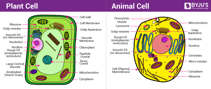
While both animal and plant cells are eukaryotic organisms containing membrane-bound nuclei and shared metabolic machinery, plant cells possess a rigid cell wall, a large central vacuole, and chloroplasts for photosynthesis. Animal cells lack cell walls and chloroplasts, utilizing smaller vacuoles, centrioles, and flexible plasma membranes to adapt to varied metabolic and motile functions.
Fundamental Similarities: The Eukaryotic Baseline
Before examining the distinct characteristics that separate animal and plant cells, it is essential to understand their shared evolutionary lineage. Both plant and animal cells belong to the domain Eukaryota. Unlike prokaryotic organisms—such as bacteria and archaea, which lack internal membrane-bound compartments—eukaryotic cells enclose their genetic material within a defined nucleus and maintain specialized subcellular compartments called organelles.
- Eukaryotic Cell
- Shared Features
- Nucleus
- Mitochondria
- ER & Golgi
- Cytoplasm
- Animal Cell
- Flexible Shape
- Centrioles
- Small Vacuoles
- Lysosomes
- Glycogen
- Plant Cell
- Rigid Shape
- Cell Wall
- Large Vacuole
- Chloroplasts
- Plasmodesmata
- Shared Features
This structural compartmentalization allows both plant and animal cells to segregate incompatible chemical reactions, maintain optimal local environments for enzyme activities, and efficiently manage intracellular transport. Both cell types share a core suite of organelle structures:
- Nucleus: Houses the cellular genome encoded in linear DNA wrapped around histone proteins. It manages gene expression, transcription, and ribosome assembly via the nucleolus.
- Plasma Membrane: A phospholipid bilayer embedded with proteins that serves as a selectively permeable barrier, controlling the flux of ions, nutrients, and waste products.
- Cytoplasm and Cytosol: The semi-fluid matrix filling the cell interior, housing soluble proteins, metabolic intermediates, and the protein filaments of the cytoskeleton.
- Mitochondria: The site of cellular respiration and oxidative phosphorylation, generating chemical energy in the form of adenosine triphosphate (ATP).
- Endoplasmic Reticulum (ER): A network of membrane-bound tubules and sacs. The Rough ER (studded with ribosomes) synthesizes and processes proteins destined for membranes or secretion, while the Smooth ER synthesizes lipids, metabolizes carbohydrates, and detoxifies metabolic byproducts.
- Golgi Apparatus: A series of flattened cisternae that modifies, sorts, and packages proteins and lipids received from the ER for secretion or transport to other organelles.
- Ribosomes: Molecular machines composed of ribosomal RNA (rRNA) and proteins that translate messenger RNA (mRNA) transcripts into polypeptide chains.
Despite these common foundations, millions of years of evolutionary divergence have shaped distinct organelle compositions and cellular architectures. These differences reflect the fundamental lifestyles of plants (autotrophic, sessile organisms) and animals (heterotrophic, motile organisms).
Key Structural Differences Between Animal and Plant Cells
The structural differences between animal and plant cells are directly tied to their biochemical requirements, physiological roles, and structural demands.
- Animal Cell
- Microvilli
- Nucleus (Center)
- Small Vacuoles
- Mitochondria
- Centrioles
- Lysosomes
- Flexible Outline
- Plant Cell
- Cell Wall
- Nucleus (Pushed)
- Central Vacuole (Turgor)
- Chloroplasts
Cell Wall vs. Plasma Membrane Only
The primary architectural distinction between plant and animal cells lies in the presence of an extracellular cell wall in plants.
- Plant Cells: Enclosed by a rigid, non-living structure called the primary cell wall, which is composed mainly of cellulose microfibrils embedded within a matrix of hemicellulose, pectin, and structural proteins. In mature woody plants, a thicker secondary cell wall impregnated with lignin is deposited inside the primary wall. The cell wall provides structural support, prevents over-expansion when water enters the cell, determines shape, and acts as a mechanical barrier against pathogens.
- Animal Cells: Completely lack a cell wall, bounded only by a flexible lipid bilayer plasma membrane. External structural support is provided instead by an extracellular matrix (ECM) made of collagen, proteoglycans, and fibronectin, anchored to the cell's cytoskeleton via transmembrane proteins called integrins. This absence of a rigid wall enables animal cells to adopt diverse shapes, migrate through tissues, and undergo rapid morphogenetic movements during development.
Chloroplasts and Plastids vs. Heterotrophic Energy Needs
Plants produce their own organic compounds from inorganic materials, whereas animals rely on consuming other organisms for energy. This nutritional distinction dictates their organelle equipment:
- Plant Cells: Contain specialized double-membrane organelles called plastids. The most prominent of these are chloroplasts, the sites of photosynthesis. Inside chloroplasts, stacked membrane structures called thylakoids (arranged in grana) contain pigment molecules, including chlorophyll a and b. These pigments harvest light energy to convert carbon dioxide and water into glucose and oxygen through light-dependent reactions and the Calvin cycle. Plant cells also feature non-pigmented plastids, such as amyloplasts (which synthesize and store starch) and chromoplasts (which store carotenoid pigments responsible for red, orange, and yellow hues in flowers and fruits).
- Animal Cells: Devoid of chloroplasts and plastids entirely. Animal cells rely exclusively on the uptake of organic nutrients (such as glucose, amino acids, and fatty acids) from their environment, converting them into energy through cytosolic glycolysis and mitochondrial respiration.
Central Vacuole System vs. Small Temporary Vacuoles
Vacuoles are membrane-bound organelles containing fluid, ions, and organic molecules, but their functional emphasis differs dramatically between plant and animal cells:
- Plant Cells: Feature a prominent large central vacuole bounded by a specialized membrane called the tonoplast. In mature plant cells, this single vacuole can occupy 30% to 90% of the total cellular volume. The central vacuole accumulates water, maintains turgor pressure against the cell wall (providing structural stiffness to leaves and non-woody stems), stores nutrients and pigment molecules (such as anthocyanins), and isolates toxic metabolic waste products or defense chemicals.
- Animal Cells: May contain one or more small, temporary vacuoles (often referred to as vesicles), but lack a central vacuole. These structures function primarily in endocytosis, exocytosis, pinocytosis, and transient storage or transport of materials across the cytoplasm.
- Plant Central Vacuole
- Single Large Tonoplast Compartment (Up to 90% Volume)
- Animal Vesicle / Vacuole
- Multiple Small Transient Vesicles
Intercellular Communication: Plasmodesmata vs. Cell Junctions
Because cell walls create a physical barrier to direct cell-to-cell contact, plants use a unique intercellular transport mechanism:
- Plant Cells: Interconnected by microscopic channels called plasmodesmata (singular: plasmodesma) that breach both the primary cell wall and the middle lamella. These channels establish direct cytoplasmic continuity between adjacent cells, enabling the movement of water, small solutes, signaling molecules, microRNAs, and transcription factors.
- Animal Cells: Communcate through direct cell-surface interactions and specialized protein structures. Specialized junctions include:
- Gap Junctions: Protein rings composed of connexins that form open pores between cells, allowing direct passage of ions and small signaling molecules (functionally analogous to plasmodesmata).
- Tight Junctions: Continuous protein seals (claudins and occludins) that prevent movement of materials through the extracellular space between adjacent epithelial cells.
- Desmosomes: Rivet-like anchoring junctions that fasten cells together into strong structural sheets via intermediate filaments.
Centrioles, Centrosomes, and Cytoskeletal Dynamics
While both cell types utilize microtubules, microfilaments (actin), and intermediate filaments to maintain internal spatial organization and orchestrate intracellular movement, their organization centers differ:
- Animal Cells: Contain a distinct centrosome located near the nucleus, which houses a pair of barrel-shaped structures called centrioles. Each centriole is composed of nine triplets of microtubules arranged in a ring. Centrioles organize the mitotic spindle during cell division, help establish cellular polarity, and act as basal bodies for the assembly of cilia and flagella—structures widely present on motile animal cells (such as sperm cells or respiratory epithelial cells).
- Plant Cells: Higher plants lack centrioles and cilia/flagella entirely (with the exception of motile male gametes in lower plants like mosses and ginkgos). Instead, plant cells organize their microtubule networks using diffuse, non-centrosomal Microtubule Organizing Centers (MTOCs) located along the nuclear envelope and beneath the plasma membrane.
Lysosomes, Glyoxysomes, and Metabolic Enzyme Storage
- Animal Cells: Contain dedicated lysosomes, acidic organelles packed with hydrolytic enzymes (such as proteases, nucleases, lipases, and phosphatases) optimized to operate at low pH (~4.5 to 5.0). Lysosomes digest engulfed extracellular materials (via phagocytosis), degrade worn-out cellular organelles (via autophagy), and facilitate programmed cell death (apoptosis).
- Plant Cells: Generally lack dedicated lysosomal compartments. Instead, the degradation of macromolecules and organelle recycling occurs within the acidic internal compartment of the large central vacuole. However, plant cells contain specialized peroxisomes called glyoxysomes, particularly in germinating seeds. Glyoxysomes contain enzymes of the glyoxylate cycle, allowing the conversion of stored seed lipids into carbohydrates to nourish the growing embryo before photosynthesis begins—a metabolic pathway absent in animal cells.
Detailed Structural Comparison
The following table summarizes the key structural and functional differences between animal and plant cells:
| Feature / Organelle | Animal Cell | Plant Cell | Primary Biological Function |
|---|---|---|---|
| Cell Wall | Absent | Present (cellulose, hemicellulose, pectin) | Structural integrity, protection, prevention of osmotic lysis |
| Cell Shape | Flexible, variable, irregular | Rigid, fixed, typically rectangular or polygonal | Structural stability vs. cellular mobility and tissue plasticity |
| Chloroplasts & Plastids | Absent | Present (chloroplasts, chromoplasts, amyloplasts) | Photosynthesis and pigment/energy storage |
| Vacuoles | Small, temporary, multiple (if present) | Single large central vacuole (tonoplast bound) | Turgor pressure maintenance, ion storage, degradation |
| Energy Storage Form | Glycogen | Starch (amylose and amylopectin) | Short- to medium-term carbohydrate reserve storage |
| Centrioles / Centrosomes | Present (pair of centrioles per centrosome) | Absent in higher plants (diffuse MTOCs instead) | Spindle formation and microtubule organization |
| Intercellular Connections | Gap junctions, tight junctions, desmosomes | Plasmodesmata | Direct cytoplasmic, mechanical, or chemical communication |
| Lysosomes | Present (abundant, membrane-enclosed) | Rare/Absent (lytic function handled by central vacuole) | Intracellular digestion, organelle recycling, apoptosis |
| Glyoxysomes | Absent | Present (especially in germinating seeds) | Conversion of stored fats to sugars via the glyoxylate cycle |
| Cilia and Flagella | Common in many cell types (e.g., sperm, epithelium) | Absent in higher plants (present only in lower plant sperm) | Motility and extracellular fluid movement |
| Cytokinesis Mechanism | Cleavage furrow formed by actin microfilaments | Cell plate formed by phragmoplast and Golgi vesicles | Physical division of single cytoplasm into two daughter cells |
Metabolic and Functional Consequences of Cellular Design
The architectural differences outlined above result in distinct cellular behaviors, metabolic strategies, and physiological outcomes.
- Cytokinesis Comparison Mechanism
- Animal Cell Cytokinesis
- Contractile Actin Ring
- Cleavage Furrow Pinches Inward
- Two Separate Flexible Cells
- Plant Cell Cytokinesis
- Phragmoplast Microtubules
- Golgi Vesicle Fusion Accumulates Middle
- Cell Plate Expands Outward to Walls
- Animal Cell Cytokinesis
Energy Production and Storage: Starch vs. Glycogen
Plant cells convert light energy into chemical energy via chloroplasts, synthesizing simple glucose molecules. Excessive glucose is polymerized into starch—a mixture of amylose (linear $\alpha$-1,4 glucan polymers) and amylopectin (branched $\alpha$-1,4 and $\alpha$-1,6 glucan polymers). Starch forms insoluble granules within amyloplasts, providing a stable, osmotic-neutral energy reserve that can be mobilized when light energy is unavailable.
Animal cells rely on external food sources. Excess glucose absorbed by animal cells is converted into glycogen, a highly branched polymer of glucose molecules structured around a core protein called glycogenin. Glycogen granules are stored directly in the cytosol, primarily within liver and muscle tissues. The hyper-branched structure of glycogen allows enzymes to rapidly cleave individual glucose units when ATP demand increases during physical activity or stress.
Osmotic Regulation and Structural Rigidity (Turgor Pressure)
Because animal cells are bounded only by a delicate plasma membrane, they are susceptible to changes in external solute concentration:
- In a hypotonic environment (low solute, high water concentration), water rapidly enters an animal cell via aquaporin channels, causing the cell to swell and potentially undergo lysis (bursting).
- In a hypertonic environment, water leaves the animal cell, leading to crenation (shriveling). Therefore, animal cells must maintain an isotonic internal state using active membrane pumps, such as the $\text{Na}^+/\text{K}^+$-ATPase pump.
Plant cells utilize osmotic pressure to maintain structural stability:
- In a hypotonic environment, water enters the central vacuole by osmosis, pushing the cytoplasm and plasma membrane tightly against the rigid cell wall. This outward hydrostatic force is called turgor pressure. The mechanical rigidity of the cell wall prevents the cell from bursting. Turgor pressure keeps non-woody plant tissue upright and expanded.
- In a hypertonic environment, water leaves the vacuole, causing the plasma membrane to pull away from the cell wall—a condition known as plasmolysis that results in leaf wilting.
| Environment | Animal Cell | Plant Cell |
|---|---|---|
| Hypotonic | Lysed (Burst) | Turgid (Normal) |
| Isotonic | Normal | Flaccid |
| Hypertonic | Shriveled (Crenated) | Plasmolyzed |
Cytokinesis: Cell Plate Formation vs. Cleavage Furrow
The presence or absence of a rigid cell wall dictates how these two eukaryotic cell types execute cytokinesis (the division of the cytoplasm during cell division):
- Animal Cell Cytokinesis: During late anaphase and telophase, a ring of actin microfilaments and myosin motor proteins forms just beneath the plasma membrane at the cell equator. This contractile ring contracts, pulling the plasma membrane inward to form a cleavage furrow. As the ring tightens, it pinches the cytoplasm in two, yielding two separate daughter cells.
- Plant Cell Cytokinesis: The presence of a rigid cell wall prevents the plasma membrane from pinching inward. Instead, a complex cytoskeletal array called the phragmoplast guides vesicles derived from the Golgi apparatus to the center of the dividing cell. These vesicles, loaded with pectins and structural polysaccharides, fuse to form a growing cell plate. The cell plate expands outward until it fuses with the existing lateral cell walls, dividing the parental cell into two distinct compartments, after which cellulose microfibrils are deposited to complete the primary cell wall.
Evolutionary Drivers Behind Cell Specialization
The structural distinctions between plant and animal cells stem from their evolutionary divergence over one billion years ago. Detailed cellular research documented in resources like the NCBI Molecular Biology of the Cell Reference Manual illustrates how early eukaryotic ancestors incorporated distinct bacterial endosymbionts, leading to different evolutionary paths:
- Primary Endosymbiosis: The common ancestor of both lineages engulfed an aerobic $\alpha$-proteobacterium, which evolved into modern mitochondria. This event conferred the capacity for high-yield aerobic respiration.
- Secondary Endosymbiosis Event: The ancestral lineage leading to plants (Archaeplastida) subsequently engulfed a photosynthetic cyanobacterium. Over evolutionary time, this endosymbiont transferred much of its genetic material to the host nucleus, evolving into the modern chloroplast.
- Ancestral Eukaryote (Heterotrophic)
- Engulfed Alpha-Proteobacterium (Evolved into Mitochondria)
- Animal Lineage (Heterotrophic, Motile)
- Retained flexible membrane
- Developed centrioles/motility
- Selective ECM structures
- Glycogen energy storage
- Plant Lineage (Autotrophic, Sessile)
- Engulfed Cyanobacterium (Evolved into Chloroplast)
- Developed rigid Cellulose Cell Wall
- Large Central Vacuole for turgor
Equipped with chloroplasts, plants adopted an autotrophic lifestyle. Because sunlight, water, and atmospheric carbon dioxide are diffuse environmental resources, plants evolved a sessile, sessile-growth strategy with high surface-area-to-volume ratios. The cell wall and central vacuole evolved to allow plants to grow vertically toward light sources without the metabolic cost of supporting heavy muscle systems or cellular locomotion networks.
Animals retained a heterotrophic lifestyle, requiring active movement to forage, hunt, escape predators, and reproduce. A rigid cellulose cell wall would inhibit movement, muscle contraction, nerve impulse propagation, and cell migration during embryonic development. Consequently, animal cells retained structural flexibility, prioritizing dynamic cytoskeletal networks, specialized extracellular matrix interactions, and rapid cell-to-cell communication mechanisms.
Frequently Asked Questions
Why do plant cells have both mitochondria and chloroplasts?
Chloroplasts use sunlight to synthesize glucose and chemical energy reserves through photosynthesis. However, chloroplasts do not directly supply ATP to power the cell's general metabolic work outside the photosynthetic pathway. Plant cells require mitochondria to oxidize the carbohydrates produced by chloroplasts via cellular respiration, generating ATP to power basic cellular processes during the night or in non-photosynthetic tissues (such as root cells).
Do any animal cells have cell walls or chloroplasts?
No animal cells naturally possess cell walls or chloroplasts. A defining biological feature of the kingdom Animalia is the absence of cell walls and plastids. While certain marine animals (such as the sea slug Elysia chlorotica) ingest algae and temporarily retain functioning chloroplasts within their digestive cells through a process called kleptoplasty, the animal cells themselves do not produce or inherit these chloroplasts.
Why do animal cells contain centrioles while higher plant cells do not?
Animal cells use centrioles within centrosomes to establish the mitotic spindle poles during nuclear division and to form the basal bodies of cilia and flagella. Higher plants lost centrioles over evolutionary time; instead, they organize their mitotic spindles using diffuse microtubule-organizing centers along the nuclear envelope. Because higher plants do not produce motile cells with cilia or flagella (with the exception of some gymnosperms and lower plants), they do not require centrioles for basal body assembly.
Can an animal cell survive in pure water?
No, an animal cell placed in pure (hypotonic) water will absorb water rapidly due to osmotic influx. Lacking a rigid cell wall to exert counter-balancing turgor pressure, the animal cell's plasma membrane will expand until it ruptures, a process known as osmotic lysis or hemolysis in red blood cells.
What is the function of plasmodesmata, and what is its animal counterpart?
Plasmodesmata are membrane-lined channels that breach plant cell walls, allowing direct cytoplasmic transport of water, nutrients, and signaling proteins between neighboring cells. The functional equivalent in animal cells is the gap junction, which consists of membrane protein complexes (connexons) that form channels allowing small ions and signaling molecules to pass directly between adjacent cell membranes.
Summary and Practical Takeaways
Understanding the differences between animal and plant cells relies on recognizing how their organelle structures support their distinct metabolic strategies:
- Plant cells are built for autotrophic energy production and mechanical support, featuring rigid cellulose cell walls, light-capturing chloroplasts, and large central vacuoles that maintain turgor pressure. They link together via plasmodesmata and execute cytokinesis by building a cell plate.
- Animal cells are structured for flexibility, motility, and heterotrophic consumption. They rely on an outer plasma membrane anchored by an extracellular matrix, organize microtubules using centrosomes with centrioles, digest waste via lysosomes, communicate through gap junctions, and divide their cytoplasm using an actin-based cleavage furrow.
These structural distinctions reflect the basic divide between autotrophic, stationary life and heterotrophic, motile life across eukaryotic biology.