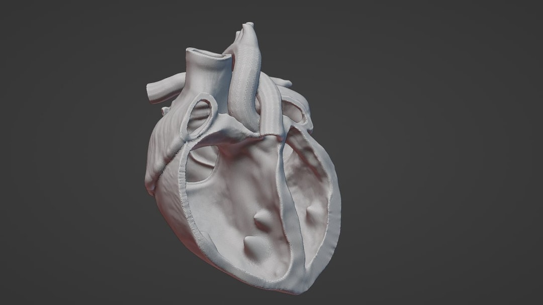
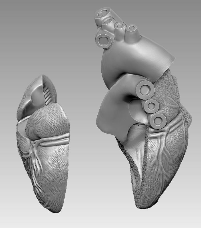
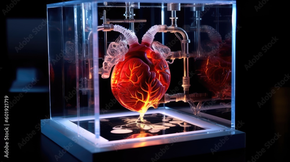
3D printing a human heart involves using advanced additive manufacturing techniques combined with biological materials, stem cells, and patient-specific imaging data to construct functional cardiac tissue or anatomical models. While complete, transplant-ready whole hearts remain in experimental stages of development, bioprinted cardiac patches, vascular networks, and precise surgical planning models are actively reshaping modern cardiovascular medicine.
Introduction to Cardiac Bioprinting and Additive Manufacturing
The intersection of additive manufacturing and cardiology represents one of the most ambitious frontiers in modern tissue engineering and regenerative medicine. For decades, the replacement of diseased or failing hearts relied exclusively on human organ donation, a scarce resource subject to severe supply constraints, immunological rejection risks, and strict geographical limitations.
The concept of 3D printing a human heart aims to bypass these limitations by generating functional cardiac tissue or entire organs on demand. However, achieving this goal requires navigating immense biological and technical complexities. Unlike traditional 3D printing, which utilizes inert plastics, resins, or metals, cardiac bioprinting involves handling living cells suspended in specialized hydrogels—collectively known as bioinks—while maintaining strict sterility, precise temperature controls, and cellular viability throughout the manufacturing process.
The Evolution from Polymers to Living Cells
Early milestones in medical 3D printing focused primarily on rigid materials. Surgeons and biomedical engineers utilized computed tomography (CT) and magnetic resonance imaging (MRI) scans to print hard, polymer-based replicas of patient hearts. These anatomical models transformed preoperative planning, allowing cardiothoracic surgeons to examine complex congenital heart defects in tangible three dimensions before ever opening a patient's chest.
As material science progressed, research shifted toward soft lithography and hydrogel-based printing. Scientists developed biocompatible inks derived from extracellular matrix (ECM) components, such as collagen, gelatin, and hyaluronic acid. These materials provide the necessary mechanical support for cells to adhere, proliferate, and organize into functional tissue structures. The integration of living human cells—including induced pluripotent stem cells (iPSCs)—into these bioinks marked the true birth of cardiac bioprinting, moving the field from mere visualization to functional tissue fabrication.
Core Technologies and Printing Methodologies
Different additive manufacturing modalities are required to address the diverse scales of a human heart, ranging from microscopic capillary networks to macro-scale muscular chambers. Selecting an appropriate printing technology depends on the desired resolution, printing speed, and the sensitivity of the bioink to mechanical and thermal stress.
Extrusion-Based Bioprinting
Extrusion-based systems are among the most widely utilized platforms in cardiac tissue engineering. These printers dispense bioinks through a nozzle using pneumatic or mechanical pressure, depositing continuous filaments layer by layer.
- Advantages: Capable of handling high cell densities and printing large-volume constructs relatively quickly.
- Limitations: Resolution is often limited compared to light-based methods, and high shear stress during extrusion can compromise cell viability if parameters are not carefully optimized.
Stereolithography (SLA) and Digital Light Processing (DLP)
Light-based bioprinting techniques employ ultraviolet (UV) or visible light to selectively crosslink and solidify photosensitive bioinks in a layer-by-layer fashion.
- Advantages: Offers exceptionally high printing resolution, enabling the fabrication of intricate microscale features and complex vascular geometries.
- Limitations: Restricted by the availability of biocompatible, photocurable hydroinks, and potential cytotoxicity associated with photoinitiators and light exposure.
Laser-Assisted Bioprinting (LAB)
Laser-assisted bioprinting utilizes a pulsed laser beam targeted at a donor ribbon coated with a bioink layer. The laser vaporizes a microscopic portion of the ribbon, propelling droplets of bioink onto a collector substrate.
- Advantages: No nozzle contact eliminates mechanical shear stress, allowing for single-cell-level precision.
- Limitations: High equipment costs, slow fabrication speeds, and difficulties scaling up for large tissue constructs.
Biological Challenges in Replicating Cardiac Tissue
Printing the macro-scale shape of a human heart is only a fraction of the challenge. The human heart is a sophisticated biological pump composed of specialized cell types operating within a highly organized extracellular matrix. Successfully printing functional cardiac tissue requires overcoming several distinct physiological hurdles.
| Challenge | Description | Current Mitigation Strategy |
|---|---|---|
| Vascularization | Living tissue requires a continuous supply of oxygen and nutrients within 100–200 micrometers of every cell to prevent necrosis. | Embedding sacrificial inks that melt away after printing to create open channels mimicking blood vessels. |
| Cell Viability | Maintaining high survival rates of fragile cardiomyocytes (heart muscle cells) during and after the printing process. | Optimizing bioink rheology, thermal management, and utilizing protective shear-thinning hydrogels. |
| Electromechanical Coupling | Cardiac muscle must beat synchronously. Cells must establish electrical connections (gap junctions) to propagate electrical signals. | Applying electrical or mechanical stimulation in bioreactors post-printing to promote cell alignment and maturation. |
| Immunogenicity | Using synthetic or xenogeneic materials can trigger severe immune responses and organ rejection. | Utilizing patient-derived induced pluripotent stem cells (iPSCs) to create autologous, personalized tissue constructs. |
The Vascularization Bottleneck
The single greatest obstacle in engineering whole organs is vascularization. While researchers can successfully print thick blocks of cardiac muscle, cells located in the interior of the construct quickly die off due to diffusion limits before blood vessels can naturally sprout into the tissue.
To solve this, advanced laboratories employ coaxial printing nozzles and sacrificial carbohydrate inks. For instance, researchers at institutions such as Tel Aviv University demonstrated the ability to print a cellularized heart complete with blood vessels by using a support bath technique (FRESH - Freeform Reversible Embedding of Suspended Hydrogels), which allows soft hydroinks to be printed inside a supportive gelatin slurry without collapsing.
Clinical Applications and Current State (2026)
As of 2026, the clinical application of 3D printing in cardiology spans several tiers, ranging from routine surgical aids to pioneering regenerative therapies. Understanding the distinct categories of clinical use helps clarify the boundary between current standard-of-care practices and long-term experimental research.
- Standard Clinical Use
- Preoperative Anatomical Models
- Surgical Guides & Devices
- Patient-Specific Prosthetics
- Experimental Research
- Whole Bioprinted Organs
- Vascularized Cardiac Patches
- Drug Screening Platforms
Anatomical Models and Preoperative Planning
Today, patient-specific 3D-printed heart models derived from real-time imaging data are standard tools in advanced pediatric and adult cardiothoracic surgery. When surgeons face complex congenital malformations—such as Tetralogy of Fallot or transposition of the great arteries—relying solely on two-dimensional scans can leave critical spatial questions unanswered.
Printing a rigid, flexible, or multi-material replica of the patient's exact heart allows surgical teams to:
- Practice complex surgical repairs on physical models prior to the operation.
- Select the precise sizing of prosthetic valves or patches.
- Educate patients and their families about upcoming surgical interventions with high clarity.
Cardiac Patches and Patch-Based Repair
For patients who have suffered a myocardial infarction (heart attack), a portion of the heart muscle typically dies and is replaced by non-functional scar tissue, leading over time to heart failure.
To address this, researchers are developing 3D-printed cardiac patches seeded with stem cells. These patches are designed to be surgically attached to the damaged ventricular wall. Rather than replacing the whole heart, these patches integrate with surviving myocardium, improve mechanical strength, secrete beneficial angiogenic growth factors, and reduce adverse ventricular remodeling.
Regulatory, Ethical, and Economic Considerations
The transition of 3D-printed cardiac products from academic laboratories to commercial clinical adoption involves rigorous regulatory oversight, ethical debate, and significant financial investment.
Regulatory Frameworks
Regulatory bodies, such as the U.S. Food and Drug Administration (FDA) and the European Medicines Agency (EMA), classify patient-matched anatomical models and bioprinted combination products under stringent medical device and regenerative medicine pathways.
- Software Validation: The software used to segment medical images and generate computer-aided design (CAD) files for printing must undergo rigorous clearance to ensure anatomical accuracy.
- Material Quality Control: Bioinks and cellular components must adhere to Good Manufacturing Practice (GMP) standards to guarantee sterility, batch-to-batch consistency, and freedom from contamination.
Ethical and Economic Access
As personalized medicine advances, questions of healthcare equity inevitably arise. The high cost of patient-specific modeling, cellular derivation, and specialized bioprinting equipment means that early access to advanced regenerative therapies may be concentrated in high-resource medical centers. Ensuring broad equitable access remains a long-term challenge for healthcare systems globally.
Frequently Asked Questions
Can doctors currently 3D print a complete, functional human heart for transplantation?
No. While researchers have successfully printed miniature, beating heart models and cellularized chambers with blood vessel networks in laboratories, fully functional, vascularized, whole human hearts suitable for human transplantation remain in the experimental research phase.
What materials are used to 3D print heart tissue?
Cardiac bioprinting relies on bioinks, which are typically composed of biocompatible hydrogels derived from extracellular matrix proteins (such as collagen, gelatin, and fibrin) mixed with living human cells, most commonly induced pluripotent stem cells (iPSCs).
How do 3D-printed heart models help surgeons?
Physicians use 3D-printed anatomical replicas of patient hearts to visualize complex congenital defects, plan surgical approaches, size medical devices, and rehearse intricate procedures before operating on the patient.
How do researchers keep printed heart cells alive?
Researchers address the oxygen and nutrient delivery challenge by engineering intricate microfluidic vascular networks within the tissue and by utilizing specialized support baths and bioreactors that provide continuous perfusion and electrical stimulation post-printing.
Practical Next Steps and Further Exploration
For researchers, clinicians, and biomedical engineers looking to engage with the field of cardiac bioprinting, progress requires deep multidisciplinary collaboration. Professionals should consult peer-reviewed literature in journals such as Acta Biomaterialia, Biomaterials, and The Journal of Heart and Lung Transplantation. Engaging with regulatory guidance documents from agencies like the FDA regarding additive manufacturing in medical devices provides essential compliance insights for translational projects.