A positive clinical sign for acute appendicitis indicates localized inflammation of the vermiform appendix and parietal peritoneal irritation. Key clinical indicators include McBurney’s point tenderness, Rovsing’s sign, the Psoas sign, the Obturator sign, and Dunphy’s sign. Clinicians interpret these maneuvers alongside inflammatory markers and clinical prediction models—such as the Alvarado and Appendicitis Inflammatory Response (AIR) scores—to determine the probability of disease and direct appropriate imaging or immediate surgical intervention.
Medical Disclaimer: This guide is structured for clinical education and professional reference. Suspected acute appendicitis is a surgical emergency requiring immediate evaluation by a qualified healthcare professional.
Pathophysiology of Appendicitis and Pain Migration
Understanding the clinical signs of acute appendicitis requires knowledge of its neuroanatomical progression. The initial pathological event is luminal obstruction of the vermiform appendix. Common etiologies include fecaliths (hardened stool impactions), lymphoid hyperplasia (often secondary to viral infections), gallstones, foreign bodies, or parasitic infections.
Once obstructed, continuous mucosal secretions elevate intraluminal pressure, compromising venous and lymphatic drainage. This leads to tissue ischemia, mucosal barrier breakdown, bacterial overgrowth (predominantly Escherichia coli and Bacteroides fragilis), and transmural inflammation.
- Luminal Obstruction (Fecalith/Lymphoid Hyperplasia)
- Elevated Intraluminal Pressure & Ischemia
- Visceral Periumbilical Pain (C-Fibers, T10 Segment)
- Transmural Inflammation & Peritoneal Contact
- Localized RLQ Somatic Pain (A-Delta Fibers)
The progression of pain follows a predictable neuroanatomical trajectory:
- Visceral Stage (Early Appendicitis): Luminal distension stimulates visceral afferent C-type nerve fibers entering the spinal cord at the T10 dermatomal level via the thoracic splanchnic nerves. This produces classic visceral pain: a dull, poorly localized, aching sensation felt around the periumbilical region. It is frequently accompanied by systemic visceral responses, including anorexia, nausea, and mild emesis.
- Somatic Stage (Advanced Appendicitis): As transmural inflammation progresses to involve the serosa, inflammatory exudate contacts the adjacent parietal peritoneum. The parietal peritoneum is innervated by somatic afferent A-delta nerve fibers, which transmit fast, precise, and sharp pain signals. This causes the pain to shift from the periumbilical region to the right lower quadrant (RLQ), specifically localizing over the inflamed appendix.
This characteristic shift—from vague visceral pain to focal somatic pain—is known as the classic pain migration pattern and represents one of the most reliable features of the patient history.
Eponymous Physical Examination Signs
During physical examination, targeted provocative maneuvers stretch or compress inflamed intra-abdominal structures, reproducing localized somatic pain.
- Physical Examination Signs
- Anterolateral
- McBurney's Point
- Rovsing's Sign
- Blumberg's Sign
- Retrocecal
- Psoas Sign
- Dunphy's Sign
- Markle Sign
- Pelvic
- Obturator Sign
- Rectal/Pelvic Tenderness
- Anterolateral
1. McBurney’s Point Tenderness
McBurney’s point is anatomically located one-third of the distance along a straight line drawn from the anterior superior iliac spine (ASIS) to the umbilicus. This point corresponds to the anatomical baseline attachment of the appendix to the cecum.
- Maneuver: The clinician applies firm, deep pressure over this exact coordinate with the patient in a supine position.
- Positive Result: Focal, sharp pain elicited directly beneath the examiner's fingers.
- Interpretation: Strong indicator of parietal peritoneal inflammation directly anterior to the cecum.
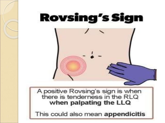
2. Rovsing’s Sign (Indirect Tenderness)
Rovsing’s sign relies on the continuous mechanical transmission of intraluminal gas within the large bowel.
- Maneuver: The examiner applies deep, continuous pressure over the left lower quadrant (LLQ) of the abdomen and slides their hand retrograde up the descending colon.
- Positive Result: Pain felt in the right lower quadrant rather than under the examining hand in the left lower quadrant.
- Interpretation: Retrograde displacement of gas increases cecal intraluminal pressure and shifts the inflamed appendiceal tissue against the right parietal peritoneum, confirming localized right-sided peritoneal irritation.
3. Psoas Sign (Cope’s Psoas Sign)
The Psoas sign evaluates inflammation adjacent to the retroperitoneal space and iliopsoas muscle complex.
- Maneuver: The patient lies on their left side (left lateral decubitus position). The examiner passively extends the patient's right hip joint backward while stabilizing the pelvis. Alternatively, the patient can flex the right hip against manual resistance while supine.
- Positive Result: Severe abdominal or flank pain triggered by hip extension or resisted flexion.
- Interpretation: Indicates a retrocecal appendix. The inflamed appendix rests directly over the right psoas major muscle; stretching or contracting the muscle causes friction against the inflamed serosa.
4. Obturator Sign
The Obturator sign identifies peritoneal inflammation deep within the pelvic cavity.
- Maneuver: With the patient supine, the examiner flexes the patient's right hip and right knee to 90-degree angles. The clinician then internally rotates the right thigh by moving the lower leg laterally while maintaining the knee position.
- Positive Result: Pain elicited in the hypogastrium or right lower quadrant during internal rotation.
- Interpretation: Indicates a pelvic appendix. Internal rotation stretches the obturator internus muscle, bringing it into contact with an inflamed appendix residing within the true pelvis.
5. Dunphy’s Sign
Dunphy’s sign is a subtle maneuver designed to detect peritoneal irritation without deep abdominal palpation.
- Maneuver: The clinician asks the patient to cough forcefully while monitoring for pain localization.
- Positive Result: Exacerbation of localized pain in the right lower quadrant upon coughing.
- Interpretation: Sudden increases in intra-abdominal pressure shift the abdominal wall abdominal muscles against the inflamed parietal peritoneum, causing acute localized somatic pain.
6. Blumberg’s Sign (Rebound Tenderness)
- Maneuver: The examiner depresses the abdominal wall slowly and deeply in the RLQ, holds the position for several seconds, and abruptly releases the manual pressure.
- Positive Result: A sharp, severe pain experienced upon sudden release of pressure, often surpassing the pain caused by initial compression.
- Interpretation: Rapid recoil of the parietal peritoneum causes sudden displacement of inflamed tissues, confirming diffuse or focal peritonitis.
7. Markle Sign (Heel Drop Test)
- Maneuver: The patient stands on their toes and drops abruptly down onto their heels, striking the floor with full weight. If the patient is bedridden, the examiner firmly taps the bottom of the patient's right heel with a fist while the leg is fully extended.
- Positive Result: Sharply localized pain in the right lower quadrant triggered by the vertical impact force.
- Interpretation: Vibrational energy travels vertically through the musculoskeletal system, jarring the abdominal organs and contacting the inflamed peritoneum.
Comparison of Clinical Signs
The table below outlines the execution, physiological mechanism, and clinical performance characteristics of common appendicitis physical signs:
| Clinical Sign | Examination Technique | Underlying Anatomical Mechanism | Diagnostic Sensitivity | Diagnostic Specificity | Diagnostic Utility & Interpretation Notes |
|---|---|---|---|---|---|
| McBurney’s Point Tenderness | Deep focal pressure applied 1/3 distance from ASIS to umbilicus. | Direct compression of inflamed appendix/parietal peritoneum. | High (50%–94%) | Moderate (75%–86%) | Most reliable early physical exam finding; less sensitive if appendix is retrocecal. |
| Rovsing’s Sign | Deep, retrograde continuous compression over LLQ. | Retrograde gas movement distends cecum, irritating right parietal peritoneum. | Low–Moderate (22%–68%) | High (58%–96%) | High specificity confirms appendicitis; negative result does not rule it out. |
| Psoas Sign | Passive right hip extension in left decubitus position or resisted hip flexion. | Focal friction between inflamed retrocecal appendix and iliopsoas muscle. | Low (16%–42%) | Very High (85%–98%) | Strong indicator of retrocecal anatomical positioning. |
| Obturator Sign | Passive internal rotation of flexed right hip at 90° angle. | Friction between inflamed pelvic appendix and obturator internus muscle. | Very Low (8%–28%) | High (80%–95%) | Indicates pelvic appendix; low sensitivity limits screening utility. |
| Dunphy’s Sign | Induced forceful coughing or table jarring. | Sudden surge in intra-abdominal pressure shifts parietal peritoneum. | Moderate (40%–65%) | Moderate–High (70%–90%) | Useful in pediatric or uncooperative patients to avoid painful palpation. |
| Rebound Tenderness (Blumberg) | Slow deep abdominal compression followed by sudden release. | Rapid mechanical recoil of inflamed parietal peritoneal surface. | High (63%–95%) | Moderate (50%–80%) | Marks progression from localized mucosal inflammation to parietal peritonitis. |
| Markle (Heel Drop) Sign | Abrupt landing on heels from toes or firm heel strike. | Vertical mechanical vibration jarring inflamed abdominal organs. | Moderate (60%–75%) | High (80%–90%) | Excellent screening maneuver before formal abdominal palpation. |
Clinical Scoring Systems and Risk Stratification
Because individual clinical signs vary in sensitivity and specificity, validated clinical scoring models combine physical signs, patient history, and basic laboratory findings to stratify risk.
1. The Alvarado Score (MANTRELS)
The Alvarado score is a widely used 10-point scoring system for evaluating suspected appendicitis.
- M - Migration of pain to RLQ (1 Point)
- A - Anorexia (1 Point)
- N - Nausea / Vomiting (1 Point)
- T - Tenderness in RLQ (2 Points)
- R - Rebound tenderness (1 Point)
- E - Elevated temperature (≥ 37.3°C/99.1°F) (1 Point)
- L - Leukocytosis (WBC > 10,000/µL) (2 Points)
- S - Shift to the left (PMN > 75%) (1 Point)
- TOTAL POSSIBLE SCORE = 10 Points
Diagnostic Risk Interpretation:
- Score 1–4 (Low Risk): Appendicitis is unlikely (negative predictive value >90%). Search for alternative diagnoses; safe for outpatient discharge with safety netting instructions.
- Score 5–6 (Intermediate Risk): Appendicitis is possible. Cross-sectional imaging (abdominal ultrasound or non-contrast/contrast CT) is indicated before surgical consultation.
- Score 7–10 (High Risk): Appendicitis is probable. Immediate surgical consultation is indicated; advanced imaging may be omitted in adult males to prevent delays in operative care, depending on institutional protocols.
2. The Appendicitis Inflammatory Response (AIR) Score
The AIR score improves diagnostic precision over the Alvarado score by using quantitative C-reactive protein (CRP) values and categorized rebound tenderness grading.
- Components: RLQ pain, light/medium/severe rebound tenderness, fever (≥38.5°C), leukocytosis (10,000–14,900/µL or ≥15,000/µL), proportion of polymorphonuclear neutrophils (70%–84% or ≥85%), and CRP elevation (10–49 mg/L or ≥50 mg/L).
- Utility: Outperforms the Alvarado score in reducing negative appendectomy rates and identifying complicated (perforated or gangrenous) appendicitis.
Presentation Nuances by Anatomical Position and Patient Population
The precision of physical signs depends on the anatomical orientation of the appendix and specific patient demographics.
- Diagnostic Variations
- Anatomical
- Retrocecal (Minimal Anterior Tenderness)
- Pelvic (Urinary/Rectal)
- Pregnancy
- Superior/Lateral Displacement
- Alder/Bryan Signs
- Demographics
- Pediatric (High Perforation)
- Geriatric (Blunted Response)
- Anatomical
Anatomical Position Variations
- Retrocecal Appendix (60%–65% of population): The appendix lies behind the cecum in the retroperitoneal space. Anterior abdominal wall tenderness is minimal or absent because the cecum cushions pressure. McBurney's point tenderness may be negative, while the Psoas sign and flank tenderness are positive.
- Pelvic Appendix (30%–35% of population): The appendix extends down into the true pelvis. Anterior abdominal wall guarding is often absent. Patients may present with dysuria, urinary frequency, diarrhea, or tenesmus due to adjacent bladder or rectal irritation. The Obturator sign is positive, and direct tenderness may only be elicited on digital rectal or pelvic examination.
- Subhepatic or Retroileal Appendix: Inflammation near the gallbladder or posterior to the terminal ileum can mimic acute cholecystitis or isolated enteritis, obscuring classic RLQ physical signs.
Special Populations
Pregnancy
As the uterus enlarges, it displaces the cecum and appendix superiorly and laterally toward the right upper quadrant (RUQ).
- Exam Findings: McBurney's point tenderness migrates upward as gestational age increases.
- Special Signs:
- Alder’s Sign: Differentiates uterine pain from extrauterine appendiceal pain. If the patient lies on her left side and pain persists at the same focal point, an extrauterine source (appendicitis) is likely.
- Bryan’s Sign: Exacerbation of pain when the uterus is shifted toward the right side.
- Risk: Appendicitis is the most common non-obstetric surgical emergency during pregnancy. Delayed diagnosis increases the risk of fetal loss and maternal morbidity.
Pediatric Patients
Infants and small children under five years old rarely present with classic pain migration due to difficulty verbalizing symptoms.
- Exam Findings: Behavioral signs, such as irritability, refusal to walk, lethargy, and hip flexion while lying down, are critical clinical indicators.
- Risk: The pediatric omentum is underdeveloped and incapable of effectively walling off infection, leading to higher rates of early perforation (up to 40%–50% at presentation).
Geriatric Patients
Older adults often present with atypical or blunted manifestations due to age-related immunosenescence and neurosensory changes.
- Exam Findings: Abdominal pain may be mild or vague; classic signs like fever, guarding, and rebound tenderness are absent in over half of elderly cases.
- Risk: Delays in seeking treatment and subtle exam findings lead to higher perforation rates and increased mortality. Maintain a low threshold for diagnostic cross-sectional imaging in elderly patients with acute abdominal complaints.
Laboratory and Imaging Integration
A positive physical examination sign provides initial diagnostic direction, but confirming acute appendicitis requires targeted laboratory tests and diagnostic imaging.
- Initial Clinical Exam (McBurney's, Rovsing's, Psoas)
- Laboratory Panel (WBC + Left Shift, CRP)
- Risk Stratification (Alvarado / AIR Score Assessment)
- Diagnostic Imaging
- Intermediate / High Risk: Abdominal CT Scan (IV Contrast)
- Pediatric / Pregnant: Abdominal Ultrasound / MRI
- Surgical Consultation / Intervention
Laboratory Biomarkers
- Leukocytosis: White blood cell (WBC) count > 10,000/µL is common. A WBC count > 15,000/µL often correlates with gangrenous or perforated appendicitis.
- Left Shift: Absolute neutrophil count (ANC) > 7,500/µL or band forms > 10% indicates an active acute inflammatory response.
- C-Reactive Protein (CRP): Elevated CRP levels (>10 mg/L) lag behind leukocytosis by 12–24 hours but show high sensitivity for complicated appendicitis.
- Negative Predictive Value: The combination of a normal WBC count and a normal CRP level has a negative predictive value of 90%–95% for acute appendicitis.
Diagnostic Imaging Modalities
- Abdominal Ultrasound: Primary initial imaging modality for pediatric patients and pregnant women to avoid ionizing radiation.
- Positive Criteria: Outer appendiceal diameter > 6 mm, non-compressible outer lumen in transverse view, target sign, appendicolith (fecalith), hyperemic appendiceal wall on color Doppler, and periappendiceal fat hyperechoic stranding or fluid collection.
- Computed Tomography (CT) of Abdomen and Pelvis: Standard imaging choice for adult males and non-pregnant females.
- Positive Criteria: Dilated appendix > 6 mm with luminal distension, wall thickening (> 2 mm), circumferential contrast wall enhancement, periappendiceal fat stranding, cecal bar sign, appendicolith, or extraluminal gas indicating perforation. CT maintains sensitivity and specificity rates above 95%.
- Magnetic Resonance Imaging (MRI): Second-line modality for pregnant patients when ultrasound findings are inconclusive.
- Positive Criteria: Appendiceal fluid-filled lumen diameter > 7 mm, wall hyperintensity on T2-weighted images, and surrounding periappendiceal soft-tissue edema.
Differential Diagnosis
When evaluating positive abdominal signs, clinicians must distinguish acute appendicitis from other intra-abdominal, pelvic, or retroperitoneal conditions:
Gastrointestinal Etiologies
- Acute Mesenteric Adenitis: Common in children; often preceded by an upper respiratory infection. Features self-limiting lymph node enlargement without focal appendiceal wall thickening on ultrasound.
- Terminal Ileitis (Crohn’s Disease): Manifests with right lower quadrant pain, chronic diarrhea, low-grade fever, and transmural bowel thickening on imaging.
- Cecal or Meckel’s Diverticulitis: Presents with focal right lower quadrant pain that can mimic acute appendicitis.
- Acute Gastroenteritis: Features diffuse cramping abdominal pain, prominent vomiting, and hyperactive bowel sounds before pain onset, without localized peritoneal signs.
Gynecological Etiologies
- Pelvic Inflammatory Disease (PID) / Salpingitis: Usually presents with bilateral lower quadrant pain, cervical motion tenderness, purulent vaginal discharge, and adnexal tenderness.
- Ruptured Ovarian Cyst: Sudden-onset sharp lower quadrant pain occurring mid-menstrual cycle; validated by pelvic ultrasound showing pelvic free fluid and absence of appendiceal wall thickening.
- Ectopic Pregnancy: Sudden lower quadrant pain with vaginal bleeding in a female of childbearing age; ruled out or confirmed using urine/serum beta-hCG testing and transvaginal ultrasound.
- Ovarian Torsion: Severe, acute lower quadrant pain with Doppler evidence of compromised adnexal arterial or venous blood flow.
Urological Etiologies
- Right Ureterolithiasis (Kidney Stone): Acute, severe colicky flank pain radiating to the groin/labia/scrotum, often accompanied by hematuria, without focal abdominal peritoneal signs.
- Pyelonephritis: Marked by flank pain, costovertebral angle tenderness, high fever, and pyuria.
Frequently Asked Questions
What is the single most reliable physical examination sign for acute appendicitis?
McBurney’s point tenderness is the most sensitive early physical examination indicator for acute appendicitis. However, no single physical exam sign is 100% diagnostic on its own. Clinicians integrate physical exam findings with localized peritoneal signs (such as Rovsing's sign or rebound tenderness), laboratory inflammation panels, and clinical prediction scores.
Can a patient have acute appendicitis with a negative McBurney’s sign?
Yes. A negative McBurney’s sign occurs frequently when the appendix sits in a retrocecal or deep pelvic anatomical position (up to 30–40% of cases). In these variations, anterior abdominal wall compression does not directly touch the inflamed organ. Clinicians must perform alternative maneuvers, such as the Psoas sign for retrocecal appendicitis or the Obturator sign for pelvic appendicitis.
How does the Psoas sign differ from the Obturator sign in clinical practice?
Both signs detect appendiceal inflammation, but they target different anatomical locations:
- The Psoas sign involves hip extension (or resisted flexion), stretching the psoas major muscle to identify a retrocecal appendix.
- The Obturator sign involves internal rotation of the flexed hip, stretching the obturator internus muscle to identify a pelvic appendix.
Why does abdominal pain shift from the periumbilical region to the right lower quadrant?
The pain shift reflects the underlying neuroanatomy. Early appendiceal lumen distension sends visceral nerve signals via C-fibers to the T10 spinal cord level, causing dull, unlocalized periumbilical discomfort. As inflammation spreads to the outer serosal surface and contacts the parietal peritoneum, sharp somatic pain signals travel via somatic A-delta fibers, localizing directly to the right lower quadrant.
What Alvarado score threshold warrants immediate surgical consultation?
An Alvarado score of 7 or higher indicates a high probability of acute appendicitis and warrants urgent surgical consultation. Scores between 5 and 6 represent intermediate probability and require cross-sectional imaging (such as an abdominal CT scan or ultrasound) to clarify the diagnosis.
Diagnostic Sequence and Action Plan
When acute appendicitis is clinically suspected based on positive signs, follow this step-by-step evaluation workflow:
Step 1: Focused Clinical History & Physical Exam
- Establish pain onset, migration pattern, systemic symptoms
- Test McBurney's, Rovsing's, Psoas, Obturator, and Dunphy's signs
Step 2: Core Laboratory Diagnostics
- Order CBC with differential (WBC, absolute neutrophil count)
- Obtain quantitative C-reactive protein (CRP)
- Perform urine pregnancy test (beta-hCG) in females of childbearing age
- Order urinalysis to rule out urolithiasis and urinary tract infection
Step 3: Diagnostic Scoring & Risk Stratification
- Calculate Alvarado or AIR score based on clinical findings and labs
Step 4: Targeted Diagnostic Imaging
- Low Risk (Alvarado 1-4): Evaluate alternative diagnoses; discharge with explicit red-flag return instructions
- Intermediate Risk (Alvarado 5-6): Obtain targeted imaging (Ultrasound for pediatric/pregnant; CT with IV contrast for adults)
- High Risk (Alvarado 7-10): Obtain urgent surgical consultation; proceed to imaging per institutional pathway
Step 5: Definitive Surgical Consultation & Management
- Maintain NPO (nothing by mouth) status and administer IV fluid resuscitation
- Provide appropriate IV analgesia (controlled analgesia does not obscure clinical examination accuracy)
- Prepare for laparoscopic or open appendectomy upon surgical confirmation
By systematically applying physical exam signs, clinical risk scoring, and targeted imaging, clinicians can rapidly confirm acute appendicitis, reduce negative appendectomy rates, and prevent complications such as gangrene or perforation.