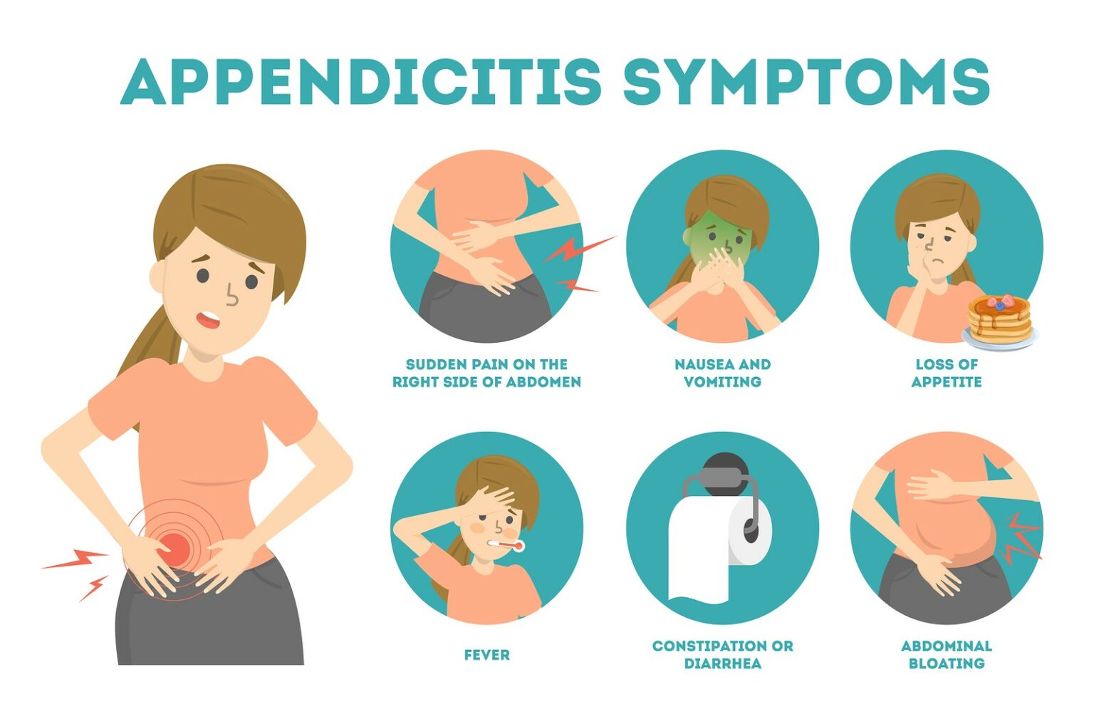
A primary nursing diagnosis for appendicitis centers on Acute Pain related to appendiceal obstruction and peritoneal inflammation, alongside Risk for Deficient Fluid Volume and Risk for Infection. Nurse-led pain identification requires tracking the progression from dull periumbilical pain to sharp right lower quadrant localization at McBurney’s point, while prevention focuses on mitigating perforation, peritonitis, and postoperative complications.
Pathophysiology of Acute Appendicitis
Acute appendicitis remains one of the most frequent causes of emergency abdominal surgery globally. The condition begins when the lumen of the vermiform appendix—a small, worm-like pouch attached to the cecum near the ileocecal valve—becomes obstructed. The primary cause of luminal obstruction in adults is a fecalith (a hardened mass of fecal matter). In children and young adults, lymphoid hyperplasia secondary to viral or bacterial infections is the dominant cause. Less common etiologies include foreign bodies, strictures, intestinal parasites (such as Enterobius vermicularis), and appendiceal tumors.
- Luminal Obstruction (Fecalith / Lymphoid Hyperplasia)
- Continued Mucus Secretion & Bacterial Proliferation
- Increased Intraluminal Pressure & Venous Stasis
- Ischemia, Transmural Necrosis & Gangrene
- Perforation & Peritonitis (if untreated within 24–72 hours)
Once luminal obstruction occurs, mucosal secretions continue unchecked behind the blockage, causing a rapid rise in intraluminal pressure. This rising pressure compresses mucosal venules and lymphatic vessels, leading to localized ischemia, venous stasis, and tissue hypoxia. Normal enteric flora—particularly anaerobic species like Bacteroides fragilis and aerobic species like Escherichia coli—overgrow rapidly within the stagnant lumen.
If the obstruction persists without surgical or medical intervention, mucosal integrity breaks down, progressing to transmural necrosis, gangrene, and eventual perforation of the appendiceal wall. Appendiceal perforation leads to the spill of purulent fluid and fecal content into the peritoneal cavity, resulting in localized abscess formation or diffuse peritonitis. Understanding this rapid pathophysiological timeline (often progressing from onset to perforation within 24 to 72 hours) is essential for nurses conducting rapid assessment, formulating timely nursing diagnoses, and prioritizing urgent care.
Pain Identification and Physical Assessment Findings
Accurate identification of abdominal pain is the hallmark of effective appendicitis assessment. Pain characteristics evolve alongside the underlying inflammation, shifting from visceral nerve pathways to parietal peritoneal irritation.
The Pain Trajectory: From Visceral to Somatic Localized Pain
Visceral Pain Phase (Early Stage): In the early hours of appendicitis, rising intraluminal pressure stretches the visceral peritoneum of the appendix. Visceral sensory innervation to the appendix travels via visceral afferent sympathetic nerve fibers to the T10 segment of the spinal cord. Because visceral innervation is diffuse and poorly localized, the patient experiences a dull, aching, vague epigastric or periumbilical discomfort. This phase is frequently accompanied by systemic manifestations such as anorexia, low-grade fever, and mild nausea or vomiting.
Somatic Pain Phase (Late Stage): As inflammatory exudate extends through the appendiceal wall to touch the parietal peritoneum, the nature of the pain alters dramatically. Parietal peritoneal innervation is mediated by somatic spinal nerves, which provide sharp, intense, and precisely localized sensation. The pain shifts from the periumbilical region to the Right Lower Quadrant (RLQ) of the abdomen, specifically settling at McBurney’s point. McBurney’s point is anatomically defined as the location two-thirds of the distance along an imaginary line drawn from the umbilicus to the right anterior superior iliac spine (ASIS).
Classic Physical Assessment Signs
Nurses must perform targeted physical examinations using standardized clinical signs to evaluate peritoneal irritation. Detailed documentation of these physical findings aids the interprofessional team in determining diagnostic urgency.
Blumberg's Sign (Rebound Tenderness): The examiner presses deeply and gently into the right lower quadrant and then releases the pressure rapidly. A sudden, sharp spike in pain upon release indicates parietal peritoneal inflammation.
Rovsing's Sign: Deep palpation of the patient’s left lower quadrant (LLQ) increases pressure within the large bowel, displacement of gas toward the cecum, and subsequent stretching of the inflamed RLQ peritoneum, causing pain in the right lower quadrant.
Psoas Sign (Cope's Psoas Test): With the patient lying on their left side, the examiner passively hyperextends the right leg backward at the hip. Alternatively, the patient actively flexes the right hip against resistance. Pain in the RLQ suggests irritation of the iliopsoas muscle, typically caused by a retrocecal appendix.
Obturator Sign: The patient lies supine with the right hip and knee flexed at 90 degrees. The examiner internally rotates the right lower extremity. Pain elicited in the hypogastrium or RLQ indicates irritation of the internal obturator muscle, often associated with a pelvic appendix position.
[Umbilicus]
- 2/3 Distance
- McBurney's Point (Maximal Somatic Pain)
- 1/3 Distance
- [Anterior Superior Iliac Spine (ASIS)]
Critical Warning Sign: Sudden Relief of Pain
A sudden, sharp drop in pain intensity reported by a patient who previously experienced severe, crescendo abdominal pain is a critical medical red flag. Sudden pain cessation usually signifies that the appendix has ruptured, relieving the acute intraluminal pressure that drove the initial pain.
However, this relief is temporary. Within hours of perforation, spilled intestinal material induces generalized peritonitis, presenting as diffuse, severe abdominal pain, board-like abdominal rigidity, high fever, tachycardia, and signs of systemic inflammatory response syndrome (SIRS) or septic shock. Nurses must notify the surgical provider immediately if a patient reports sudden pain resolution.
Formulating Appendicitis Nursing Diagnoses (NANDA-I Aligned)
Nursing diagnoses structure clinical decision-making, direct nursing interventions, and establish clear criteria for evaluating patient outcomes. According to NANDA-International (NANDA-I) terminology, several primary nursing diagnoses apply to patients with acute appendicitis in both preoperative and postoperative settings.
1. Acute Pain
- Related to: Inflammation and swelling of the appendiceal wall, parietal peritoneal irritation, surgical tissue trauma, and abdominal incision.
- Evidenced by: Patient self-report of pain at McBurney's point (numeric rating scale score of 7/10 or higher), guarding behavior, facial grimacing, restlessness, tachycardia, and elevated blood pressure.
- Desired Outcomes: Patient will report a decrease in pain intensity to a manageable level (e.g., ≤3/10), demonstrate relaxed body posture, and engage comfortably in deep breathing exercises and mobilization postoperatively.
2. Risk for Deficient Fluid Volume
- Related to: Preoperative NPO (nothing by mouth) restriction, fluid loss through vomiting, elevated metabolic rate associated with fever, and potential fluid sequestration (third-spacing) in the peritoneal cavity.
- Risk Factors: Persistent nausea, decreased oral intake, hyperthermia, and inflammatory exudate accumulation.
- Desired Outcomes: Patient will maintain adequate hydration as evidenced by balanced fluid intake and output, moist mucous membranes, stable blood pressure, normal skin turgor, and a urine output of at least 0.5 mL/kg/hour.
3. Risk for Infection
- Related to: Impaired tissue integrity secondary to appendiceal rupture, spill of enteric contents into the peritoneal cavity, invasive surgical incisions (laparoscopic trocars or open laparotomy), and retained fluid collections/abscesses.
- Risk Factors: Ruptured appendix, operative site contamination, delayed surgical intervention, and compromised immune response.
- Desired Outcomes: Patient will remain free from localized incision infection or systemic intra-abdominal infection, evidenced by normal body temperature (<38.0°C / 100.4°F), normal white blood cell (WBC) counts, and surgical wounds showing clean, healing edges without purulent drainage.
4. Deficient Knowledge (Perioperative Care)
- Related to: Lack of exposure to surgical procedures, emergency nature of the diagnosis, and unfamiliarity with post-discharge activity and wound care instructions.
- Evidenced by: Frequent questions regarding the procedure, expressed anxiety, or verbalization of misconceptions regarding post-surgical recovery.
- Desired Outcomes: Patient and caregivers will articulate the rationale for surgical intervention, explain post-discharge wound care procedures, identify red-flag symptoms requiring emergency medical evaluation, and follow lifting and activity restrictions.
Nursing Care Plan Matrix for Appendicitis
The following structured nursing care plan details standardized diagnoses, evidence-based nursing interventions, rationales, and evaluation criteria across the preoperative and postoperative continuum.
| Nursing Diagnosis | Clinical Etiology / Related Factors | Diagnostic Indicators / Evidence | Targeted Interventions & Rationales | Outcome Evaluation Criteria |
|---|---|---|---|---|
| Acute Pain | Appendiceal distension, peritoneal irritation, surgical incision. | Numeric pain score >6/10, RLQ guarding, facial grimacing, tachycardia. | • Assess pain location and intensity every 2–4 hours using validated scales.• Place patient in semi-Fowler's position with knees flexed to reduce abdominal wall tension.• Administer IV analgesics (e.g., acetaminophen, opioids) as ordered post-diagnosis.• Avoid applying heat to the abdomen to prevent rupture. | Patient reports pain level ≤3/10 within 45 minutes of analgesic administration and exhibits relaxed facial expression. |
| Risk for Deficient Fluid Volume | Preoperative NPO status, repeated emesis, fever, third-spacing. | Dry mucous membranes, oliguria (<30 mL/hr), elevated urine specific gravity, tachycardia. | • Establish IV access and infuse isotonic crystalloids (0.9% Normal Saline or Lactated Ringer's) as ordered.• Maintain strict Intake and Output (I&O) recording.• Administer antiemetics (e.g., ondansetron) to control emesis.• Monitor serum electrolytes and hematocrit levels. | Urine output maintained >0.5 mL/kg/hr; heart rate 60–100 bpm; skin turgor elastic; mucous membranes moist. |
| Risk for Infection | Appendiceal rupture, peritoneal contamination, surgical incision. | Fever, leukocytosis (>12,000/mcL), purulent incisional drainage, worsening diffuse abdominal pain. | • Administer prophylactic broad-spectrum IV antibiotics (e.g., cefoxitin, ampicillin-sulbactam) as ordered.• Perform wound dressing changes using strict aseptic technique.• Monitor surgical site for erythema, warmth, and purulent discharge.• Monitor vital signs and WBC trend daily. | Surgical incision remains intact without erythema or purulent drainage; body temperature remains within normal limits (<38.0°C). |
| Risk for Impaired Gastrointestinal Motility | Anesthesia, abdominal surgery, peritoneal inflammation, bowel handling. | Absent or hypoactive bowel sounds, abdominal distension, failure to pass flatus or stool, nausea. | • Auscultate bowel sounds in all 4 quadrants every 4–8 hours.• Encourage early ambulation postoperatively to stimulate peristalsis.• Maintain NPO status until bowel sounds return or flatus is passed.• Progress diet gradually from clear liquids to regular food as tolerated. | Patient passes flatus and soft formed stool; tolerates regular diet without nausea, vomiting, or abdominal distension. |
| Deficient Knowledge | Unfamiliarity with surgical recovery, emergency admission. | Expression of confusion, improper wound handling, anxiety about activity restrictions. | • Provide concise written and verbal instruction on incisional wound care.• Educate on red-flag symptoms: high fever, increasing pain, redness, or drainage.• Review activity limits (e.g., no heavy lifting >10 lbs for 2–4 weeks).• Verify understanding using the "teach-back" technique. | Patient accurately demonstrates wound care procedures and correctly identifies 4 signs requiring medical re-evaluation. |
Preoperative and Postoperative Nursing Interventions
Managing an appendicitis patient requires attention to safety protocols before surgery, followed by post-operative strategies to avoid complications.
Preoperative Safety Protocols and Absolute Contraindications
When a patient is admitted with suspected acute appendicitis, specific precautions must be implemented to reduce the risk of appendiceal rupture:
Strict NPO Status: Keep the patient strictly nothing-by-mouth to prepare for emergency appendectomy under general anesthesia and to rest the gastrointestinal tract.
Contraindication of Heat Application: Never apply hot packs or heating pads to the patient’s abdomen. Heat causes local vasodilation, increases blood flow and intraluminal pressure within the hyperemic appendix, and significantly heightens the risk of appendiceal rupture. Cold packs may be applied for comfort if cleared by the clinical team, though repositioning is preferred.
Avoidance of Laxatives and Enemas: Never administer laxatives, cathartics, or enemas to a patient with suspected appendicitis. Stimulating intestinal peristalsis significantly increases the mechanical risk of appendix perforation.
Cautious Analgesic Timing: Historically, analgesics were withheld prior to surgical evaluation out of concern that masking pain would delay diagnosis. Modern clinical practice, backed by evidence summarized in resources such as the National Center for Biotechnology Information (NCBI) StatPearls, supports administering titrated IV analgesia once an initial evaluation is under way, as it improves patient comfort without compromising diagnostic accuracy.
Intravenous Access and Hydration: Insert a large-bore peripheral IV catheter (18–20 gauge) for rapid fluid resuscitation with IV crystalloids to correct fluid deficits from fever and emesis.
PREOPERATIVE CONTRAINDICATIONS & ALERTS
- NO Heating Pads on Abdomen: Risk of vasodilation & rupture
- NO Laxatives or Enemas: Risk of peristaltic rupture
- NO Oral Intake (Strict NPO): Prepares for anesthesia
- [!] ALERT ON Pain Sudden Loss: Sign of appendiceal rupture
Postoperative Nursing Care and Complication Prevention
Postoperative care focuses on physiological recovery, wound healing, pain management, and early detection of postoperative complications such as pelvic abscesses, wound infection, or paralytic ileus.
1. Surgical Site Care
- For laparoscopic appendectomy (the primary approach in uncomplicated cases), inspect 3 to 4 small port incisions for signs of bleeding, hematoma, or infection.
- For open appendectomy (often required for severe rupture or widespread peritonitis via a McBurney's or lower midline incision), monitor abdominal dressings closely. If drains (such as a Jackson-Pratt drain) are placed to clear purulent exudate, document output quality and color, and record drainage volume every 8 hours.
2. Gastrointestinal Function Recovery
- Monitor bowel sounds across all four quadrants.
- Encourage early ambulation within 6 to 12 hours after surgery as tolerated. Early walking promotes bowel peristalsis, helps clear trapped intraperitoneal CO2 gas used during laparoscopy (which can cause shoulder tip pain), and reduces the risk of paralytic ileus.
- Advance diet slowly from clear liquids to a regular diet once bowel sounds return and the patient passes flatus.
3. Pulmonary Hygiene
- Teach the patient to use an incentive spirometer (10 breaths every hour while awake) and practice splinting their abdominal incision with a pillow while coughing. This prevents postoperative atelectasis and pneumonia.
Appendicitis Prevention: Primary and Secondary Clinical Insights
Patients and families frequently ask whether appendicitis can be prevented. Clear educational guidance requires distinguishing between primary prevention (preventing the initial onset) and secondary/tertiary prevention (preventing severe complications once disease begins).
Primary Prevention
Acute appendicitis cannot be prevented with certainty because factors such as lymphoid tissue enlargement, unexpected foreign bodies, or anatomical variations are largely uncontrollable. However, epidemiological research published through public health agencies like the National Institute of Diabetes and Digestive and Kidney Diseases (NIDDK) highlights a strong association between dietary fiber and appendiceal health:
- High-Fiber Diets: Consuming a diet rich in dietary fiber—including fresh fruits, leafy vegetables, legumes, whole grains, and nuts—improves overall stool consistency and speeds gastrointestinal transit time.
- Reducing Fecalith Formation: Enhanced bowel motility reduces the likelihood of hard, dry stool formation and constipation, directly lowering the probability of fecalith development—the primary mechanical cause of appendiceal luminal obstruction.
Secondary and Tertiary Prevention
In clinical nursing, prevention shifts toward avoiding adverse complications through vigilant care:
- Preventing Rupture: Timely recognition of evolving symptoms, immediate surgical referral, and strict avoidance of heat packs or laxatives minimize the risk of luminal rupture.
- Preventing Surgical Site Infection (SSI): Administering prophylactic broad-spectrum IV antibiotics within 60 minutes prior to surgical incision, utilizing sterile technique during wound dressing changes, and providing clear home-care instructions prevent bacterial colonization.
- Preventing Sepsis and Abscess Formation: In cases of perforated appendicitis, ensuring complete peritoneal irrigation intraoperatively and maintaining appropriate postoperative IV antibiotic therapy prevents intra-abdominal abscess formation and systemic sepsis.
Frequently Asked Questions
What is the single most important nursing priority when acute appendicitis is suspected?
The top priority is maintaining strict NPO status while avoiding interventions that risk perforation, such as applying heat to the abdomen or administering laxatives. The nurse must promptly assess vital signs and abdominal characteristics, secure IV access, and notify the surgical provider to expedite formal evaluation.
How does a nurse tell the difference between appendicitis pain and other causes of acute abdominal pain?
Appendicitis pain typically follows a characteristic progression: it starts as a dull, poorly localized periumbilical or epigastric ache (visceral phase) and shifts over 12 to 24 hours to a sharp, intense pain localized to the right lower quadrant at McBurney's point (somatic phase). Pain that worsens with movement, coughing, or light percussion, combined with rebound tenderness and positive Rovsing or Psoas signs, strongly points toward appendicitis rather than general gastroenteritis or gynecological pain.
Why are heating pads strictly contraindicated for appendicitis pain management?
Applying heat to the abdomen causes localized vascular dilation and increased blood flow to the inflamed appendix. This increases fluid accumulation, hyperemic swelling, and intraluminal pressure, significantly raising the risk that the appendix wall will rupture and spill infectious contents into the peritoneal cavity.
What should a nurse do if a patient reports that their appendicitis pain suddenly vanished?
The nurse must immediately re-assess the patient's vital signs and notify the attending surgeon. A sudden loss of severe pain typically indicates that the appendix has ruptured, relieving the pressure that caused the initial pain. This is a surgical emergency; without rapid intervention, the patient will develop diffuse peritonitis and potential septic shock.
Can eating a high-fiber diet completely prevent appendicitis?
While a high-fiber diet cannot completely eliminate the risk of appendicitis, epidemiological studies show that populations with high dietary fiber intake have a significantly lower incidence of the condition. Fiber adds bulk and moisture to stool, reducing constipation and decreasing the formation of fecaliths—the main mechanical cause of appendiceal obstruction.
Actionable Nursing Practice Guidance
To optimize patient outcomes when managing appendicitis, nurses should follow this clear operational framework:
- Perform Continuous Pain & Abdominal Re-Assessments: Evaluate pain characteristics, location, and intensity every 2 to 4 hours. Document whether pain remains localized to McBurney's point or becomes diffuse, and report any sudden relief or sharp increases in abdominal rigidity immediately.
- Enforce Strict Preoperative Safety: Maintain NPO status, establish IV access for fluid resuscitation, ensure no heating pads or laxatives are used, and prepare the patient for imaging (ultrasound or contrast-enhanced CT scan) as ordered.
- Deliver Comprehensive Postoperative Care: Prioritize early ambulation to restore bowel motility and prevent venous thromboembolism. Instruct the patient on incentive spirometry and incision splinting, monitor wound dressing sites for infection, and ensure pain is adequately controlled to support mobility.
- Ensure Effective Discharge Education: Teach the patient and family how to care for surgical wounds, emphasize avoiding heavy lifting (>10 lbs) for 2 to 4 weeks, and outline red-flag symptoms requiring immediate emergency care, including fever >38.0°C (100.4°F), worsening abdominal pain, persistent vomiting, or purulent wound drainage.