Active transport is a vital cellular mechanism that moves ions and molecules across biological membranes against their electrochemical or concentration gradients. By utilizing cellular energy—typically in the form of adenosine triphosphate (ATP)—cells maintain homeostatic internal environments, regulate nutrient uptake, and generate the necessary electrical potential for signal transduction and muscle contraction.
The Energetic Necessity of Active Transport
Cells exist in a state of dynamic equilibrium with their external environment. While many small, non-polar molecules can traverse the lipid bilayer via passive diffusion, larger molecules and ions require assistance. Passive transport relies on the random kinetic motion of particles moving from areas of high concentration to low concentration. However, biological survival often dictates that a cell must accumulate specific substances to levels significantly higher than those found in the interstitial fluid or bloodstream.
Without active transport, cells would be unable to concentrate essential nutrients like glucose or amino acids, nor could they discard toxic waste products once their concentrations inside the cell matched those outside. By coupling transport to the hydrolysis of ATP—or, in secondary instances, to the established gradient of another molecule—cells override the natural tendency toward entropy within the cytoplasmic space. This energy-dependent process is the primary reason cells can maintain distinct chemical environments that differ radically from their surroundings.
Primary vs. Secondary Active Transport
Active transport is categorized based on the source of energy utilized to drive the movement of solutes. Understanding this distinction is fundamental to grasping how complex tissues, such as neurons and renal tubules, function under varying physiological demands.
Primary Active Transport
In primary active transport, the protein transporter itself acts as an enzyme, binding and hydrolyzing ATP directly to provide the mechanical energy required for translocation. The most prominent example is the sodium-potassium pump (Na⁺/K⁺-ATPase). This protein is ubiquitous in animal cells; it pumps three sodium ions out of the cell and two potassium ions into the cell for every ATP molecule consumed. This action not only manages cell volume but also creates a significant electrochemical gradient across the plasma membrane, which serves as a reservoir of potential energy for other cellular tasks.
Secondary Active Transport
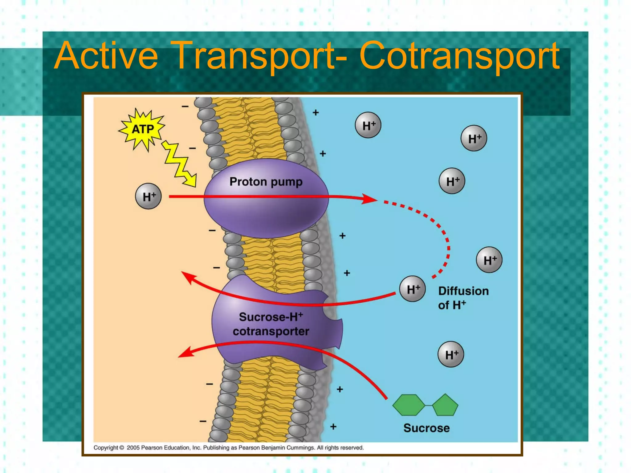
Secondary active transport, or co-transport, does not use ATP directly. Instead, it leverages the potential energy stored in an electrochemical gradient created by a primary active transport process. Transporters called symporters move two substances in the same direction, while antiporters move them in opposite directions. For instance, the SGLT (sodium-glucose linked transporter) allows sodium to move down its steep concentration gradient into the cell, using the energy of that motion to pull glucose molecules against their concentration gradient into the cytoplasm.
Functional Mechanisms of Transport Proteins
Transport proteins, often referred to as pumps, act as molecular "gates" that undergo conformational changes to move cargo. These proteins are highly specific, featuring binding sites with precisely shaped chemical environments that accept only certain substrates.
- Substrate Binding: The target molecule binds to a specific site on the protein’s extracellular or intracellular domain.
- Conformational Change: Upon binding (and often triggered by phosphorylation via ATP), the protein shifts its 3D architecture. This shift effectively "flips" the binding site from one side of the membrane to the other.
- Release: The substrate is deposited on the opposite side of the membrane.
- Resetting: The protein returns to its original shape, ready to repeat the process.
This mechanism ensures that the membrane remains impermeable to the cargo while the transporter is closed, preventing the "leakage" of ions that would otherwise dissipate the carefully maintained gradients.
Comparison of Transport Mechanisms
| Feature | Passive Transport | Primary Active Transport | Secondary Active Transport |
|---|---|---|---|
| Energy Source | Kinetic/Concentration Gradient | ATP Hydrolysis | Electrochemical Gradient |
| Direction | Downhill (High to Low) | Uphill (Low to High) | Uphill (Low to High) |
| Protein Required | Yes (if facilitated) | Yes (Pumps) | Yes (Co-transporters) |
| Efficiency | High (Entropy-driven) | Variable (ATP-limited) | Variable (Gradient-limited) |
Biological Significance and Homeostasis
The implications of active transport extend far beyond basic cellular survival. In the human body, specific systems rely entirely on the precise function of these pumps. For example, in the kidneys, secondary active transport in the proximal convoluted tubule is essential for reabsorbing glucose, ensuring that valuable fuel is not lost in urine. Similarly, the nerve impulses that govern everything from heartbeats to conscious thought are dependent on the gradients established by the Na⁺/K⁺-ATPase pump. If this pump fails—due to metabolic inhibition or hypoxia—neurons lose their resting membrane potential, leading to a cessation of neural activity.
Furthermore, active transport plays a crucial role in organelle function. Lysosomes, for instance, utilize proton pumps (H⁺-ATPase) to move hydrogen ions into their interior. This creates the acidic environment required for the digestive enzymes within the lysosome to function, protecting the rest of the cell from being broken down by these same enzymes.
Common Clinical and Biological Challenges
Dysfunction in active transport mechanisms is linked to numerous human pathologies. When transport proteins are genetically mutated or inhibited, the physiological consequences can be systemic:
- Cystic Fibrosis: This condition involves a defective chloride ion channel (CFTR). While this is technically a facilitated diffusion channel, it illustrates how the disruption of membrane protein integrity causes severe imbalances in mucus viscosity and respiratory health.
- Cardiac Glycosides: Medications such as digoxin, used in treating heart failure, function by inhibiting the Na⁺/K⁺-ATPase pump. By slowing this pump, intracellular sodium levels rise, which eventually increases intracellular calcium, leading to a stronger heart contraction.
- Metabolic Exhaustion: During periods of starvation or extreme physical stress, the cell's inability to produce sufficient ATP causes a failure of active transport. This leads to cell swelling and edema, as the internal concentration of ions can no longer be regulated.
Troubleshooting and Verification in Laboratory Settings
In research environments, scientists verify active transport activity through several standard methodologies. If a study suggests an active transport process, researchers must confirm it is not merely passive diffusion or facilitated transport.
- Metabolic Inhibition: Researchers treat the cells with substances that stop ATP production, such as cyanide or dinitrophenol. If the transport of the molecule ceases, it is confirmed to be ATP-dependent active transport.
- Ion-Dependency Tests: By altering the external concentration of ions (like removing sodium from the medium), researchers can verify if the transport process relies on secondary co-transport.
- Radiolabeled Tracers: Researchers use isotopes to track the movement of specific molecules across the membrane, allowing them to measure the rate of transport precisely over time.
- Patch-Clamping: This electrophysiological technique allows researchers to measure the tiny currents generated by ion pumps, providing a real-time observation of pump kinetics.
Frequently Asked Questions
Why does active transport require ATP? Active transport moves molecules against their natural diffusion gradient. Since this decreases the entropy of the system, it is thermodynamically unfavorable. ATP hydrolysis provides the necessary Gibbs free energy to drive the reaction forward.
Can secondary active transport function without primary active transport? No. Secondary active transport relies entirely on the concentration gradients established by primary active transport. If the primary pumps fail, the electrochemical gradients dissipate, and secondary transport mechanisms stop functioning shortly thereafter.
Does all active transport move molecules into the cell? No. Active transport is bidirectional. While it is often discussed in terms of nutrient uptake, it is equally responsible for exporting metabolic waste, signaling molecules, and excess ions out of the cell to prevent toxic buildup.
Are there limits to how fast active transport can occur? Yes. Unlike simple diffusion, which is limited only by the concentration gradient, active transport is limited by the number of available carrier proteins (saturation kinetics) and the availability of ATP. Once all transport proteins are occupied, the transport rate reaches a Vmax (maximum velocity).
How do cells prevent the "backflow" of transported molecules? Cells use gated conformational changes to prevent backflow. The transporter undergoes structural shifts that ensure the binding site is never simultaneously open to both the extracellular and intracellular environments, maintaining the integrity of the barrier.
Next Steps for Students and Researchers
To further your understanding of active transport, focus on the specific structural biology of the P-type ATPases. Reviewing the crystal structures of these proteins reveals how they change shape to perform their work. For those interested in the clinical application, studying the pharmacology of transport inhibitors provides insight into how modern medicine manipulates these fundamental biological gates to treat complex cardiovascular and neurological diseases. Experimentally, using radioactive isotope tracing remains the gold standard for quantifying these fluxes in a controlled laboratory setting.