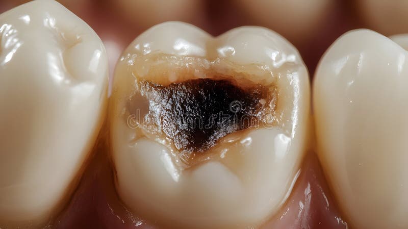
Discovering a molar with a cavity requires prompt professional attention to prevent the progression of tooth decay into the deeper structures of the tooth. When a molar—a large, multi-rooted tooth responsible for heavy grinding—develops a lesion, the integrity of your bite, the health of the dental pulp, and the structural longevity of the tooth are at immediate risk.
The Nature of Cavities in Molars
A molar with a cavity is the result of demineralization caused by acid-producing bacteria that thrive on sugar and starch. Because molars possess deep grooves and fissures on their biting surfaces, they are particularly susceptible to plaque accumulation. If this plaque is not removed through consistent mechanical cleaning, it turns into tartar and creates an environment where enamel begins to dissolve.
The progression of a cavity typically follows a predictable sequence. It begins in the enamel, the hardest substance in the human body. At this stage, the process may be reversible with professional fluoride treatment and enhanced oral hygiene. Once the decay penetrates the enamel and reaches the underlying dentin, the process becomes irreversible, and a restorative procedure is required to halt further destruction.
Recognizing Symptoms and Warning Signs
Identifying a cavity in a molar often occurs before physical pain manifests. However, as the decay deepens, the tooth will begin to communicate its distress.
- Sensitivity: You may notice sudden, sharp pain when consuming hot, cold, or sugary beverages. This occurs because the protective layer of the enamel has been compromised, leaving the dentin—which contains microscopic tubules connected to the tooth’s nerves—exposed to external stimuli.
- Visible Alterations: While many cavities occur in the crevices of the chewing surface (occlusal), they can also appear as brown, black, or chalky white spots.
- Persistent Discomfort: A dull ache or pressure in the molar area, especially when biting down, often indicates that the cavity has compromised the structural integrity of the tooth or reached a depth that causes inflammation of the pulp.
- Food Impaction: If a cavity develops between two molars (interproximal), food may frequently become stuck in the space, causing pressure and localized gingival inflammation.
Diagnostic Procedures
When you consult a dentist, they will perform several diagnostic steps to assess the extent of the cavity. A physical examination using a dental explorer and high-intensity lighting is the first step. Because molar decay often hides in the deep fissures of the crown, visual inspection is rarely sufficient on its own.
Dental radiography, or X-rays, is the primary tool for visualizing the "hidden" aspects of the tooth. Bitewing X-rays are specifically designed to show the crowns of the upper and lower molars in a single image, allowing the dentist to see the extent of decay between teeth and under existing fillings. In 2026, many dental practices utilize digital radiography, which reduces radiation exposure and provides high-resolution images that can be enlarged on a monitor to show patients exactly where the structural compromise exists.
Treatment Options Based on Severity
The treatment approach for a molar with a cavity is dictated entirely by how much healthy tooth structure remains.
1. Dental Sealants (Preventive/Early Stage)
If the cavity is in the very early stages of enamel demineralization, a dentist may opt to clean the surface and apply a sealant. This is a thin, protective resin coating that is bonded to the chewing surface of the molar, effectively sealing off the deep fissures and preventing further bacterial infiltration.
2. Composite Resin Fillings
For small to moderate cavities that have entered the dentin, a composite resin filling is the standard of care. After the dentist removes the decayed portion of the tooth, they etch the surface to create a microscopic texture for bonding. The composite resin is applied in layers, hardened with a specialized light, and shaped to match the original anatomy of your molar.
3. Dental Inlays and Onlays
When the decay is too extensive for a standard filling but does not yet require a full crown, an onlay or inlay may be recommended. These are custom-made restorations fabricated in a lab or via CAD/CAM milling machines. An onlay covers one or more of the "cusps" (the raised points) of the molar, providing the structural reinforcement necessary to withstand the high forces of mastication.
4. Dental Crowns
If a significant portion of the molar is decayed or fractured, a crown (or "cap") is necessary. A crown covers the entire visible portion of the tooth, acting as a structural shield. This restoration is critical for molars, as it prevents the weakened remaining tooth structure from splitting under pressure.
Comparison of Common Restorative Procedures
| Procedure | Scope of Restoration | Material Used | Best For |
|---|---|---|---|
| Filling | Fills the void left by decay | Composite Resin | Minor to moderate damage |
| Inlay | Fits inside the grooves | Porcelain/Ceramic | Moderate decay within cusps |
| Onlay | Covers one or more cusps | Porcelain/Ceramic | Extensive surface damage |
| Crown | Encases entire tooth crown | Zirconia/Porcelain | Severe decay/structural loss |
The Consequences of Neglect
Ignoring a molar with a cavity is a common mistake that leads to significant financial and health complications. If the decay is left to advance unchecked, it will eventually breach the pulp chamber—the center of the tooth housing nerves and blood vessels.
Once the pulp is infected, the condition progresses from a simple restorative matter to a surgical one. You will likely require a root canal procedure, where the infected tissue is removed, the interior is disinfected, and the tooth is sealed. If even this is ignored, an abscess may form, potentially leading to systemic infection and the eventual extraction of the molar. Replacing a lost molar requires an implant or bridge, both of which represent a substantially higher financial investment and a longer treatment timeline than treating the original cavity.
Maintaining Oral Hygiene Post-Treatment
After your molar has been treated, the goal is to prevent a recurrence of decay in that tooth or its neighbors. Restorations do not have an infinite lifespan, and the junction between the filling material and your natural tooth remains a vulnerable site for secondary decay.
- Interdental Cleaning: Use dental floss, interdental brushes, or water flossers daily. Decay frequently begins between molars because standard bristles cannot reach these tight spaces.
- Fluoride Utilization: Use a fluoride-containing toothpaste twice daily to assist in the remineralization of enamel.
- Controlled Sugar Intake: Bacteria thrive on refined sugars. Limiting the frequency of sugary snacks reduces the "acid attack" duration on your dental enamel.
- Routine Professional Care: Regular check-ups allow for the early detection of any wear or leakage around your restorations, often allowing for minor repairs before major work becomes necessary.
Expert Guidance and Next Actions
If you suspect you have a molar with a cavity, your first step is to schedule an appointment with a licensed dentist. Do not attempt "at-home" remedies, such as filling kits or topical numbing agents meant for temporary relief, as these do not remove the underlying infection and may allow the decay to progress further into the tooth structure.
When you arrive at your appointment, ask your dentist to show you the X-rays and explain the classification of the decay. Understanding whether the cavity is in the enamel or the dentin will help you make an informed decision regarding the treatment plan, particularly if you are comparing different restorative materials for an onlay or crown.
Frequently Asked Questions
How quickly does a molar cavity progress? The rate of progression varies significantly based on your diet, saliva chemistry, and oral hygiene routine. Some cavities progress over several years, while others, particularly those in the deep fissures of molars, can reach the pulp within months if hygiene is poor.
Is it painful to get a cavity filled in a molar? With modern local anesthesia, the procedure is generally painless. You will feel pressure during the removal of the decay, but the numbing agents are highly effective at blocking pain signals. If you experience anxiety, discuss sedation options with your dentist.
Do all cavities in molars need to be filled? Not necessarily. Very incipient, shallow cavities confined to the enamel layer can sometimes be arrested or remineralized using high-concentration fluoride treatments and improved daily hygiene, provided the dentist determines the decay has not breached the enamel surface.
How long do molar fillings last? Composite resin fillings typically last between 5 and 10 years, though this depends on the size of the cavity and your biting force. Crowns and onlays often have a longer lifespan, frequently exceeding 15 years with proper maintenance.
Can a cavity in a molar cause bad breath? Yes. A cavity provides a protected environment for bacteria to accumulate and produce sulfur compounds. Furthermore, the decay process can cause food particles to become trapped and decompose within the tooth, contributing to halitosis.
What is the cost range for treating a molar cavity? Costs fluctuate based on geography, the severity of the decay, and the type of insurance coverage. In 2026, a basic resin filling is the least expensive, while a full ceramic crown represents the highest cost. Always request an itemized treatment plan from your provider before beginning the procedure.