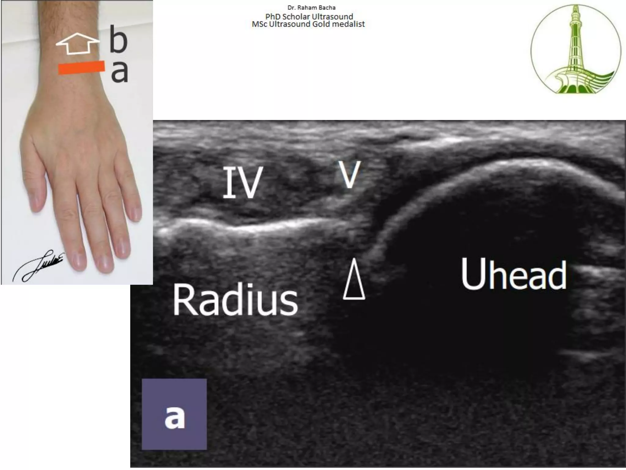
Abnormal wrist ultrasound findings typically reveal localized musculoskeletal pathology such as tendonitis, tenosynovitis, ganglion cysts, ligament tears, or nerve entrapments like carpal tunnel syndrome. High-resolution ultrasonography provides real-time, dynamic imaging of these structures, allowing clinicians to visualize inflammation, structural defects, and fluid collections with high precision.
Introduction to Wrist Ultrasound Imaging
Musculoskeletal ultrasound has evolved into a primary diagnostic tool for evaluating wrist pain, swelling, and mechanical symptoms. Unlike static modalities such as standard radiography, ultrasound offers dynamic evaluation, meaning the radiologist or sonographer can examine the wrist while the patient moves their fingers, hand, or wrist joint. This capability is essential for assessing tendon subluxation, dynamic nerve impingement, and the real-time movement of fluid within synovial sheaths.
When a patient undergoes this non-invasive imaging procedure, high-frequency sound waves create detailed cross-sectional images of superficial soft tissues, including tendons, ligaments, nerves, blood vessels, and the outer contours of the carpal and metacarpal bones. While magnetic resonance imaging (MRI) remains the gold standard for deep osseous structures and internal carpal ligaments, ultrasound is frequently preferred for its accessibility, cost-effectiveness, lack of radiation, and superior spatial resolution for superficial soft-tissue pathology. Understanding what constitutes an abnormal wrist ultrasound finding helps patients and clinicians interpret diagnostic reports and formulate targeted treatment plans.
Common Pathological Findings in Wrist Ultrasound
Abnormalities discovered during a wrist ultrasound generally fall into distinct anatomical and pathological categories. Because the wrist is a complex anatomical crossroads housing multiple tendons, nerves, and intricate ligaments packed into a tight space, distinct patterns of pathology emerge regularly.
Tendon Abnormalities: Tendonitis and Tenosynovitis
Tendons are among the most frequently evaluated structures in wrist sonography. Inflammation of a tendon (tendonitis) or its surrounding synovial sheath (tenosynovitis) produces characteristic ultrasound features.
- Tenosynovitis: Appears as an abnormal anechoic (dark) or hypoechoic fluid accumulation surrounding the tendon within the sheath. The tendon itself may appear thickened and rounded. Color Doppler or power Doppler imaging often reveals hypervascularity (increased blood flow) within the inflamed synovial tissue, indicating active inflammation.
- Tendinosis: Characterized by internal degeneration of the tendon fibers, showing loss of the normal fibrillar pattern, hypoechogenicity, and generalized thickening.
- Tendon Tears: Can be partial or complete. A partial tear manifests as a focal hypoechoic defect within the tendon substance, while a complete tear shows a complete discontinuity of the tendon ends, often accompanied by retraction and a fluid-filled gap (the "empty sheath sign").
Ganglion Cysts and Other Soft-Tissue Masses
Soft-tissue lumps and bumps on the dorsal or volar wrist are common reasons for ordering an ultrasound.
- Ganglion Cysts: The most common mass lesion of the wrist. On ultrasound, a ganglion cyst appears as a well-circumscribed, anechoic (completely dark) or hypoechoic structure with posterior acoustic enhancement (increased brightness of the tissues directly behind the cyst). They are frequently multiloculated and typically originate from a nearby joint capsule or tendon sheath.
- Giant Cell Tumors of the Tendon Sheath: The second most common soft-tissue mass in the hand and wrist. These appear as solid, hypoechoic masses closely associated with a tendon sheath, often showing internal vascularity on Doppler interrogation.
Nerve Compression Syndromes: Carpal Tunnel and Guyon's Canal
Ultrasound is increasingly used to diagnose peripheral nerve entrapments, most notably carpal tunnel syndrome affecting the median nerve at the wrist.
- Median Nerve Pathology: An abnormal ultrasound finding for carpal tunnel syndrome typically demonstrates an increased cross-sectional area of the median nerve at the level of the pisiform or hamate bone (usually exceeding 10 to 12 square millimeters). Other findings include flattening of the nerve as it passes under the transverse carpal ligament, hypoechogenicity (swelling and edema of the nerve fascicles), and bowing of the transverse carpal ligament itself.
- Ulnar Nerve Pathology: Evaluated within Guyon's canal, where abnormalities may include compression by an accessory muscle, ganglion cyst, or localized tenosynovitis of the adjacent flexor tendons.
Ligamentous Injuries and Dynamic Instability
While deep intrinsic ligaments like the scapholunate and lunotriquetral interosseous ligaments are best visualized via MRI or arthroscopy, high-resolution ultrasound can identify cortical avulsions, disruption of dorsal ligamentous fibers, and dynamic joint instability during stress maneuvers.
Summary of Common Abnormal Wrist Ultrasound Findings
| Finding Category | Specific Pathology | Sonographic Appearance | Clinical Significance |
|---|---|---|---|
| Tendinous | De Quervain's Tenosynovitis | Thickening of the abductor pollicis longus and extensor pollicis brevis tendons with surrounding fluid and hypervascularity. | Radial-sided wrist pain, positive Finkelstein test, localized inflammation. |
| Cystic | Dorsal Wrist Ganglion | Well-defined, anechoic, multiloculated cystic structure with posterior acoustic enhancement attached to the scapholunate ligament. | Dorsal wrist lump, localized aching, potential pressure on adjacent nerves. |
| Neurological | Carpal Tunnel Syndrome | Enlarged median nerve cross-sectional area ($>10 \text{ mm}^2$) proximal to the carpal tunnel with flattening at the tunnel inlet. | Numbness, tingling, and weakness in the thumb, index, and middle fingers. |
| Traumatic | Partial/Complete Tendon Tear | Focal hypoechoic defect or total discontinuity of fibrillar pattern with fluid-filled retraction gap. | Loss of active movement, mechanical weakness, history of acute trauma or chronic wear. |
Technical Aspects and Limitations of Wrist Sonography
Interpreting abnormal wrist ultrasound findings requires an understanding of both the capabilities and inherent limitations of the modality. Ultrasound is highly operator-dependent; the quality of the examination relies heavily on the skill, experience, and ergonomic technique of the sonographer or physician performing the scan.
Anisotropy: A Critical Artifact
Anisotropy is an artifact unique to ultrasound imaging of fibrillar structures like tendons and ligaments. If the ultrasound beam does not strike the tendon at a perpendicular angle (90 degrees), the structure appears artifactually dark (hypoechoic), which can mimic tendinosis or a partial tear. Skilled operators constantly adjust the probe angle (heel-toe maneuver) to ensure true pathology is differentiated from directional artifacts.
Diagnostic Limitations
- Deep Osseous Structures: Ultrasound cannot evaluate the interior architecture of carpal bones. Occult scaphoid fractures or deep bone bruises require radiography, CT scans, or MRI.
- Intrinsic Ligaments: While dorsal and volar portions of superficial ligaments can be seen, deep interosseous ligaments are obscured by overlying carpal bones.
- Body Habitus and Scarring: Extensive soft-tissue scarring, severe edema, or complex post-surgical anatomy can degrade image quality and obscure deep pathology.
Clinical Correlation and Next Actions
An abnormal wrist ultrasound finding should never be interpreted in isolation. Diagnostic imaging results must be correlated directly with the patient's clinical history, physical examination findings, and occupational or athletic demands.
Next Steps After Receiving Abnormal Results
- Consult the Ordering Physician: Bring the radiology report and images to the primary care provider, rheumatologist, orthopedic specialist, or hand surgeon who ordered the study.
- Determine Functional Impact: Assess whether the findings explain the patient's symptoms (e.g., pain, numbness, locking, weakness).
- Formulate a Treatment Plan: Depending on the specific abnormality, management may range from conservative measures (rest, splinting, non-steroidal anti-inflammatory drugs, targeted physical therapy, or ultrasound-guided corticosteroid injections) to surgical intervention (such as carpal tunnel release or ganglion excision).
- Consider Further Imaging: If clinical suspicion remains high despite a negative or inconclusive ultrasound, or if deep ligamentous or osseous injury is suspected, the clinician may order a magnetic resonance imaging (MRI) scan or computed tomography (CT).
Medical Disclaimer: The information provided in this article is for educational and informational purposes only and does not constitute medical advice, diagnosis, or treatment. Always consult a qualified healthcare provider regarding any medical condition or diagnostic test results.
Frequently Asked Questions
What does a hypoechoic finding mean on a wrist ultrasound?
A hypoechoic finding means that a specific tissue reflects fewer sound waves than normal, appearing darker gray on the monitor. In the wrist, hypoechogenicity often indicates fluid accumulation, edema, inflammation, or structural degradation within tendons, nerves, or soft tissues. While frequently associated with pathology such as tenosynovitis or nerve swelling, hypoechogenicity can also be an artifact if the ultrasound beam is not perpendicular to the tissue structure.
Can an ultrasound detect a fractured wrist bone?
Ultrasound can detect cortical breaks (surface irregularities or breaks in the outer bone line) and associated soft-tissue swelling or hematomas. However, it is not the primary imaging modality for evaluating carpal bone fractures. Standard X-rays are the initial standard for suspected fractures, and computed tomography (CT) or magnetic resonance imaging (MRI) is utilized for complex or occult fractures that are not visible on radiographs.
Is wrist ultrasound painful?
No, a standard diagnostic wrist ultrasound is a non-invasive, painless procedure. The sonographer applies a water-based acoustic gel to the skin and gently moves a handheld transducer (probe) across the wrist. Patients may feel mild, temporary discomfort if the transducer presses directly over an acutely inflamed tendon, compressed nerve, or tender ganglion cyst.
How long does a wrist ultrasound take?
A diagnostic wrist ultrasound typically takes between 15 and 30 minutes to complete. The duration depends on whether a single specific structure (such as the median nerve or a known lump) is evaluated, or if a comprehensive survey of all dorsal and volar tendons, ligaments, and joints of the wrist is required.
What is the difference between an ultrasound and an MRI for wrist pain?
Ultrasound uses high-frequency sound waves to provide real-time, dynamic imaging of superficial soft tissues, tendons, and nerves, and it is cost-effective and non-invasive. Magnetic resonance imaging (MRI) uses a powerful magnetic field and radio waves to create comprehensive, three-dimensional images of all internal structures, including deep bone marrow, intricate interosseous ligaments, and cartilage, making MRI better suited for complex internal derangements of the wrist.