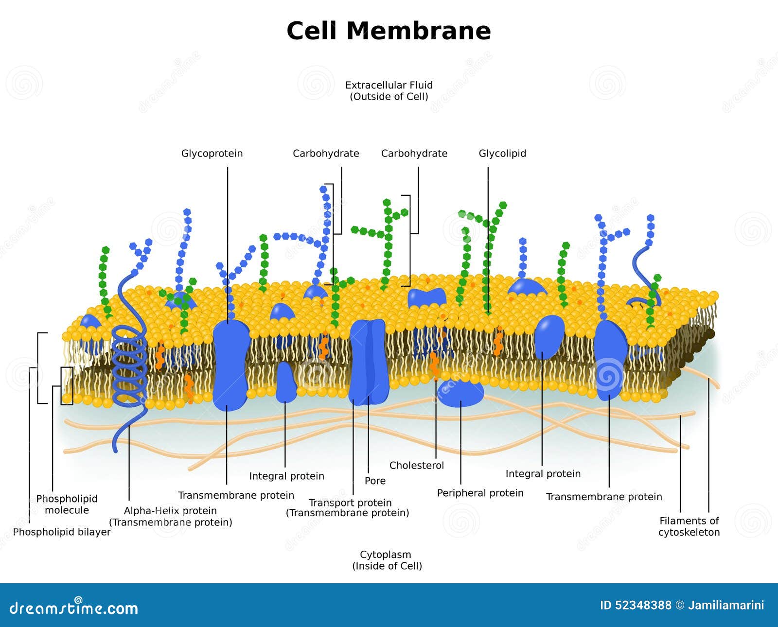
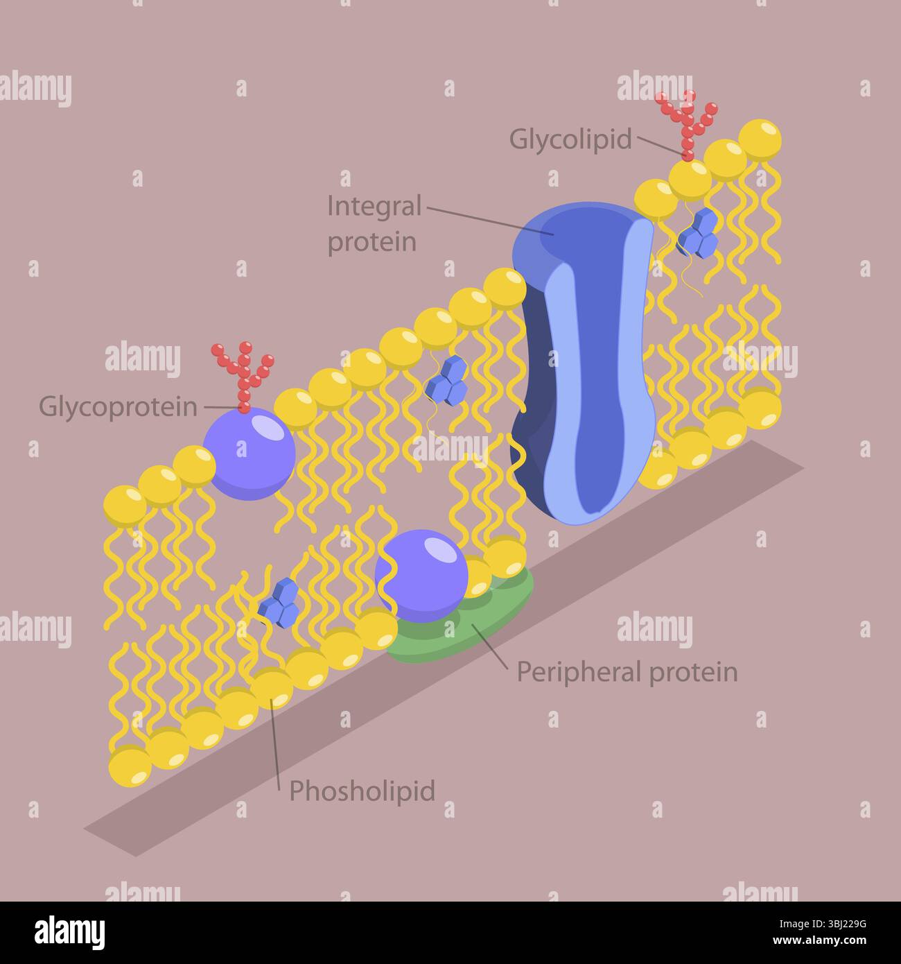
The most definitive example of plasma membrane structure is the Fluid Mosaic Model, which describes the membrane as a dynamic, double layer of phospholipids interspersed with a diverse array of proteins, cholesterol, and carbohydrates. This complex arrangement creates a semi-permeable barrier that regulates the passage of substances into and out of the cell.
The plasma membrane is far more than a simple boundary or "skin" for the cell. It is a sophisticated, heterogeneous, and highly active biological interface. By understanding its structure through the lens of the Fluid Mosaic Model—first proposed by S.J. Singer and Garth L. Nicolson in 1972 and refined through decades of research leading into 2026—we can appreciate how life maintains order at a microscopic level. This article examines the specific components, chemical properties, and functional examples of this critical cellular architecture.
The Phospholipid Bilayer: The Structural Matrix
The fundamental scaffold of the plasma membrane is the phospholipid bilayer. Phospholipids are amphipathic molecules, meaning they possess both a hydrophilic (water-loving) head and a hydrophobic (water-fearing) tail. In the aqueous environment of a living organism, these molecules spontaneously organize themselves into two layers.
The Hydrophilic Heads
The "heads" of the phospholipids are composed of a phosphate group attached to a glycerol backbone. Because the phosphate group carries a negative charge, it is polar and attracted to water molecules. In the membrane structure, these heads point outward toward the extracellular fluid and inward toward the intracellular cytoplasm, acting as the interface with the watery surroundings.
The Hydrophobic Tails
The "tails" consist of two long-chain fatty acids. These are non-polar and repel water. In the bilayer, the tails of both layers point toward each other, creating a fatty, oily core that is shielded from the water on either side. This hydrophobic interior is the primary reason the membrane acts as a barrier; it prevents water-soluble (polar) substances, such as ions and glucose, from simply drifting through the membrane without assistance.
Fluidity and Fatty Acid Saturation
The "fluid" part of the model refers to the fact that these lipid molecules are not fixed in place. They can rotate and move laterally within their layer. The degree of fluidity is heavily influenced by the type of fatty acids present:
- Saturated Fatty Acids: These have straight tails that can pack tightly together, making the membrane more rigid.
- Unsaturated Fatty Acids: These contain "kinks" (double bonds) that prevent tight packing, ensuring the membrane remains fluid even at lower temperatures.
The "Mosaic" of Membrane Proteins
While the lipid bilayer provides the basic structure, the "mosaic" aspect comes from the diverse proteins embedded within or attached to the bilayer. These proteins are responsible for the vast majority of the membrane's specialized functions.
Integral (Transmembrane) Proteins
Integral proteins are permanently attached to the biological membrane. Most integral proteins span the entire width of the bilayer, known as transmembrane proteins. These proteins often have hydrophobic middle regions that nestle among the lipid tails and hydrophilic ends that expose themselves to the water on either side.
- Channel Proteins: These act like tunnels, allowing specific ions or molecules (like water through aquaporins) to bypass the hydrophobic core.
- Carrier Proteins: These change shape to "carry" substances across the membrane, often using energy in the form of ATP.
Peripheral Proteins
Peripheral proteins do not extend into the hydrophobic core. Instead, they are loosely attached to the exterior or interior surfaces of the membrane, often anchored to integral proteins or the cytoskeleton. They play vital roles in cell signaling, maintaining the cell's shape, and anchoring the cell to neighboring tissues.
Cholesterol: The Fluidity Buffer
In animal cells, cholesterol is a critical structural component found tucked between the hydrophobic tails of the phospholipids. In the context of 2026 cellular biology standards, cholesterol is recognized as a "fluidity buffer."
Its role is dual-purpose:
- At High Temperatures: It stabilizes the membrane by restraining the movement of phospholipids, preventing the membrane from becoming too fluid or "leaky."
- At Low Temperatures: It prevents the phospholipids from packing too tightly and crystallizing, which would make the membrane brittle and prone to breaking.
Without cholesterol, human cells would struggle to maintain the precise mechanical integrity required to survive fluctuating internal environments.
Carbohydrates and the Glycocalyx
On the exterior surface of the plasma membrane, you will find short chains of carbohydrates attached to either proteins (glycoproteins) or lipids (glycolipids). These carbohydrate chains vary significantly between species, individuals, and even different cell types within the same body.
Collectively, these carbohydrates form the glycocalyx, a "sugar coating" that serves several functions:
- Cell Recognition: It acts as a molecular ID tag. For example, the ABO blood groups are determined by the specific carbohydrate patterns on the surface of red blood cells.
- Immune Response: The immune system uses these markers to distinguish between "self" cells and foreign invaders like bacteria or viruses.
- Cell Adhesion: These molecules help cells stick to each other to form tissues.
Detailed Comparison of Plasma Membrane Components
To better understand how these parts interact, the following table summarizes the primary components found in an example of a human plasma membrane.
| Component | Chemical Nature | Primary Location | Key Function |
|---|---|---|---|
| Phospholipids | Amphipathic Lipids | Main fabric of the membrane | Provides a semi-permeable barrier; creates the bilayer. |
| Integral Proteins | Large Polypeptides | Embedded through the bilayer | Transport (channels/pumps), receptors for hormones. |
| Peripheral Proteins | Small/Medium Polypeptides | Surface of the membrane | Cell signaling, enzymatic activity, structural support. |
| Cholesterol | Steroid (Lipid) | Between phospholipid tails | Regulates membrane fluidity and mechanical stability. |
| Glycoproteins | Carbohydrate + Protein | Extracellular surface | Cell-to-cell recognition and immune signaling. |
| Glycolipids | Carbohydrate + Lipid | Extracellular surface | Maintains membrane stability and facilitates cell bonding. |
A Practical Example: The Human Erythrocyte Membrane
One of the most thoroughly studied examples of plasma membrane structure is that of the human erythrocyte (red blood cell). Because red blood cells lack internal organelles and a nucleus, their plasma membrane is easily isolated and analyzed.
The erythrocyte membrane must be incredibly flexible yet resilient to survive being squeezed through narrow capillaries. This is achieved through a specific structural arrangement:
- The Lipid Composition: It contains almost equal parts cholesterol and phospholipids, providing the necessary stiffness to withstand blood pressure.
- Spectrin Network: Beneath the lipid bilayer lies a meshwork of a protein called spectrin. This protein acts like a biological spring, tethering the membrane to the cytoskeleton and allowing the cell to deform and سپس snap back into its original biconcave shape.
- Band 3 Protein: This is a major integral protein in the red blood cell that facilitates the exchange of bicarbonate and chloride ions, which is essential for carbon dioxide transport in the blood.
This specific example demonstrates that while the Fluid Mosaic Model provides a general blueprint, each cell type "customizes" its membrane structure to meet its physiological demands.
How Structure Dictates Function: Selective Permeability
The specific arrangement of the plasma membrane results in selective permeability. This means the membrane allows some substances to pass through while blocking others. This is not a random process but a direct consequence of the chemical properties we have discussed.
- Small, Non-polar Molecules: Oxygen ($O_2$) and Carbon Dioxide ($CO_2$) can dissolve in the lipid bilayer and pass through easily via simple diffusion.
- Small, Polar Molecules: Water ($H_2O$) is polar but small enough to slowly diffuse through the lipids or move quickly through specialized aquaporin channels.
- Large or Charged Molecules: Ions (like $Na^+$ or $K^+$) and large molecules (like glucose) cannot pass through the oily interior of the membrane. They require specific integral proteins (pumps or channels) to cross.
By controlling which proteins are present and active, the cell can precisely regulate its internal environment, a state known as homeostasis.
Common Misconceptions in Membrane Structure
As of 2026, educational models emphasize correcting several common misunderstandings regarding the plasma membrane:
- The Membrane is Not a Rigid Shell: Many students visualize the membrane as a solid wall. In reality, it has the consistency of light vegetable oil.
- Proteins Are Not Randomly Scattered: While called a "mosaic," proteins often cluster in functional groups known as lipid rafts. These rafts are specialized microdomains that facilitate specific signaling pathways.
- The Two Sides Are Not Identical: The membrane is asymmetric. The types of phospholipids and the presence of carbohydrates differ significantly between the inner and outer leaflets of the bilayer.
Frequently Asked Questions
What is the most important part of the plasma membrane structure?
While all components are essential, the phospholipid bilayer is the most critical structural element. It provides the fundamental barrier that defines the cell's boundaries. Without the bilayer, the proteins and carbohydrates would have no framework to occupy, and the cell would lose its integrity.
Why is the Fluid Mosaic Model still used as an example today?
The Fluid Mosaic Model remains the standard because it accurately describes the dual nature of the membrane: its liquid-like flexibility and its complex, tile-like composition of various molecules. Even with advanced imaging technologies in 2026, the core tenets of the model—fluidity and mosaicism—have been validated and refined rather than replaced.
How does the plasma membrane structure change in response to the environment?
Cells can actively change their membrane composition. For example, if a cell is moved to a colder environment, it may increase the proportion of unsaturated fatty acids in its phospholipids to prevent the membrane from freezing or becoming too rigid. This process is known as homeoviscous adaptation.
Do all living things have the same plasma membrane structure?
All known life forms (Bacteria, Archaea, and Eukarya) use a plasma membrane, but the chemistry varies. For instance, Archaea often have membranes made of ether-linked lipids (instead of ester-linked) and may even have a lipid monolayer, which allows them to survive in extreme environments like volcanic vents.
Can the plasma membrane repair itself?
Yes, to a limited extent. Because phospholipids are attracted to each other and repelled by water, small tears in the membrane will often spontaneously "seal" themselves as the molecules rearrange to hide their hydrophobic tails from the surrounding water. This self-sealing property is vital for cell survival after minor mechanical stress.
Further Exploration and Next Actions
To gain a deeper understanding of plasma membrane dynamics, students and researchers should look into the following areas:
- Signal Transduction: Study how integral proteins act as receptors to communicate environmental changes to the cell interior.
- Active vs. Passive Transport: Explore the energetic costs associated with moving molecules against their concentration gradient using membrane pumps.
- Microscopy: Review recent Cryo-Electron Microscopy (Cryo-EM) studies that provide near-atomic resolution of membrane protein structures.
Understanding the plasma membrane is the first step toward mastering cell biology. Whether you are studying for an exam or conducting biological research, keep the Fluid Mosaic Model at the center of your conceptual framework.