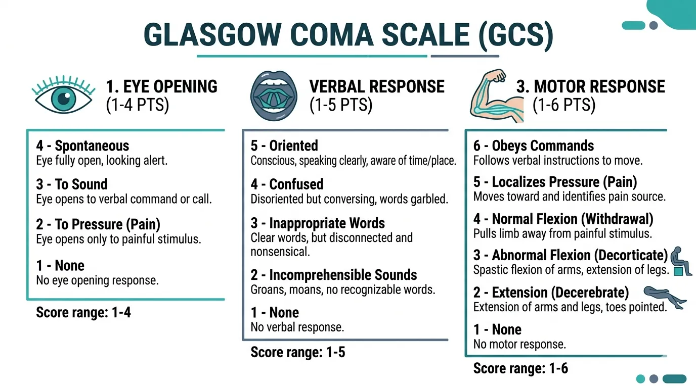
A Glasgow Coma Scale (GCS) score of 3 represents the deepest level of unresponsiveness on the standard neurological assessment scale, indicating that a patient is entirely unresponsive to eye, verbal, and motor stimuli. Recovery from a GCS 3 state requires a multidisciplinary, highly structured clinical approach focused on acute stabilization, neuro-rehabilitation, and long-term supportive management.
Understanding the Glasgow Coma Scale (GCS) and a Score of 3
The Glasgow Coma Scale is the universal clinical standard used by medical professionals to assess and document the level of consciousness in patients with acute brain injury. Developed in 1974 by Bryan Teasdale and Graham Jennett at the University of Glasgow, the scale evaluates neurological function across three distinct behavioral domains:
- Eye Opening (E): Ranging from spontaneous opening (4) to no response at all (1).
- Verbal Response (V): Ranging from fully oriented conversation (5) to no vocal sounds (1).
- Motor Response (M): Ranging from obeying commands (6) to no motor movement or flaccid paralysis (1).
A score of 3 is the absolute minimum score achievable on the scale, resulting from a sub-score of 1 in every single category ($E_1V_1M_1$). Clinically, this denotes a deep coma. Patients presenting with a GCS of 3 typically show no purposeful response to painful stimuli, do not open their eyes spontaneously or to noxious stimuli, and produce no intelligible words or sounds.
Etiology and Common Causes
A GCS 3 presentation rarely occurs in isolation; it is a symptom of severe structural, metabolic, or toxic insult to the central nervous system. Common etiologies include:
- Traumatic Brain Injury (TBI): Severe motor vehicle collisions, high-velocity impacts, falls from significant heights, or penetrating head trauma resulting in diffuse axonal injury (DAI), severe intracranial hemorrhage, or extensive cerebral contusions.
- Hypoxic-Ischemic Encephalopathy (HIE): Global oxygen deprivation to the brain, commonly resulting from cardiac arrest, severe respiratory failure, near-drowning, or asphyxiation.
- Acute Cerebrovascular Events: Massive ischemic strokes, large intracranial hemorrhages, or brainstem strokes that rapidly compromise the reticular activating system.
- Central Nervous System Infections and Toxicity: Severe meningoencephalitis, status epilepticus, or acute pharmaceutical/toxin overdoses that suppress central nervous system activity.
Understanding the underlying cause is the foundational step in determining the trajectory of recovery, as a hypoxic-ischemic event often carries a distinct prognostic profile compared to a traumatic injury of equivalent neurological depth.
Strategy 1: Acute Physiological Stabilization and Brain Protection
The immediate priority for any patient presenting with a GCS of 3 is the preservation of life and the prevention of secondary brain injury. Primary brain injury occurs at the exact moment of trauma or insult and cannot be reversed. Secondary brain injury, however, unfolds over subsequent hours and days due to systemic and intracranial complications such as hypoxia, hypotension, intracranial hypertension, and cerebral ischemia. Medical and surgical teams focus heavily on mitigating these secondary insults.
Advanced Airway Management and Hemodynamic Optimization
Because a patient with a GCS of 3 cannot protect their own airway, endotracheal intubation and mechanical ventilation are initiated immediately to ensure adequate oxygenation and prevent aspiration pneumonia. Hypoxemia (low blood oxygen) is catastrophic for an already compromised brain.
Simultaneously, healthcare providers maintain strict control over systemic blood pressure. Cerebral Perfusion Pressure (CPP) is defined as the difference between Mean Arterial Pressure (MAP) and Intracranial Pressure ($\text{CPP} = \text{MAP} - \text{ICP}$). Maintaining an adequate MAP is critical to ensure that blood flows against elevated intracranial pressures to supply oxygen and glucose to surviving neuronal tissue.
Intracranial Pressure (ICP) Monitoring and Surgical Intervention
In cases of severe traumatic brain injury or expansive stroke, brain swelling can drastically increase intracranial pressure. Elevated ICP reduces cerebral perfusion and can lead to brain herniation—a fatal mechanical displacement of brain tissue.
- Surgical Decompression: Neurosurgeons may perform an emergency craniectomy (removal of a portion of the skull) to allow the swollen brain room to expand without crushing vital structures.
- Osmotherapy: Administration of hypertonic saline or mannitol to draw fluid out of the brain parenchyma and reduce ICP.
- Cerebrospinal Fluid Drainage: Placement of an external ventricular drain (EVD) to drain excess CSF and monitor real-time ICP.
Strategy 2: Comprehensive Neuromonitoring and Multimodal Prognostication
Once the patient is stabilized in an intensive care unit (ICU), the clinical focus shifts toward continuous monitoring of brain function and rigorous prognostication. Predicting recovery from a GCS 3 state is notoriously complex, and clinicians rely on multimodal testing to avoid premature withdrawal of care while remaining realistic about long-term outcomes.
Advanced Neurological Monitoring Technologies
Modern neuro-critical care utilizes sophisticated technologies to assess brain activity beyond standard physical examinations:
- Continuous Electroencephalography (cEEG): Detects subclinical seizures, non-convulsive status epilepticus, and evaluates baseline cortical background reactivity. A reactive EEG background is generally associated with a more favorable prognosis than an unreactive or suppressed background.
- Multimodality Monitoring: Incorporates brain tissue oxygen tension ($PbtO_2$) monitors and cerebral microdialysis to measure metabolic byproducts like lactate-to-pyruvate ratios, indicating cellular distress.
- Somatosensory Evoked Potentials (SSEP): Evaluates the integrity of sensory pathways from peripheral nerves up to the cerebral cortex. The bilateral absence of cortical N20 potentials following cardiac arrest is a strong predictor of poor neurological recovery.
Biomarkers and Imaging Modalities
Advanced neuroimaging, such as high-resolution magnetic resonance imaging (MRI) utilizing diffusion tensor imaging (DTI), can identify microstructural damage in white matter tracts like the corpus callosum. Additionally, serum biomarker assays measuring neuron-specific enolase (NSE) or neurofilament light chain (NfL) provide quantifiable data regarding the extent of neuronal injury.
| Diagnostic Tool | Primary Clinical Purpose | Value in GCS 3 Assessment |
|---|---|---|
| cEEG | Detects seizures and background reactivity | Assesses cortical electrical function and reactivity |
| SSEP | Evaluates sensory nerve pathway integrity | High predictive value for poor motor recovery in anoxia |
| Brain MRI (DTI) | Visualizes white matter tracts and structural damage | Identifies diffuse axonal injury not seen on standard CT |
| ICP Monitoring | Measures real-time intracranial pressure | Guides medical and surgical treatment of brain swelling |
Strategy 3: Early Neuro-Rehabilitation and Specialized Supportive Care
If a patient survives the acute phase and begins showing signs of neurological transition—such as moving from a GCS 3 to a higher score, exhibiting sleep-wake cycles, or demonstrating visual tracking—the strategy shifts entirely toward specialized neuro-rehabilitation and secondary prevention of complications.
Multidisciplinary Rehabilitation Framework
Recovery from severe coma is a protracted marathon that requires a coordinated team of specialists. Rehabilitation does not wait until a patient is fully conscious; early mobilization and sensory stimulation protocols begin in the ICU.
- Physical Therapy: Focuses on passive range-of-motion exercises, prevention of severe muscle contractures, serial casting, and early verticalization (tilt-table protocols) to stimulate orthostatic reflexes and autonomic stability.
- Occupational and Speech Therapy: Addresses oral-motor function, early assessment for tracheostomy weaning, and management of swallowing disorders (dysphagia) to prevent aspiration.
- Cognitive and Psychological Support: Engages patients as they emerge into Minimally Conscious States (MCS) or emerge from coma entirely, addressing post-traumatic amnesia, cognitive deficits, and emotional dysregulation.
Managing Systemic Complications of Prolonged Immobility
Prolonged immobilization in patients recovering from deep coma introduces severe medical vulnerabilities that require rigorous preventative nursing and medical care:
- Pressure Injuries (Bedsores): Mitigated through specialized alternating-pressure mattresses, scheduled turning regimens every two hours, and meticulous skin hygiene.
- Deep Vein Thrombosis (DVT) and Pulmonary Embolism (PE): Prevented using sequential compression devices (SCDs), low-molecular-weight heparin, or inferior vena cava (IVC) filters when pharmacological anticoagulation is contraindicated.
- Infectious Complications: Ventilator-associated pneumonia (VAP) and catheter-associated urinary tract infections (CAUTIs) are managed through strict sterile protocols, oral hygiene bundles, and timely removal of invasive lines.
- Nutrition and Metabolism: Catabolic breakdown is countered early via enteral nutrition (nasogastric or percutaneous endoscopic gastrostomy tubes) to supply adequate caloric and protein intake for tissue repair.
Comparative Overview of Recovery Pathways
The trajectory of recovery from a GCS 3 state depends heavily on the underlying mechanism of injury. While individual outcomes vary widely, broad categories help frame clinical expectations.
- Initial GCS 3 Presentation
- Acute ICU Stabilization & ICP Control
- Hypoxic-Ischemic Event
- SSEP & EEG Prognostication
- Traumatic Brain Injury
- Surgical Decompression
- Massive Stroke / Hemorrhage
- Vascular Management
- Emergence into Minimally Conscious State
- Long-Term Multidisciplinary Rehabilitation
- Hypoxic-Ischemic Event
- Acute ICU Stabilization & ICP Control
- Traumatic Brain Injury (TBI): Often exhibits patchy structural damage. Younger patients with isolated TBI and managed intracranial pressure can sometimes achieve surprising functional recoveries over months or years, although cognitive and behavioral sequelae often persist.
- Hypoxic-Ischemic Encephalopathy (HIE): Characterized by global, diffuse cortical damage. The prognosis for functional recovery following a prolonged GCS 3 score caused by cardiac arrest is generally guarded, with many patients transitioning to a chronic vegetative state or minimally conscious state.
Frequently Asked Questions
Can a patient fully recover from a GCS score of 3?
Full recovery is possible, but it is statistically rare and depends heavily on the patient's age, baseline health, and the underlying cause of the coma. Traumatic brain injuries occasionally yield remarkable recoveries in younger patients, whereas global anoxia (lack of oxygen) resulting in a GCS 3 lasting more than a few days typically carries a poor prognosis for independent functional recovery.
How long do patients typically remain at a GCS 3?
The duration varies widely. Some patients transition to higher GCS scores (such as 4 or 5) within hours or days as acute intoxication wears off or swelling subsides. Others remain at a GCS 3 for weeks, at which point clinical definitions may shift to chronic disorders of consciousness, such as unresponsive wakefulness syndrome (UWS), formerly known as a vegetative state.
What is the difference between a coma and brain death?
A coma (such as a GCS 3) is a state of profound unconsciousness where the patient's brainstem may still retain certain reflex functions (such as breathing drives, pupillary reflexes, or gag reflexes). Brain death is the irreversible cessation of all brain function, including the brainstem, confirmed through rigorous clinical apnea testing and legally recognized as death.
How do doctors test brainstem reflexes in a GCS 3 patient?
Doctors test brainstem integrity using cranial nerve examinations. This includes checking pupillary light reflexes (optic and oculomotor nerves), corneal reflexes (trigeminal and facial nerves), vestibulo-ocular reflexes (doll's eyes test or caloric testing), and the gag or cough reflex (glossopharyngeal and vagus nerves).
Are there experimental treatments for severe coma recovery?
Clinical research investigates various neuromodulatory techniques, such as amantadine administration to boost dopaminergic transmission, vagus nerve stimulation (VNS), and deep brain stimulation (DBS) for patients in minimally conscious states. However, standard evidence-based care remains rooted in meticulous physiological stabilization and comprehensive neuro-rehabilitation.
Next Actions and Clinical Guidance
For families and practitioners navigating a GCS 3 scenario, the immediate focus must remain on partnering closely with neuro-intensive care specialists to ensure optimal physiological stability and accurate prognostic testing. Avoid relying on single indicators; insist on comprehensive multimodal evaluations. As the patient stabilizes, transition planning toward specialized inpatient rehabilitation facilities experienced in disorders of consciousness offers the best environment for maximizing long-term recovery potential.
Note: This article is for informational and educational purposes only and does not constitute formal medical advice, diagnosis, or prognosis. Decisions regarding acute medical care and goals of treatment should be made in consultation with qualified neuro-intensivists and medical specialists.