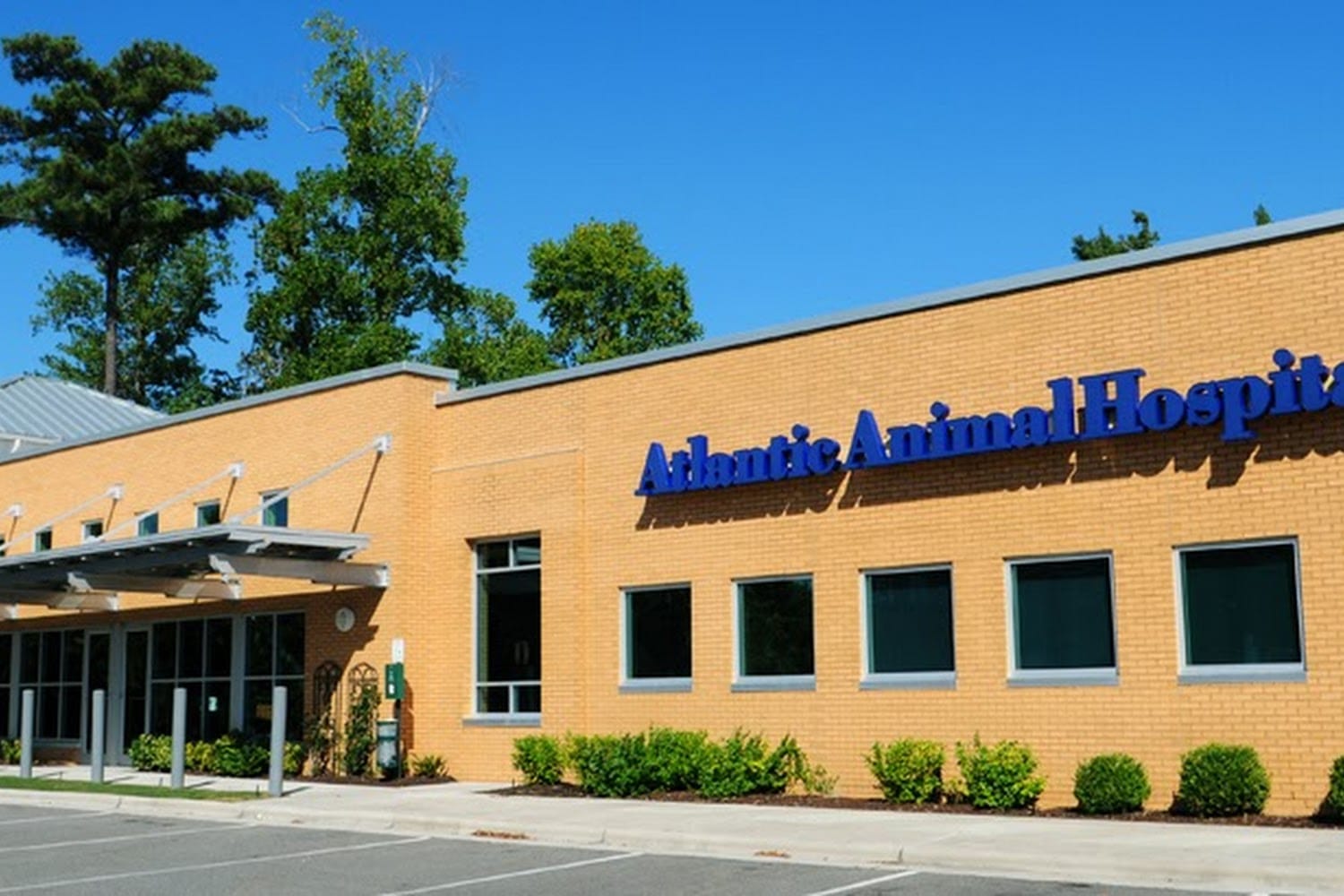
Animal eye care in Wilmington, NC encompasses both primary veterinary services for routine issues and specialized care from board-certified veterinary ophthalmologists for complex ocular diseases. Local pet owners can access diagnostics like tonometry and fluorescein staining through primary clinics, while severe conditions—such as deep corneal ulcers, acute glaucoma, or cataracts—may require referral to dedicated regional veterinary specialists.
The Spectrum of Veterinary Eye Care in Wilmington, NC
Vision is critical to an animal's quality of life. Whether managing an active hunting dog in the Cape Fear region, a companion cat in historic downtown Wilmington, or an equine athlete in New Hanover County, maintaining ocular health requires prompt recognition and appropriate medical intervention.
Veterinary eye care operates across two main tiers: general veterinary practice and board-certified veterinary ophthalmology.
- Pet Owner Notices Ocular Symptoms
- Primary Care Veterinarian (Wilmington)
- Initial Ocular Exam (Fluorescein, Tonometry, STT)
- Diagnoses & Treats Routine Issues (e.g., Superficial Ulcers, Conjunctivitis)
- Resolved / Managed
- Complex / Refractory Case
- Veterinary Ophthalmologist (DACVO)
- Advanced Diagnostics (Slit-lamp, ERG, Ocular Ultrasound)
- Microsurgery (Phacoemulsification, Corneal Flaps)
- Veterinary Ophthalmologist (DACVO)
Primary Veterinary Care
Primary care veterinarians in Wilmington serve as the first line of defense for animal eye health. During a routine or urgent physical examination, a general practitioner evaluates the external structures of the eye, including the eyelids, conjunctiva, cornea, and iris. Primary clinics perform essential diagnostic procedures to identify straightforward issues such as superficial corneal abrasions, bacterial conjunctivitis, mild allergic reactions, and early tear film deficiencies.
Specialty Veterinary Ophthalmology (DACVO)
When an eye condition is severe, vision-threatening, refractory to standard treatment, or requires advanced microsurgery, patients are referred to a specialist. A veterinary ophthalmologist is a veterinarian who has completed four years of veterinary school, a one-year internship, and a rigorous three-to-four-year residency program in ophthalmology, culminating in board certification by the American College of Veterinary Ophthalmologists (DACVO).
These specialists utilize advanced diagnostic tools—such as slit-lamp biomicroscopy, electroretinography, and high-frequency ocular ultrasound—and perform delicate surgeries using operating microscopes.
Coastal Environmental Factors Impacting Pet Eye Health in Wilmington
Pets living in Wilmington, Wrightsville Beach, Carolina Beach, and surrounding coastal communities face distinct environmental pressures that influence ocular health.
| Coastal Environmental Risk | Impact |
|---|---|
| 1. Airborne Sand & Wind | Mechanical Abrasions & Ulcers |
| 2. Marine Salt Spray | Tear Film Irritation & Dryness |
| 3. High UV Exposure | Chronic Superficial Keratitis |
| 4. Heavy Pine/Pollen Season | Allergic Conjunctivitis & Rubbing |
Airborne Sand and Shell Debris
Coastal winds carry micro-particles of sand and crushed sea shells along local beaches and waterways. Active dogs running along the shoreline frequently experience foreign body entry into the conjunctival sac or mechanical scratching of the cornea. Micro-abrasions from sand can rapidly progress to infected corneal ulcers if bacteria enter the compromised epithelial layer.
Marine Salt Spray and Sun Reflection
High salinity in windborne ocean spray acts as a mild desiccant, stripping moisture from the pre-corneal tear film. Additionally, intense solar radiation reflected off ocean waters and white sand increases UV exposure. In susceptible breeds, prolonged sunlight exposure exacerbates conditions such as Pannus (chronic superficial keratitis), an immune-mediated disease affecting the cornea.
High Seasonal Pollen Levels
Southeastern North Carolina experiences heavy pollen seasons, particularly from yellow pine, oak, and coastal grasses. Airborne allergens trigger allergic conjunctivitis in dogs and cats, leading to intense itching (pruritus). Pets attempting to relieve discomfort by rubbing their faces against carpet or furniture often inflict self-trauma, causing secondary corneal lacerations.
Common Ocular Diseases in Dogs, Cats, and Exotic Pets
Understanding common animal eye diseases helps pet owners identify subtle clinical changes before irreversible vision loss occurs.
1. Corneal Ulcers and Abrasions
A corneal ulcer is a breach in the outermost epithelial layer of the clear front surface of the eye, exposing the underlying corneal stroma.
- Superficial Ulcers: Involve only the top epithelial layer. Usually heal rapidly within 3 to 7 days with topical antibiotic therapy.
- Deep and Melting Ulcers: Involve deep stromal layers. Bacterial enzymes break down corneal collagen, causing the tissue to literally "melt." These are medical emergencies that risk corneal perforation.
- Indolent (Non-Healing) Ulcers: Also known as Spontaneous Chronic Corneal Epithelial Defects (SCCEDs), these occur when the epithelium fails to adhere properly to the underlying stroma. Common in middle-aged dogs like Boxers and Corgis, they require specialized procedures like diamond burr debridement or grid keratotomy to heal.
2. Cataracts versus Lenticular Sclerosis
Pet owners often report a "cloudy" or "bluish" tint in their older pet's eyes. Distinguishing between normal aging and pathology is critical.
- Lenticular (Nuclear) Sclerosis: A normal aging change caused by the compression of lens fibers over time. It typically appears after age 6 or 7 in dogs. While the lens appears hazy or bluish, light still penetrates to the retina, and functional vision is preserved. No treatment is needed.
- Cataracts: An opacity of the lens or its capsule that blocks light from reaching the retina. Cataracts can be hereditary, secondary to trauma, or caused by metabolic diseases such as diabetes mellitus. Unlike nuclear sclerosis, cataracts impair vision and can lead to complete blindness, phacolytic uveitis, or secondary glaucoma.
3. Glaucoma
Glaucoma is an increase in intraocular pressure (IOP) caused by impaired drainage of aqueous humor, the fluid inside the eye. Normal canine and feline IOP ranges between 10 and 25 mmHg. In acute glaucoma, pressures can spike above 40 to 60 mmHg.
- Primary Glaucoma: An inherited anatomical defect in the iridocorneal drainage angle, common in breeds such as Cocker Spaniels, Basset Hounds, and Huskies.
- Secondary Glaucoma: Caused by underlying ocular pathology, such as anterior lens luxation, chronic uveitis, intraocular tumors, or trauma.
Caution: Acute glaucoma causes rapid, permanent damage to the optic nerve and retina. Irreversible optic nerve death can occur within 24 to 48 hours of sustained high pressure.
- Normal Eye
- Fluid Outflow = Inflow
- (Normal Pressure: 10-25 mmHg)
- Healthy Retina & Nerve
- Glaucoma
- Drainage Angle Blocked
- (High Pressure: >30-60 mmHg)
- Optic Nerve Damage
- Permanent Blindness
4. Keratoconjunctivitis Sicca (KCS or "Dry Eye")
KCS is an immune-mediated condition in which the body's immune system attacks the lacrimal (tear-producing) glands. Lacking adequate aqueous tear film, the eye becomes chronically inflamed, painful, and prone to thick, mucoid discharge. Left untreated, KCS causes corneal pigmentation, scarring, vascularization, and eventual blindness.
5. Feline Herpesvirus-1 (FHV-1) Ocular Manifestations
In cats, ocular disease is overwhelmingly linked to Feline Herpesvirus-1. Up to 90% of cats are exposed to FHV-1 during their lifetimes. The virus remains latent in the trigeminal ganglion and recrudesces during times of stress or illness.
Clinical manifestations include recurrent conjunctivitis, dendritic corneal ulcers (branch-like lesions), corneal sequestra (necrotic corneal tissue), and symblepharon (adhesion of conjunctival tissue to the cornea).
6. Anterior Uveitis
Uveitis is inflammation of the uveal tract (iris, ciliary body, and choroid). In Southeastern North Carolina, uveitis is frequently linked to systemic infectious diseases, including tick-borne illnesses like Ehrlichia and Rickettsia rickettsii (Rocky Mountain Spotted Fever), fungal infections like Blastomyces, or feline infectious peritonitis (FIP). Symptoms include severe pain, miosis (constricted pupil), photophobia, low intraocular pressure, and flare (cloudiness in the anterior chamber).
Standard Ophthalmic Diagnostics in Veterinary Medicine
Accurate diagnosis of animal eye disease relies on specialized, non-invasive diagnostic tests performed in sequence during an exam.
| Diagnostic Test | Clinical Purpose & Target Values |
|---|---|
| Schirmer Tear Test | Measures aqueous tear volume (Normal: 15–25 mm/min) |
| Fluorescein Staining | Identifies corneal defects; tests tear duct patency |
| Tonometry | Measures intraocular pressure (Normal: 10–25 mmHg) |
| Slit-Lamp Bio. | Magnifies anterior chamber structures (cornea, iris, lens) |
| Ophthalmoscopy | Examines posterior segment (vitreous, retina, optic nerve) |
Schirmer Tear Test (STT)
The Schirmer Tear Test measures aqueous tear production. A small, sterile strip of calibrated filter paper is inserted into the lower eyelid pouch (lower conjunctival fornix) for exactly 60 seconds. Moisture travels down the strip by capillary action.
- Normal (Canine): 15 to 25 mm of wetting per minute.
- Borderline KCS: 11 to 14 mm per minute.
- Severe KCS: Under 10 mm per minute.
Diagnostic Sequence Note: The STT must always be performed before applying topical drops, flushes, or stains, as exogenous liquids invalidate tear measurement.
Fluorescein Stain Testing
Fluorescein is a water-soluble, hydrophilic fluorescent dye. The outer layer of the cornea (the epithelium) is lipophilic and repels water-soluble stains. If the epithelium is damaged, the dye penetrates the exposed stroma beneath and binds to its hydrophilic collagen fibers.
Under a Cobalt blue light source, the damaged area glows bright green, delineating the exact size, depth, and margin of a corneal ulcer.
Fluorescein is also used in the Jones Test, assessing whether tear drainage ducts (nasolacrimal ducts) are clear. Dye placed on the eye should pass through the tear duct and exit the nostrils within a few minutes.
Tonometry
Tonometry measures intraocular pressure (IOP). Modern veterinary practices use electronic tonometers:
- Rebound Tonometry (TonoVet): Uses a lightweight plastic-tipped probe that briefly contacts the cornea, recording the rebound velocity. It does not require topical anesthetic drops.
- Applanation Tonometry (Tono-Pen): Measures the micro-force required to flatten a small area of the cornea. Requires a drop of topical anesthetic (proparacaine) prior to measurement.
Elevated IOP (>25 mmHg) indicates glaucoma, while abnormally low IOP (<10 mmHg) combined with ocular inflammation indicates anterior uveitis.
Slit-Lamp Biomicroscopy and Ophthalmoscopy
- Slit-Lamp Biomicroscopy: Delivers a focused, high-magnification beam of light to evaluate the anterior segment (cornea, anterior chamber, iris, lens) in three dimensions. It allows the clinician to spot sub-millimeter foreign bodies, faint corneal edema, and microscopic protein leakage (aqueous flare).
- Indirect Ophthalmoscopy: Uses a headset light source and a handheld condensing lens to visualize the fundus, providing a wide-field view of the retina, retinal vasculature, and optic nerve head.
Ophthalmic Surgery and Advanced Treatment Procedures
When medical management with topical drops or oral medications is insufficient, surgical intervention is required to save vision or relieve pain.
- Medical Management Failure
- Surgical Assessment & Plan
- Cataract: Phacoemulsification
- Deep Ulcer: Conjunctival Flap
- End-Stage: Enucleation
Phacoemulsification for Cataract Removal
Phacoemulsification is the gold standard procedure for restoring vision lost to cataracts. Performed under general anesthesia using an operating microscope, an ophthalmic surgeon makes a microscopic incision into the cornea and lens capsule.
An ultrasonic handpiece vibrates at high frequencies to break apart (emulsify) the opaque lens contents, which are simultaneously aspirated out. An artificial folded Intraocular Lens (IOL) is then inserted into the remaining capsular bag, restoring sharp focusing power.
- Corneal Incision
- Capsulorhexis
- Ultrasonic Emulsification
- IOL Insertion
Conjunctival Pedicle Flaps and Grafting
For deep, structurally compromised corneal ulcers or perforations, a surgeon creates a delicate flap of conjunctival tissue (the vascular membrane lining the sclera) and sutures it directly over the corneal defect using ultra-fine suture material (often 8-0 to 10-0 monofilament). The flap provides structural support and supplies direct blood flow to accelerate healing and prevent eye loss.
Surgical Correction of Entropion and Cherry Eye
- Entropion Repair: Entropion is an inward rolling of the eyelid margins, causing eyelashes and fur to rub against the delicate cornea. Surgical blepharoplasty removes a precise crescent of skin below or above the eyelid to roll the margin outward into a normal anatomic position.
- Cherry Eye Repair (Pocket Technique): Cherry eye refers to a prolapsed gland of the third eyelid (nictitating membrane). Because this gland produces roughly 30% to 50% of the eye's tear film, it should never be surgically excised. Instead, surgeons use the Morgan Pocket Technique to create a tissue pocket and tuck the gland back into its normal anatomic position.
Enucleation
In cases of end-stage glaucoma, intraocular neoplasia (cancer), or severe ocular trauma where vision is completely lost and pain cannot be controlled medically, enucleation (surgical removal of the eyeball) is performed. The eyelids are permanently closed over the orbital space. Pets generally adapt exceptionally well to enucleation and experience immediate relief from chronic pain.
Triage Guide: Recognizing Ocular Emergencies
Ocular conditions can deteriorate rapidly. Delaying treatment by even a few hours can mean the difference between saving and losing a pet's eye.
| Category | Clinical Symptoms & Actions |
|---|---|
| IMMEDIATE EMERGENCY (Seek Care Within 1–2 Hours) | - Proptosis (Eyeball displaced out of socket)- Acute Trauma, Lacerations, or Chemical Exposure- Sudden Loss of Sight accompanied by severe pain or pupil dilation- High Intraocular Pressure Signs (Bulging eye, cloudy cornea, extreme lethargy) |
| URGENT CARE (Seek Care Within 12–24 Hours) | - Moderate to severe squinting (blepharospasm) or rubbing- Sudden onset of cloudiness, blue haze, or redness- Yellow or green discharge (mucopurulent exudate) |
| ROUTINE / NON-URGENT (Schedule Regular Appointment) | - Mild clear tear discharge without pain or redness- Gradual, symmetric cloudiness in older dogs (suspected lenticular sclerosis)- Small, non-painful eyelid growths without corneal contact |
Red Flag Symptoms
If a pet exhibits any of the following symptoms, seek immediate evaluation at a primary veterinary clinic or 24/7 emergency veterinary hospital:
- Blepharospasm: Severe squinting, holding the eye shut, or continuous blinking.
- Epiphora and Discharge: Profuse tearing or thick yellow/green discharge.
- Corneal Haze or Color Change: A blue, white, or opaque spot on the clear corneal surface.
- Hyphema: Visible blood inside the front chamber of the eye.
- Anisocoria: Asymmetrical pupil sizes (one pupil abnormally large or small).
- Proptosis: Traumatic displacement of the eyeball out of the orbital socket.
Comparative Overview of Animal Eye Conditions
The following table summarizes common animal eye conditions, key presenting signs, diagnostic methods, urgency levels, and primary treatment pathways:
| Condition | Primary Symptoms | Diagnostic Test | Urgency Level | Typical Treatment Approach |
|---|---|---|---|---|
| Superficial Corneal Ulcer | Squinting, tearing, redness, focal cloudiness | Fluorescein Stain Positive | Urgent (12–24 hrs) | Topical broad-spectrum antibiotic drops, analgesic oral medication, Elizabethan collar (E-collar). |
| Melting / Deep Ulcer | Severe pain, gelatinous corneal softening, discharge | Fluorescein Stain + Slit-Lamp | Immediate Emergency | Intensive topical anti-collagenase therapy, targeted antibiotics, structural conjunctival flap surgery. |
| Acute Glaucoma | Bulging eye, dilated pupil, cloudiness, intense pain | Elevated IOP via Tonometry (>25–60 mmHg) | Immediate Emergency | Topical/systemic carbonic anhydrase inhibitors, hyperosmotic agents, emergency surgical referral. |
| Dry Eye (KCS) | Thick mucoid discharge, dull cornea, redness | Low Schirmer Tear Test (<10 mm/min) | Urgent (24–48 hrs) | Life-long topical cyclosporine or tacrolimus, tear stimulants, frequent artificial tear lubrication. |
| Mature Cataracts | Dense white opacity in lens, bumping into objects | Indirect Ophthalmoscopy & Slit-Lamp | Non-Emergency / Referral | Surgical Phacoemulsification with Intraocular Lens (IOL) placement; anti-inflammatory drops. |
| Anterior Uveitis | Redness, pupil constriction (miosis), cloudy fluid | Low IOP via Tonometry + Slit-Lamp | Urgent (12–24 hrs) | Topical anti-inflammatory drops (prednisolone/dexamethasone), treatment of underlying infection. |
| Cherry Eye | Red mass protruding from inner corner of eye | Visual Examination & Ophthalmic Exam | Non-Emergency (Prompt) | Surgical replacement/anchoring of tear gland using Morgan Pocket Technique (do NOT excise). |
| Entropion | Eyelids rolling inward, eyelash contact, tearing | Visual & Biomicroscopic Exam | Routine to Urgent | Surgical blepharoplasty to remove excess tissue and roll eyelid margin outward. |
Practical Guidance: Daily Preventive Eye Care for Coastal Pets
Pet owners in Wilmington can implement several protective measures to minimize environmental risks to vision:
- Preventive Care Routine
- Post-Beach Flush
- Isotonic Saline
- Rinse Sand Out
- Protective Gear
- Rex Specs Goggles
- For UV/Sand Protection
- Grooming Care
- Keep Fur Trimmed
- Keep Soaps Out
- Post-Beach Flush
1. Post-Beach Saline Rinses
After visits to Wrightsville Beach, Fort Fisher, or local dog parks, flush your dog's eyes with a sterile, isotonic ophthalmic saline flush. Gently squirt the solution from the inner corner of the eye outward to wash away trapped sand, salt, and debris before micro-abrasions occur.
Caution: Never use contact lens cleaning solutions, hydrogen peroxide, or human medicated eye drops, which can cause chemical burns to animal corneas.
2. UV and Wind Protection
Active dogs that spend extended time on boats, in open truck beds, or at the beach can benefit from protective eye gear such as Rex Specs. These UV-blocking goggles protect vulnerable eyes from solar radiation, windborne sand, and high-velocity bug strikes.
3. Proper Grooming Safety
Keep long facial fur trimmed away from the eyes to prevent chronic mechanical irritation. When bathing pets at home, apply a drop of sterile mineral oil or protective ophthalmic ointment to each eye prior to washing to guard against soap and shampoo burns.
4. E-Collar Compliance
If your veterinarian diagnoses an eye issue and supplies an Elizabethan collar (cone), keep it on 24/7 until cleared. A single paw swipe can rupture an already compromised cornea, transforming a manageable ulcer into a permanent loss of the eye.
Navigating Referrals and Specialty Care in Wilmington and Regional NC
When a pet in Wilmington requires advanced diagnostics or surgical interventions beyond general practice capabilities, the referral process follows established veterinary protocols.
- Local Wilmington Primary Practice
- Diagnostic Findings & Medical History Shared
- Regional Veterinary Ophthalmology Specialist (e.g., Regional Referral Centers or NC State CVM)
- Advanced Microsurgery / Specialized Diagnostic Workup
- Re-check & Continued Maintenance back at Primary Clinic
The Referral Workflow
- Initial Assessment: Your primary veterinarian conducts baseline diagnostics (STT, Fluorescein, Tonometry) and stabilizes acute symptoms.
- Specialist Referral: If the condition requires advanced tools (like ultrasound or phacoemulsification) or is unresponsive to initial treatment, your primary vet submits a referral packet to a board-certified veterinary ophthalmologist.
- Specialized Consultation: The veterinary ophthalmologist conducts a comprehensive eye exam, discusses advanced surgical or medical treatment plans, and outlines prognosis.
- Co-Management: Following specialized treatment or surgery, routine follow-ups and long-term medication refills are often coordinated between the specialist and your local Wilmington primary veterinarian.
For complex academic referrals or rare systemic diseases affecting vision, pet owners in Southeastern North Carolina may also access tertiary care centers such as the NC State College of Veterinary Medicine in Raleigh.
Frequently Asked Questions
How can I tell if my dog's eye cloudiness is a cataract or just aging?
Nuclear sclerosis (normal aging) appears as a diffuse, bluish-grey haze deep within both lenses simultaneously in dogs over 6 or 7 years old. The eye remains functional, and light passes through to the retina. Cataracts, by contrast, are opaque, white or marble-like structures that block light, impair vision, and can affect one or both eyes unevenly. A primary veterinarian or ophthalmologist uses a slit-lamp or direct ophthalmoscope to definitively differentiate the two.
Can a general veterinarian in Wilmington perform cataract surgery?
No. Cataract surgery (phacoemulsification) requires micro-surgical techniques, specialized operating microscopes, ultrasound equipment, and advanced training available exclusively to board-certified veterinary ophthalmologists (DACVO). Primary veterinarians can diagnose cataracts, evaluate general health for surgery, and manage referral to a qualified specialist.
How do I safely clean sand out of my dog's eyes after playing on the beach?
Use only over-the-counter sterile ophthalmic saline solution (labeled specifically for eyes). Tilt your dog's head up gently, hold the eyelids open, and flush across the surface from the nose toward the outer corner. Allow the dog to blink naturally. Never scrub the eye with a towel, tissue, or cotton swab, as dragging micro-sand particles across the cornea causes severe scratches.
Is cataract surgery permanent for animals?
Yes. During phacoemulsification, the cloudy natural lens is permanently removed and replaced with an artificial intraocular lens (IOL). Cataracts cannot regrow on the new artificial lens. In some cases, mild scarring of the remaining lens capsule (posterior capsular opacification) can occur over time, but true cataract recurrence is impossible.
What should I do if my cat is squinting and has green eye discharge?
Squinting indicates ocular pain, and green discharge signals a secondary bacterial infection, frequently secondary to underlying Feline Herpesvirus-1 (FHV-1) or corneal ulceration. Schedule an urgent veterinary exam within 12 to 24 hours. Do not use leftover drops from other pets, as topical steroids applied to an active herpes flare or corneal ulcer can cause corneal breakdown and perforation.
Next Steps for Wilmington Pet Owners
If you observe changes in your pet's eyes—such as redness, cloudiness, squinting, unusual discharge, or vision changes—prompt action is essential to protect their sight.
- Schedule an Immediate Exam: Contact your primary Wilmington veterinarian for an initial ocular assessment including fluorescein staining and tonometry.
- Prevent Self-Trauma: Fit your pet with an Elizabethan collar (cone) immediately to stop them from rubbing or scratching the eye while awaiting care.
- Prepare Medical Records: Gather details regarding when symptoms started, exposure to sand/water, and any current medications to assist your veterinary team in formulating an accurate treatment plan.
Disclaimer: This guide is for educational purposes only and does not replace professional veterinary medical advice, diagnosis, or treatment. Always consult a licensed veterinarian or veterinary ophthalmologist regarding your animal's health.