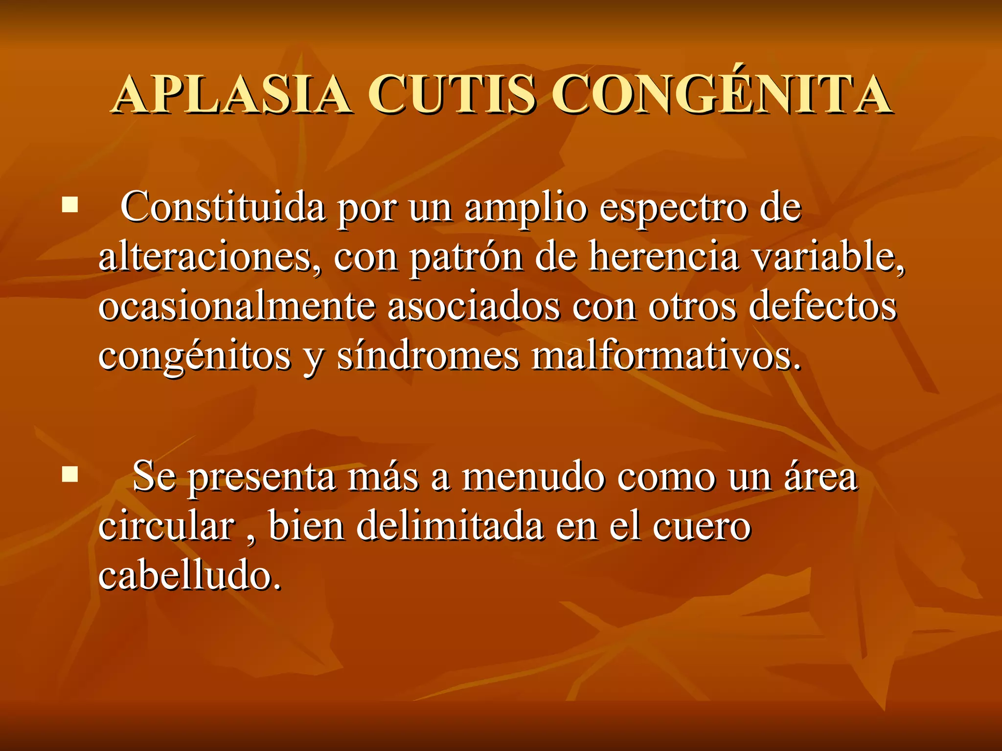
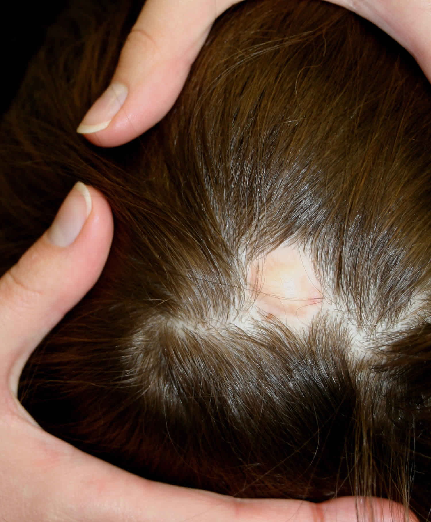
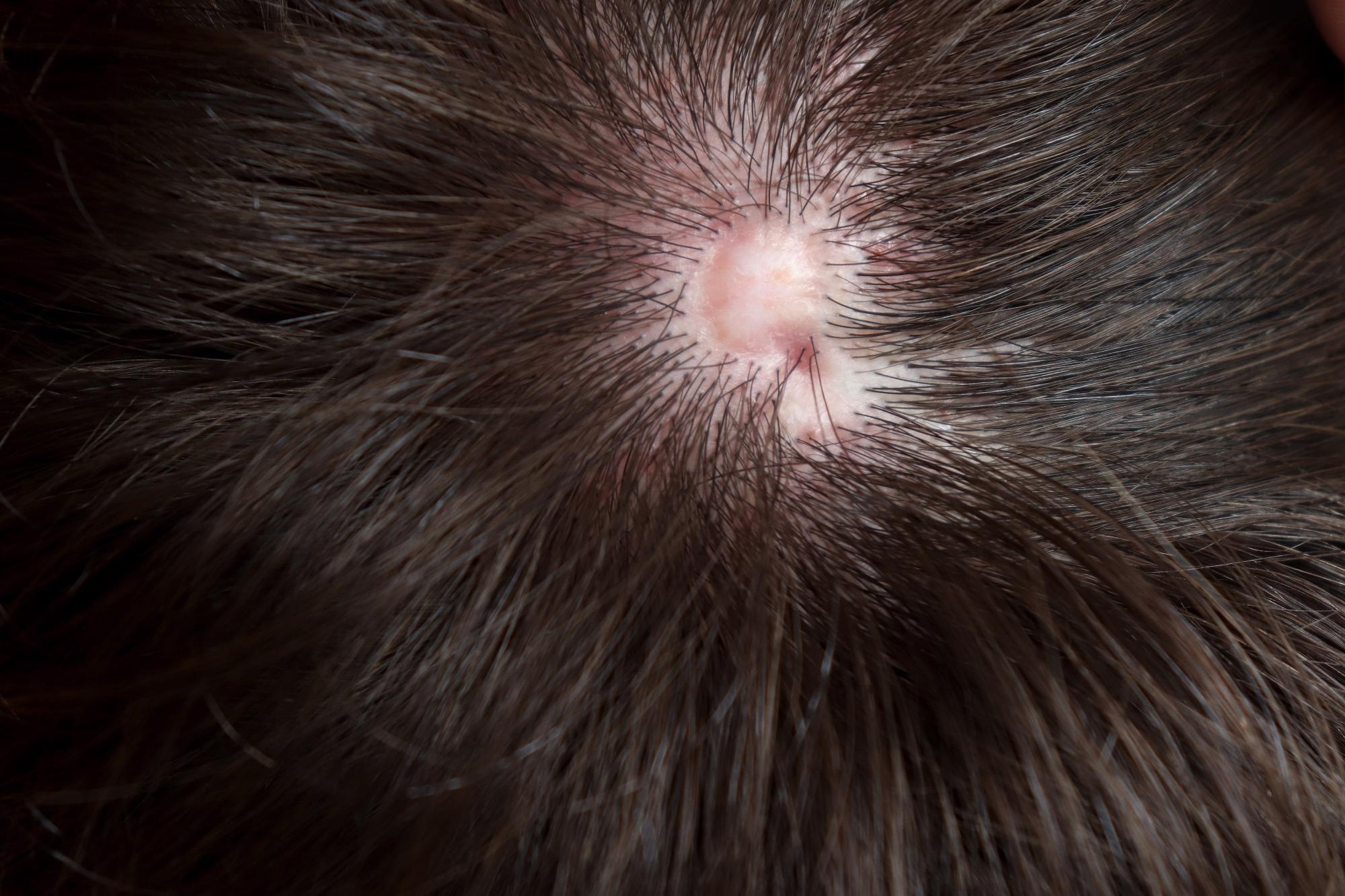
Aplasia cutis congenita is a rare congenital disorder characterized by the localized absence of skin at birth, most commonly occurring on the scalp. The causes of aplasia cutis congenita are multifactorial and heterogeneous, ranging from specific genetic gene mutations and hereditary syndromes to intrauterine vascular disruption, teratogenic drug exposures (such as methimazole), mechanical trauma, and fetal infections.
Medical Disclaimer: This article is intended solely for informational and educational purposes and does not constitute medical advice, diagnosis, or treatment. Clinical presentation of congenital skin lesions requires immediate evaluation by qualified healthcare professionals, including pediatric dermatologists and geneticists.
Overview of Aplasia Cutis Congenita
Aplasia cutis congenita (ACC) was first documented in medical literature by Cordon in 1767. It represents a focal deficiency of dermis, epidermis, and occasionally underlying subcutaneous tissue, skull bone, or dura mater. Although ACC can appear anywhere on the human body, approximately 84% of all presenting lesions are localized to the scalp, particularly near the vertex or parietal regions.
The birth prevalence of ACC is estimated to be between 1 and 3 per 10,000 live births. Lesions are typically solitary, circular, or oval, ranging in size from a few millimeters to several centimeters. At birth, the affected area may present as an open ulceration, a membrane-covered defect, a fragile scar, or a bullous lesion. While many isolated, superficial lesions heal spontaneously with conservative wound care, larger or deeper defects present significant medical risks, including sagittal sinus hemorrhage, severe bacterial infection, and meningitis.
Understanding the causes of aplasia cutis congenita is essential for establishing an accurate diagnosis, directing clinical workups, identifying associated systemic anomalies, and offering informed genetic counseling to families.
Primary Etiological Mechanisms of ACC
The precise developmental pathogenesis of aplasia cutis congenita remains an area of ongoing investigation. However, clinical and molecular studies confirm that ACC is not driven by a single universal pathology. Instead, it arises from multiple distinct developmental insults that disrupt normal embryonic skin fusion and vascularization during early gestation.
1. Genetic Mutations and Inheritance Patterns
Genetic alterations play a prominent role in both isolated (non-syndromic) and syndromic forms of aplasia cutis congenita. Sporadic cases are common, but autosomal dominant transmission with variable expressivity and incomplete penetrance is frequently observed across multi-generational families. Autosomal recessive inheritance also occurs, particularly in specific complex syndromes.
Several key genes and pathways have been identified in the pathogenesis of ACC:
- BMS1 Mutation: Research published in genetic literature identified heterozygous mutations in the BMS1 gene (which encodes a ribosome biogenesis factor) on chromosome 10q11 as a causative agent for autosomal dominant isolated scalp ACC. Disruption of BMS1 function leads to impaired ribosomal RNA processing and cell cycle arrest in embryonic epidermal cells.
- Notch Signaling Pathway (NOTCH1, DLL4, RBPJ): Mutations in genes encoding components of the Notch signaling pathway are primary drivers of Adams-Oliver syndrome, a classic genetic condition featuring scalp ACC and terminal limb defects. Notch signaling is vital for embryonic vascular formation and cell fate determination; disruption impairs localized skin and blood vessel development.
- GTPase Regulatory Genes (ARHGAP31, DOCK6): Mutations in ARHGAP31 (autosomal dominant) and DOCK6 (autosomal recessive) alter the function of Rho GTPases (Cdc42 and Rac1). These proteins regulate cellular cytoskeleton dynamics, cell migration, and blood vessel integrity during embryogenesis. Failure of these processes leads to localized cutaneous and osseous defects.
- KCTD1 Gene: Heterozygous loss-of-function mutations in KCTD1 cause Scalp-Ear-Nipple (SEN) syndrome, characterized by scalp aplasia cutis alongside ear abnormalities and hypoplastic nipples.
2. Intrauterine Vascular Disruption
Vascular hypoperfusion or focal ischemia during early fetal development is a well-established cause of ACC. When embryonic cutaneous tissues experience transient blood flow cessation or microvascular thrombosis, tissue necrosis occurs, leading to localized skin loss.
Vascular-mediated ACC occurs under several key prenatal conditions:
- Fetus Papyraceus and Twin-to-Twin Transfusion: In twin gestations where one fetus dies in utero (becoming a fetus papyraceus), thromboplastic material or emboli from the deceased twin can transfer to the surviving twin through placental vascular anastomoses. This embolization or sudden hemodynamic shift causes transient ischemia in the surviving fetus's skin and subcutaneous tissues, generating extensive trunk or limb ACC (often classified as Frieden Type V).
- Placental Infarction: Localized placental thrombosis or vascular compromise can reduce oxygenation to specific embryonic dermatomes, resulting in localized tissue atrophy or complete failure of skin closure.
3. Teratogenic and Environmental Exposures
Maternal exposure to specific pharmaceutical agents and substances during the first trimester of pregnancy—the critical window for cutaneous embryogenesis—can disrupt normal tissue formation and lead to aplasia cutis congenita.
- Maternal Exposure / Teratogen
- Embryonic Disruption
- Focal Skin Repair Failure
- ACC Presentation at Birth
- Antithyroid Drugs (Methimazole and Carbimazole): Methimazole (and its prodrug carbimazole), used to treat maternal hyperthyroidism, is the classic drug associated with ACC. Teratogen-induced ACC typically manifests as solitary or multiple circular lesions on the vertex of the scalp. The risk has led to clinical guidelines recommending propylthiouracil (PTU) over methimazole during the first trimester of pregnancy.
- Valproic Acid: Used as an anticonvulsant and mood stabilizer, valproate exposure in early pregnancy is linked to neural tube defects, facial dysmorphisms, and occasional cutaneous aplasia.
- Misoprostol: Maternal ingestion of misoprostol for pregnancy termination can induce violent uterine contractions and intense fetal hypoperfusion, leading to vascular disruption injuries including ACC and Moebius syndrome.
- Illegal Drugs and Alcohol: Maternal cocaine use induces severe vasoconstriction in placental and fetal blood vessels, leading to ischemic skin breakdown in utero.
4. Intrauterine Infections
Congenital viral infections can cause localized inflammation, tissue necrosis, and scarring in developing embryonic skin. Key viral agents linked to intrauterine infection-mediated ACC include:
- Herpes Simplex Virus (HSV): Intrauterine HSV infection can trigger focal cutaneous necrosis that manifests at birth as aplasia cutis or scarring ulcers.
- Varicella-Zoster Virus (VZV): Congenital varicella syndrome features dermatomal skin scarring, hypoplastic limbs, and structural defects secondary to neurotropic viral destruction.
5. Mechanical Amniotic Traumas and Banding
Rupture of the amnion early in pregnancy can lead to the formation of fibrous amniotic bands. These bands float within the amniotic cavity and can wrap tightly around developing fetal structures, such as limbs, digits, or the scalp. The mechanical constriction impairs localized blood supply and mechanically tears the delicate fetal epithelium, producing amniotic band sequence defects that present as linear or ring-like aplasia cutis congenita.
Frieden Classification of Aplasia Cutis Congenita
Because aplasia cutis congenita presents with diverse patterns and etiologies, Marion B. Frieden established a definitive classification system in 1986. This system categorizes ACC into nine distinct types based on lesion location, presence of associated anomalies, genetic inheritance, and causative factors.
| Frieden Type | Category Name | Underlying Etiology & Causes | Typical Clinical Features & Associated Anomalies |
|---|---|---|---|
| Type 1 | Isolated Scalp ACC | Sporadic or Autosomal Dominant (BMS1 mutations) | Vertex scalp lesions; no associated systemic or organ malformations. |
| Type 2 | Scalp ACC with Limb Anomalies | Genetic (NOTCH1, DLL4, ARHGAP31, DOCK6); Adams-Oliver syndrome | Scalp lesions accompanied by terminal transverse limb reduction, syndactyly, or oligodactyly. |
| Type 3 | Scalp ACC with Epidermal Nevi | Sporadic mosaicism; somatic mutations | Scalp aplasia associated with epidermal or organoid nevi; may involve neurological signs. |
| Type 4 | ACC Overlying Embryologic Defects | Developmental closure failure | Lesions overlying spinal dysraphism, meningomyelocele, encephalocele, or cranial defects. |
| Type 5 | ACC Associated with Fetus Papyraceus | Vascular disruption; twin embolization / hypoperfusion | Extensive linear or stellate trunk/extremity skin defects following early in utero demise of a twin. |
| Type 6 | ACC with Epidermolysis Bullosa | Genetic mutations (COL7A1, ITGA6, ITGB4); Bart syndrome | Widespread extremity aplasia cutis associated with congenital blistering and mucosal involvement. |
| Type 7 | ACC of Extremities Without EB | Sporadic or local vascular occlusion | Isolated localized defects on hand, foot, or leg without skin fragility disorders. |
| Type 8 | ACC Caused by Specific Teratogens / Infections | External exposures (Methimazole, Valproate, intrauterine HSV) | Scalp or body lesions directly linked to maternal medication exposure or intrauterine viral infection. |
| Type 9 | ACC Associated with Malformation Syndromes | Chromosomal abnormalities; complex multi-gene mutations | Scalp or body defects in syndromes like Trisomy 13, Johanson-Blizzard, or Scalp-Ear-Nipple syndrome. |
Syndromes Associated with Aplasia Cutis Congenita
When aplasia cutis congenita is accompanied by extracutaneous congenital anomalies, it is designated as syndromic ACC. Recognizing associated syndromes is vital, as internal organ involvement often dictates patient prognosis.
Adams-Oliver Syndrome (AOS)
Adams-Oliver syndrome is the primary genetic syndrome associated with ACC. Characterized by vertex scalp defects (often with underlying skull bone hypoplasia) and distal limb reduction anomalies (such as short digits, missing phalanges, or hypoplastic nails), AOS can be inherited in autosomal dominant or recessive patterns.
Mutations causing AOS occur in genes critical for cellular signaling and structural integrity, including ARHGAP31, DOCK6, RBPJ, NOTCH1, DLL4, and EOGT. Structural congenital heart defects (such as ventricular septal defects, tetralogy of Fallot, or coarctation of the aorta) and pulmonary hypertension occur in up to 20% of AOS patients, requiring early screening.
Bart Syndrome
First described by Bart in 1966, this condition features congenital aplasia cutis of the lower extremities combined with epidermolysis bullosa (EB) and mucosal involvement or nail dystrophy. The condition follows an autosomal dominant inheritance pattern and is tied to mutations in the collagen type VII alpha 1 chain gene (COL7A1), which anchors the epidermis to the dermis. The skin defect typically manifests as a well-demarcated absence of skin stretching from the feet up the lower legs.
Johanson-Blizzard Syndrome
Johanson-Blizzard syndrome is a rare autosomal recessive genetic disorder caused by mutations in the UBR1 gene on chromosome 15q15. In addition to scalp aplasia cutis, affected individuals display severe exocrine pancreatic insufficiency, hypoplasia of the nasal alae, sensorineural hearing loss, dental anomalies, growth failure, and intellectual disability.
Trisomy 13 (Patau Syndrome)
Trisomy 13 is a severe chromosomal abnormality resulting from an extra copy of chromosome 13. Scalp aplasia cutis, often multiple and deep, is a characteristic physical finding in infants with Trisomy 13. Other manifestations include holoprosencephaly, microphthalmia, cleft lip and palate, polydactyly, and severe congenital cardiac defects.
Scalp-Ear-Nipple (SEN) Syndrome
Also known as Finlay-Marks syndrome, SEN syndrome is caused by heterozygous mutations in the KCTD1 gene. Features include congenital aplasia cutis of the scalp, dysplastic pinnae (ears), hypoplastic or absent nipples and breast tissue, and dental anomalies such as microdontia.
Diagnostic Evaluation and Risk Assessment
Evaluating an infant born with aplasia cutis congenita requires a thorough, multi-system clinical approach to identify the underlying cause, evaluate structural depth, and screen for associated malformations.
- Infant Presentation with ACC
- Detailed Perinatal History (Family history, maternal medications, twin gestation)
- Physical Examination (Location, depth, limb defects, dysmorphisms)
- Non-Invasive Imaging (Ultrasound / MRI to evaluate dura & sagittal sinus)
- Molecular Genetic Testing (Targeted gene panels or chromosomal microarray)
1. Maternal and Perinatal History
Clinicians must record a detailed pregnancy and exposure history:
- Maternal medication usage during the first trimester (specifically antithyroid medications like methimazole, anticonvulsants, or ACE inhibitors).
- Evidence of early twin gestation or vanished twin (fetus papyraceus).
- Maternal viral exposures or febrile illnesses during pregnancy.
- Family history of scalp defects, missing digits, cardiac malformations, or unusual skin fragility.
2. Clinical Physical Examination
The location, count, size, shape, and depth of the lesions must be documented. Scalp lesions positioned directly over the sagittal suture or posterior fontanelle demand heightened vigilance due to the danger of underlying calvarial defects and proximity to the superior sagittal sinus. Clinicians carefully inspect the limbs, nails, ears, face, and nipples to evaluate for syndromic features like Adams-Oliver or SEN syndrome.
3. Diagnostic Imaging
Imaging is essential for scalp lesions to rule out underlying osseous defects, encephaloceles, or vascular anomalies:
- Scalp Ultrasonography: A high-frequency non-invasive first-line tool to evaluate skin thickness and confirm bone continuity.
- Magnetic Resonance Imaging (MRI): Warranted when lesions are deep, large, or located along the midline scalp. MRI clearly visualizes intracranial extension, dural integrity, cerebral parenchymal involvement, and superior sagittal sinus patency.
- Computed Tomography (CT): Used selectively to delineate three-dimensional cranial bone defects when surgical reconstruction of the skull is considered.
4. Genetic Evaluation
When ACC is accompanied by structural anomalies, positive family history, or multiple defects, consultation with a medical geneticist is recommended. Chromosomal microarray (CMA) analysis, targeted gene sequencing panels (covering BMS1, NOTCH1, DLL4, ARHGAP31, DOCK6, COL7A1), or whole-exome sequencing (WES) help pinpoint the molecular etiology, providing accurate prognostic information and recurrence risk estimates for future pregnancies.
Clinical Management Overview
While management strategies depend on lesion depth and underlying cause, treatment falls into two general categories: conservative conservative wound management and surgical intervention.
- Aplasia Cutis Congenita Assessment
- Small / Superficial Lesion (Intact dura, bone preserved)
- Conservative Management
- Gentle cleansing & petrolatum
- Topical antibiotic ointment
- Protective headwear / dressings
- Conservative Management
- Large / Deep Lesion (Dural defect, sinus exposure)
- Surgical Reconstruction
- Full-thickness skin grafts
- Local scalp rotation flaps
- Dural repair / cranial bone graft
- Surgical Reconstruction
- Small / Superficial Lesion (Intact dura, bone preserved)
Conservative Management
For small (under 1–2 cm), superficial defects where the cranial bone and dura remain intact, conservative management is the standard of care. Interventions include:
- Keeping the lesion clean with sterile saline.
- Applying topical petroleum jelly or moist antibacterial ointments to promote secondary-intention healing and prevent dressing adherence.
- Protecting the area from physical trauma or friction.
- Monitoring closely for signs of localized infection (erythema, purulent discharge, warmth).
Superficial lesions typically re-epithelialize over several weeks to months, leaving behind a hypertrophic or smooth atrophic scar with localized alopecia (hair loss).
Surgical Intervention
Surgical management is indicated for large scalp defects (often >3–4 cm), defects with complete loss of underlying skull bone, or cases where the superior sagittal sinus is exposed. Exposed vascular structures carry a life-threatening risk of sagittal sinus thrombosis or massive hemorrhage if the membrane dries out and ruptures.
Surgical options executed by pediatric neurosurgeons and plastic surgeons include:
- Early Soft Tissue Coverage: Utilizing local scalp rotation flaps or full-thickness skin grafts to cover vulnerable vascular channels.
- Dural Reconstruction: Repairing underlying dural defects using synthetic materials or pericranial autogenous grafts.
- Delayed Cranioplasty: Repairing persistent structural bone defects with bone grafts or custom prosthetic plates when the child reaches 2 to 5 years of age.
Frequently Asked Questions
Is aplasia cutis congenita a hereditary condition?
Aplasia cutis congenita can be hereditary, but it also occurs sporadically. Hereditary forms are frequently transmitted in an autosomal dominant pattern with variable expression, meaning an affected parent has a 50% chance of passing the genetic variant to their offspring. Genes like BMS1 and NOTCH1 have been implicated in familial cases. Sporadic cases often stem from non-inherited intrauterine events, vascular disruptions, or environmental teratogens.
What medications taken during pregnancy are known to cause aplasia cutis congenita?
The most well-documented teratogenic medication linked to ACC is methimazole (and its prodrug carbimazole), used to manage hyperthyroidism. Exposure during the first trimester increases the risk of scalp aplasia cutis in the newborn. Other medications linked to cutaneous defects include valproic acid (an anticonvulsant) and misoprostol. Pregnant women taking thyroid or anti-seizure medications should consult their physicians to optimize drug selection prior to conception.
Can aplasia cutis congenita heal naturally on its own?
Yes, small and superficial aplasia cutis congenita defects frequently heal spontaneously through secondary intention. With proper wound hygiene and protective ointment application, epidermal tissue gradually covers the area over several weeks, leaving a hairless scar. However, deep or large lesions involving underlying skull bone loss do not safely heal on their own and require specialized surgical reconstruction to prevent life-threatening hemorrhage or infection.
How does an intrauterine vascular event cause aplasia cutis congenita?
During embryonic development, skin cells require a continuous blood supply to proliferate and fuse across the midline. If localized blood flow is interrupted—due to blood clot formation, placental infarction, or hemodynamic shifts following the death of a twin fetus (fetus papyraceus)—the ischemic skin tissue undergoes necrosis. The dead tissue resorbs in utero, resulting in a localized absence of skin at birth.
How is aplasia cutis congenita differentiated from birth trauma or an encephalocele?
ACC is differentiated from birth trauma (such as scalp lacerations from vacuum extraction or forceps) by its clinical appearance, clear demarcated borders, histological features, and lack of active mechanical bruising. It is differentiated from an encephalocele (herniation of brain tissue through a skull defect) using high-resolution ultrasound or MRI, which confirms whether intracranial contents or cerebral spinal fluid extend through the cutaneous lesion.
Next Steps and Recommended Actions
When an infant is born with aplasia cutis congenita, systematic clinical coordination ensures patient safety and optimal recovery:
- Immediate Lesion Protection: Keep the defect moist with sterile petrolatum gauze or non-adherent dressings to prevent eschar drying, tissue tearing, or sagittal sinus rupture.
- Structural Imaging: Obtain a non-invasive scalp ultrasound or head MRI for lesions located near the scalp midline to evaluate dural and calvarial integrity before discharge.
- Comprehensive Pediatric Examination: Perform a focused physical examination examining digits, limbs, ears, heart sounds, and dysmorphic features to evaluate for syndromic forms like Adams-Oliver syndrome.
- Pediatric Dermatology and Genetic Consultation: Request specialist evaluations to guide long-term scar and alopecia management, perform molecular genetic testing if multi-system involvement exists, and provide genetic counseling regarding future pregnancy risks.