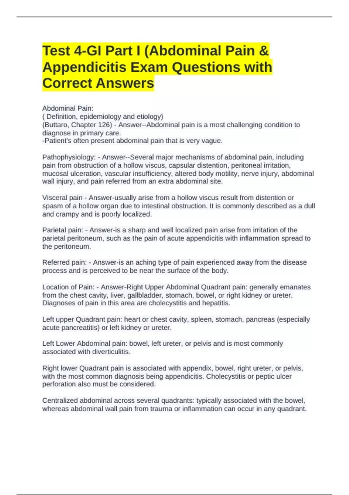
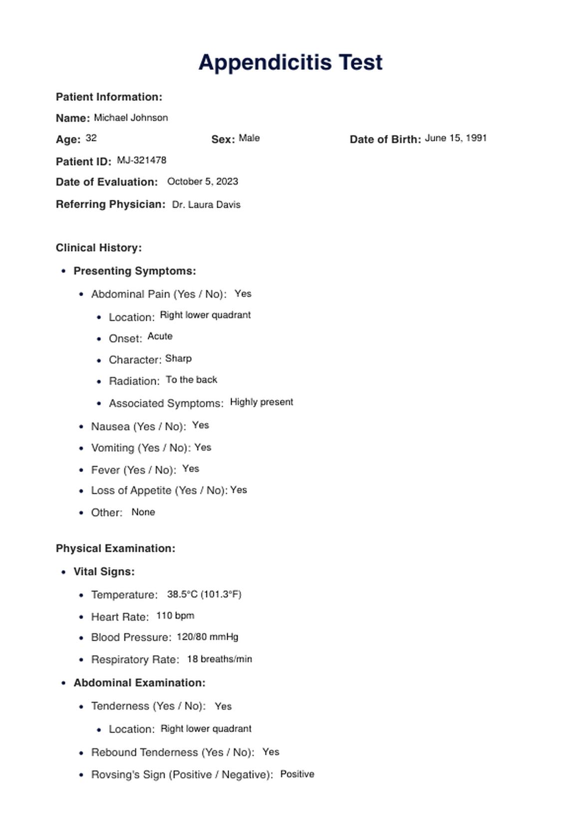
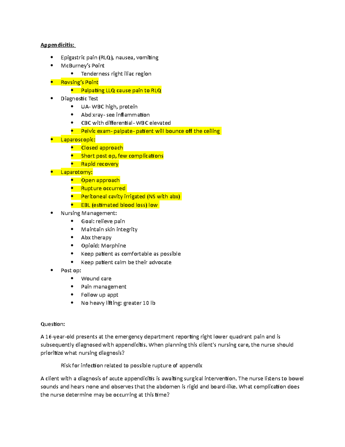
An "appendicitis test foot" search usually refers to physical examination maneuvers involving the lower body, most notably the Markle sign (heel-drop test) or heel-tap test. These physical assessments direct a minor shockwave through the pelvis and abdomen. If the appendix is inflamed and irritating the abdominal wall, this sudden motion triggers sharp pain in the right lower abdomen. While these maneuvers assist clinicians in identifying abdominal lining irritation, they are screening tools rather than definitive home diagnostics.
Disclaimer: Appendicitis is a potentially life-threatening medical emergency. Self-testing cannot rule out or diagnose appendicitis. If you or someone in your care experiences severe, unexplained abdominal pain, seek emergency medical evaluation immediately.
What Is the "Appendicitis Foot Test"?
When people look for a foot or heel test for appendicitis, they are typically seeking information on physical exam techniques used to detect peritoneal irritation (peritonitis). The primary maneuver associated with the foot is known clinically as the Markle sign, often referred to colloquially as the heel-drop test or heel-tap test.
First described by Dr. George B. Markle IV in 1973, the Markle sign relies on creating a sudden, gentle impact through the axial skeleton. When a person drops from their tiptoes onto their heels or receives a firm tap on the bottom of the foot while lying flat, vibrations travel up the leg bones, through the pelvis, and into the abdominal cavity.
If the appendix is healthy, this vibration causes minimal to no discomfort. However, if the appendix is acute, swollen, or inflamed, the sudden displacement of the abdominal organs causes the inflamed organ to contact or stretch the parietal peritoneum (the sensitive inner lining of the abdominal wall). This contact produces a sharp, localized surge of abdominal pain, particularly in the lower right quadrant.
In addition to the Markle sign, clinicians evaluate leg movements through maneuvers such as the Psoas sign and the Obturator sign. These physical maneuvers do not test foot health itself; rather, they use the foot, heel, and leg as levers to move pelvic muscles that rest directly adjacent to the appendix.
The Anatomical Mechanism: Why Foot Impact Triggers Abdominal Pain
To understand why a test performed on the foot or heel elicits abdominal pain, it is necessary to examine the anatomical relationship between the appendix, the peritoneum, and the surrounding musculature.
- Axial Vibration / Impact at Heel
- Transmitted Up Femur to Pelvis
- Movement of Parietal Peritoneum
- Sudden Contact with Inflamed Appendix (McBurney's Point)
- Sharp Pain Reaction in Right Lower Quadrant
1. Visceral vs. Parietal Pain
In the early stages of appendicitis, obstruction of the appendiceal lumen leads to swelling and stretching of the organ tissue. This initial phase produces visceral pain—a dull, diffuse, aching sensation felt around the navel (periumbilical area). Visceral nerves are poor at pinpointing exact locations.
As inflammation progresses over 12 to 24 hours, the infection spreads to the outer layers of the appendix and begins irritating the parietal peritoneum. Unlike visceral tissues, the parietal peritoneum is richly supplied with somatic sensory nerves. These nerves transmit sharp, precise pain signals. Once parietal irritation occurs, pain shifts from the navel to the right lower abdomen, centered near McBurney’s point (located two-thirds of the distance between the navel and the right hip bone).
2. Jarring Motion and Rebound Tenderness
Any movement that rapidly shifts or jolts the abdominal contents will cause the sensitized parietal peritoneum to rub against the inflamed appendix. This mechanism underlies standard clinical tests for peritoneal signs:
- Rebound Tenderness (Blumberg Sign): Deep pressure is applied to the abdomen and quickly released. The sudden recoil causes sharp pain.
- Cough Test: The patient is asked to cough forcefully; the sudden rise in intra-abdominal pressure causes localized pain.
- Markle Heel-Drop Sign: The impact of dropping onto the heels causes the abdominal organs to move downward under gravity and then suddenly halt, creating a internal recoil against the peritoneal wall.
3. Anatomical Variations of the Appendix
The exact position of the appendix varies among individuals. While the base of the appendix is consistently at the cecum, the tip can lie in several anatomical positions:
- Retrocecal (65% of cases): Tucked behind the cecum. Leg maneuvers like the Psoas sign are often more revealing than standard abdominal palpation in these cases.
- Pelvic (31% of cases): Hanging down into the pelvis. The Obturator sign is frequently positive when the appendix rests against the internal obturator muscle.
- Subcecal or Pre-ileal: Positioned below or in front of the lower small intestine.
Because these variations affect how physical movement triggers pain, clinicians use a combination of foot, heel, and leg maneuvers during a physical exam.
How Physical Examination Maneuvers Are Conducted
In clinical environments, emergency physicians and general surgeons use precise protocols to perform lower-extremity physical maneuvers. Understanding how these tests are performed clarifies their role in clinical diagnosis.
The Markle Sign (Heel-Drop Test)
- Standing Technique: The individual stands upright with feet together and raises their body up onto their tiptoes. On command, they relax their muscles and allow their body weight to fall abruptly onto their heels.
- Interpretation: A positive Markle sign occurs when the landing impact produces a sudden, sharp pain in the right lower quadrant of the abdomen. If the patient refuses to complete the drop due to severe pain, or if even light walking or jumping induces severe right lower quadrant pain, peritoneal irritation is strongly indicated.
The Bedside Heel-Tap Test
- Supine Technique: For patients who are too ill, weak, or unsteady to stand, the patient lies flat on their back (supine) on an examination table. The clinician elevates the patient’s right leg slightly while keeping the knee straight. Using the palm of their hand or a soft fist, the clinician taps firmly on the bottom of the patient's heel.
- Interpretation: The force transmits up the leg to the hip and pelvis. Sharp abdominal pain upon impact indicates positive peritoneal irritation.
The Psoas Sign
- Technique: The patient lies on their left side. The clinician gently extends the patient’s right leg backward behind the torso, stretching the iliopsoas muscle group. Alternatively, while the patient lies flat on their back, the clinician asks them to raise their straight right leg against downward manual resistance.
- Interpretation: If an inflamed appendix lies behind the cecum (retrocecal position), it sits directly atop the psoas muscle. Stretching or contracting the psoas muscle rubs against the inflamed organ, causing significant abdominal pain.
The Obturator Sign
- Technique: The patient lies on their back. The examiner flexes the patient's right hip and right knee to a 90-degree angle, then rotates the lower leg outward (which internally rotates the hip joint).
- Interpretation: This rotation stretches the internal obturator muscle in the pelvis. If an inflamed pelvic appendix is in contact with this muscle, internal rotation causes deep pelvic or lower abdominal pain.
Diagnostic Accuracy and Clinical Limitations
While physical maneuvers like the heel-drop and heel-tap tests provide useful bedside information, they are not infallible. Understanding their sensitivity, specificity, and limitations explains why physicians never rely on physical tests alone.
Clinical Metrics of Physical Signs
Research published in emergency medicine literature highlights the variable accuracy of lower-body appendicitis physical signs:
- Markle Sign (Heel Drop): Studies indicate the Markle sign has a sensitivity ranging between 74% and 85% for acute appendicitis, making it somewhat more sensitive than standard rebound tenderness in certain populations. However, its specificity varies, as any condition causing pelvic or abdominal peritonitis can trigger a positive result.
- Psoas Sign: Has a relatively high specificity (around 85% to 95%), meaning a positive test strongly suggests appendicitis or localized retroperitoneal inflammation. However, its sensitivity is low (16% to 40%), because it is primarily positive only when the appendix is retrocecal.
- Obturator Sign: Similar to the Psoas sign, it demonstrates high specificity but very low sensitivity (often under 20%), registering positive mainly in pelvic appendicitis cases.
Risk of False Positives and False Negatives
Physical tests involving the foot and leg can yield inaccurate conclusions if interpreted in isolation:
Potential Causes of False Positives
A positive heel-drop test indicates peritoneal irritation, but appendicitis is not the only cause of abdominal inflammation. Other conditions that can produce a positive heel-drop or heel-tap test include:
- Pelvic Inflammatory Disease (PID) or ovarian cyst rupture in females.
- Diverticulitis or severe inflammatory bowel disease (IBD) flare-ups.
- Perforated peptic ulcer or acute gastroenteritis with localized bowel wall inflammation.
- Renal colic (kidney stones passing through the ureter).
Potential Causes of False Negatives
A patient may have active appendicitis even if the heel-drop or heel-tap test appears negative:
- Early Appendicitis: If the inflammation remains confined inside the appendiceal wall and has not yet reached the parietal peritoneum, physical movement may not trigger sharp localized pain.
- Use of Pain Medication: Analgesics or anti-inflammatory drugs can mute the pain reflex during impact maneuvers.
- Retrocecal Position: An appendix shielded deep behind a gas-filled cecum may not contact the abdominal wall directly during a heel shockwave.
- High Pain Tolerance or Neuropathy: Patients with altered pain perception (e.g., advanced diabetes) may not register localized pain responses clearly.
Diagnostic Method Comparison
Because physical signs like the Markle test serve as screening tools rather than definitive diagnoses, clinical teams combine physical exams with laboratory and imaging studies.
| Diagnostic Method | Primary Objective | Key Advantage | Sensitivity / Specificity Profile | Clinical Role |
|---|---|---|---|---|
| Markle Sign (Heel-Drop Test) | Detect peritoneal irritation via axial vibration | Fast, non-invasive bedside screening tool | Sensitivity ~74-85%; Variable specificity | Initial clinical examination and triage indicator |
| Psoas / Obturator Signs | Evaluate pain caused by specific pelvic muscle movement | Helps identify retrocecal or pelvic appendix positions | High specificity (~85-95%); Low sensitivity (~15-40%) | Focused physical exam to localize appendiceal anatomy |
| Abdominal Ultrasound | Visualize appendiceal diameter and wall thickening | No ionizing radiation; safe for pediatric and pregnant patients | Sensitivity ~85%; Specificity ~90% (operator dependent) | First-line imaging choice for children and pregnant individuals |
| Contrast-Enhanced Abdominal CT | Provide definitive anatomical visualization of abdomen/pelvis | Highly accurate; identifies alternative diagnoses and complications | Sensitivity >95%; Specificity >95% | Gold-standard definitive imaging in adults |
| Complete Blood Count (CBC) & CRP | Detect systemic signs of infection and inflammation | Quantitative measurement of white blood cell count and inflammatory markers | High sensitivity; Low specificity | Supporting laboratory evidence within scoring models (e.g., Alvarado Score) |
Medical research indexed in the National Center for Biotechnology Information (NCBI) Bookshelf confirms that combining clinical examination maneuvers with structured risk scoring systems (such as the Alvarado Score or Appendicitis Inflammatory Response Score) improves diagnostic accuracy prior to imaging.
Standard Symptoms of Appendicitis
Evaluating a suspected case of appendicitis requires looking beyond physical foot maneuvers. The condition follows a characteristic timeline of symptoms that helps distinguish it from minor digestive issues.
- Early Phase (0-12 hrs): Dull pain around navel, loss of appetite, mild nausea
- Progressing (12-24 hrs): Pain migrates to Right Lower Quadrant (McBurney's Point)
- Advanced (24-48+ hrs): Low-grade fever, extreme tenderness, positive heel/cough signs
- Perforation / Rupture: Temporary pain drop, followed by severe generalized abdominal pain
1. Migratory Abdominal Pain
The hallmark of acute appendicitis is the classic progression of pain:
- Onset: Starts as a vague, dull, aching discomfort centered around the belly button or upper abdomen.
- Migration: Over 6 to 24 hours, the pain shifts down and to the right side of the abdomen.
- Localization: Settles into a constant, sharp, intense pain located in the lower right quadrant. The area becomes extremely sensitive to light pressure, coughing, walking, or bumpy car rides.
2. Associated Digestive Symptoms
- Anorexia: A sudden, total loss of appetite almost universally accompanies appendicitis. If a patient feels hungry and requests a full meal, appendicitis becomes less likely (though not impossible).
- Nausea and Vomiting: Usually develops after the onset of abdominal pain. If severe vomiting precedes abdominal pain, acute gastroenteritis is more frequently the cause.
- Bowel Changes: Mild diarrhea, constipation, or a feeling of incomplete bowel evacuation may occur.
3. Systemic Infection Signs
- Low-Grade Fever: Body temperature often rises to between 99.5°F (37.5°C) and 101°F (38.3°C).
- Malaise and Fatigue: A general sense of illness, lethargy, and inability to perform standard daily tasks.
When Suspected Appendicitis Is a Medical Emergency
Appendicitis is a time-sensitive medical condition. Left untreated, an inflamed appendix can undergo ischemia (loss of blood supply), gangrene, and eventual perforation (rupture).
According to emergency medicine guidelines published by the American College of Emergency Physicians (ACEP), delayed presentation or diagnosis increases the risk of appendiceal rupture, which can lead to severe peritonitis, pelvic abscess formation, and sepsis.
Critical Red Flag Symptoms
Seek immediate emergency services (call 911 or visit the nearest emergency department) if any of the following signs appear:
- Sudden Relapses after Brief Relief: A temporary reduction in pain followed by rapid, worsening pain throughout the entire abdomen often signals that the appendix has ruptured, spreading infectious material into the peritoneal cavity.
- Rigid, Board-Like Abdomen: The muscles of the abdominal wall involuntarily contract into a hard, rigid state to protect inflamed internal organs.
- High-Grade Fever and Chills: Temperatures exceeding 101°F (38.3°C) accompanied by shaking chills suggest an advancing infection or abscess.
- Signs of Systemic Shock or Sepsis: Confusion, extreme lethargy, rapid heart rate, lightheadedness, cold clammy skin, or difficulty breathing.
- Inability to Retain Liquids: Persistent vomiting that prevents keeping down water or oral rehydration fluids.
Common Diagnostic Mistakes to Avoid
When individuals attempt to assess abdominal pain at home using foot or heel tests, several critical mistakes can delay proper medical care:
- Delaying Evaluation to "Wait and See": Appendicitis symptoms can progress from mild discomfort to full rupture within 24 to 72 hours. Waiting several days to see if symptoms improve increases the risk of surgical complications.
- Taking Laxatives or Enemas: Applying laxatives or enemas to treat perceived constipation when abdominal pain is present can stimulate bowel contractions and dramatically increase the risk of appendiceal rupture.
- Applying Direct Heat to the Abdomen: Using heating pads or hot water bottles on the right lower quadrant increases local blood flow and tissue edema, potentially accelerating inflammation and rupture risk.
- Administering Strong Pain Relievers Before Medical Exam: Taking narcotic analgesics or heavy pain medications prior to an emergency department assessment can mask physical exam signs (including the Markle heel sign), making accurate clinical evaluation more difficult.
- Relying Exclusively on Home Tests: A negative heel-drop test does not guarantee a normal appendix. If clinical symptoms (pain migration, nausea, fever) align with appendicitis, immediate emergency care is necessary regardless of home test results.
Frequently Asked Questions
What does a positive heel-drop test feel like?
A positive heel-drop test causes a sharp, localized jolt of pain in the lower right side of the abdomen when the heels hit the floor. The pain feels distinctly localized to the right lower quadrant rather than a generalized bodily jar or mild muscle ache in the legs or back.
Can I do the appendicitis foot test on a child?
While you can ask a older child to jump up and down or drop onto their heels, children often struggle to describe or localize physical pain accurately. Pediatricians frequently observe whether a child refuses to jump, walks bent over, or cries when bumped during transport. In children with acute abdominal pain, rely on medical evaluation rather than home physical tests.
What is the difference between the Markle sign and the Heel Tap test?
Both tests use the same anatomical principle: transmitting force up the skeleton to vibrate the peritoneum. The Markle sign typically refers to the patient standing on tiptoes and dropping down onto their heels. The bedside heel tap involves the clinician tapping the bottom of the patient's foot while the patient lies flat on an exam table.
Does a negative foot test completely rule out appendicitis?
No. A negative foot or heel test cannot rule out appendicitis. In early stages of inflammation, or when the appendix is positioned behind the cecum or deep within the pelvis, impact maneuvers may not trigger significant pain. If classic symptoms like migrating abdominal pain, loss of appetite, and nausea are present, seek emergency medical care regardless of the foot test outcome.
What other conditions cause right lower quadrant abdominal pain?
Several conditions mimic appendicitis symptoms, including acute mesenteric adenitis (swollen lymph nodes in the abdomen, common in children), ovarian cysts, ectopic pregnancy, pelvic inflammatory disease, kidney stones, Crohn's disease, cecal diverticulitis, and right-sided testicular torsion in males. A medical workup including imaging and lab tests is required to differentiate these conditions.
Recommended Action Plan
If you suspect that you or someone near you has acute appendicitis, take the following structured steps:
- Cease All Physical Testing: Stop performing jumping, heel-drop, or deep palpation tests once peritoneal signs are suspected. Repeated jarring can cause unnecessary pain and discomfort.
- Avoid Food, Drink, and Medications: Do not eat, drink, or take pain relievers, laxatives, or antacids. Keeping the stomach empty is essential if emergency surgery (appendectomy) or anesthesia is required.
- Seek Immediate Medical Attention: Go to the nearest Emergency Department or Urgent Care facility equipped with diagnostic imaging (ultrasound or CT scanner). Do not drive yourself if you are experiencing severe pain or dizziness; arrange transport or call emergency services (911).
- Inform the Triage Team: Upon arrival, clearly state the duration of abdominal pain, whether the pain migrated from the navel to the lower right side, and if movements like walking, coughing, or bumping heels aggravate the pain.