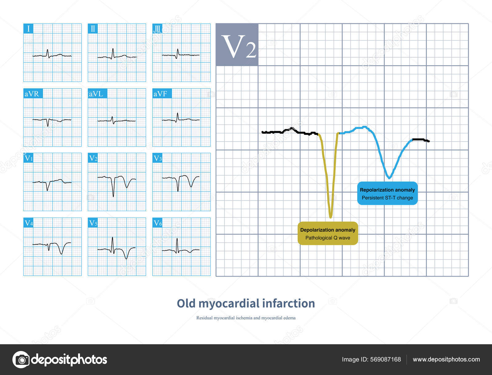
An anterior infarct, or anterior wall myocardial infarction, is a significant medical event occurring when the left anterior descending artery—the heart’s primary blood vessel—becomes blocked. This obstruction deprives the heart's large anterior wall of oxygenated blood, requiring immediate emergency intervention to restore circulation, minimize permanent muscle damage, and preserve the long-term pumping function of the heart.
Anatomy of an Anterior Infarct
To understand an anterior infarct, one must first recognize the anatomy of the coronary circulation. The left coronary artery branches into the left anterior descending (LAD) artery, often referred to as the "widow-maker" due to the critical volume of heart muscle it supplies. The LAD travels down the front (anterior) surface of the heart, providing oxygenated blood to the left ventricle, which is the heart’s main pumping chamber.
When a plaque rupture or thrombus (blood clot) obstructs the LAD, the tissue downstream begins to suffer from ischemia—a state of oxygen deprivation. If the blockage is not cleared quickly, the tissue progresses to necrosis, or cell death. Because the left ventricle is responsible for pumping blood to the entire systemic circulation (the rest of the body), damage to this area is particularly concerning. The size of the infarct depends heavily on the precise location of the blockage; a proximal occlusion (near the start of the vessel) affects a larger area of the heart than a distal occlusion (near the end of the vessel).
Recognizing Symptoms and Early Indicators
Recognizing an anterior myocardial infarction is a race against time. The symptoms often mirror those of other cardiac events but may feel more intense due to the significance of the area involved.
- Chest Pain (Angina): This is the most common symptom. It is frequently described as a crushing pressure, tightness, or a heavy weight on the chest. Unlike minor muscle strain, this pain typically does not improve with rest or change when the patient presses on the chest wall.
- Radiating Pain: The discomfort often migrates to the left arm, neck, jaw, or back. This occurs because the nerves supplying the heart and those supplying the skin and muscles of the upper body share common spinal pathways.
- Autonomic Symptoms: Many individuals experience nausea, vomiting, or cold sweats (diaphoresis). These occur because the stress of the infarct triggers the body’s sympathetic nervous system.
- Shortness of Breath: Dyspnea, or difficulty breathing, is a hallmark of significant anterior involvement. As the left ventricle weakens, it struggles to move blood forward, causing fluid to back up into the lungs (pulmonary congestion).
Immediate Medical Response and Diagnostics
If an anterior infarct is suspected, medical professionals operate under the mantra that "time is muscle." Upon arrival at an emergency department or via emergency medical services (EMS), several steps are taken simultaneously to verify the condition and stabilize the patient.
Diagnostic Verification
The most critical diagnostic tool is the 12-lead electrocardiogram (ECG). In an anterior infarct, the ECG will typically show ST-segment elevation in leads V1 through V4. These leads represent the electrical activity of the anterior portion of the heart. Additionally, clinicians order blood tests to detect cardiac troponins. Troponins are proteins released into the bloodstream when heart muscle cells are damaged. Elevated levels of these markers serve as biochemical evidence that a myocardial infarction has occurred.
Reperfusion Therapy
The gold standard for treating an anterior infarct is immediate reperfusion—re-establishing blood flow. The preferred method is primary percutaneous coronary intervention (PCI), commonly known as angioplasty and stenting. During this procedure, a cardiologist threads a catheter through a blood vessel to the site of the blockage. A balloon is inflated to open the vessel, and a stent is placed to keep it open. If PCI is not available within a reasonable timeframe (typically 90 to 120 minutes), fibrinolytic (clot-dissolving) therapy may be administered to break down the obstruction.
Comparative Overview of Myocardial Infarction Types
While all heart attacks involve restricted blood flow, the location and extent of the damage vary significantly. The following table highlights differences based on the affected anatomy and typical clinical considerations.
| Infarction Type | Affected Artery | Primary Concern | Common ECG Changes |
|---|---|---|---|
| Anterior | Left Anterior Descending | Left ventricle pump failure | Leads V1–V4 (ST elevation) |
| Inferior | Right Coronary Artery | Conduction system damage | Leads II, III, aVF |
| Lateral | Left Circumflex | Electrical arrhythmias | Leads I, aVL, V5, V6 |
| Posterior | Posterior Descending | Often accompanies other infarcts | Reciprocal changes in V1–V3 |
Challenges and Potential Complications
The recovery phase and the immediate post-infarct period carry specific risks. Because the left ventricle is heavily impacted, patients are at a higher risk for developing complications during the healing process.
Heart Failure and Remodeling
Following an infarct, the heart undergoes a process called ventricular remodeling. The heart muscle attempts to compensate for the dead tissue by stretching and changing shape. While this helps initially, it often leads to thinning of the ventricle walls, which can result in heart failure—a condition where the heart no longer pumps blood as efficiently as the body requires. Modern medication protocols, including ACE inhibitors and beta-blockers, are specifically prescribed to slow or prevent this maladaptive remodeling.
Arrhythmias
The damage to the myocardium (heart muscle) can disrupt the electrical pathways that coordinate the heartbeat. Anterior wall damage can specifically trigger ventricular arrhythmias, which are dangerous heart rhythms originating in the lower chambers of the heart. Continuous cardiac monitoring during the initial hospital stay is essential to detect and treat these rhythm disturbances before they become life-threatening.
Mechanical Complications
Rarely, the necrotic tissue in the anterior wall can become weakened to the point of rupture. This includes rupture of the ventricular septum or the free wall of the heart. While these are catastrophic events, their incidence has decreased significantly in recent years due to rapid reperfusion strategies and advancements in surgical support.
Rehabilitation and Lifestyle Management
Recovery from an anterior infarct is a lifelong process. Once the acute phase is over, the focus shifts to secondary prevention—preventing a recurrence.
- Cardiac Rehabilitation: This is a structured program of exercise and education. Supervised physical activity helps strengthen the heart muscle without overtaxing it, while educational sessions provide the tools needed to manage diet, stress, and medication adherence.
- Pharmacological Therapy: Most patients are placed on a "cocktail" of heart-protective medications. This typically includes dual antiplatelet therapy (DAPT) to prevent further clots, statins for cholesterol management, and blood pressure-lowering agents.
- Dietary Adjustments: Adopting a heart-healthy diet—rich in fiber, vegetables, and lean proteins, and low in saturated fats and sodium—is fundamental. Reducing sodium intake is particularly important for patients who have experienced any degree of heart failure to prevent fluid retention.
- Risk Factor Modification: Smoking cessation is the single most effective lifestyle change one can make after an infarct. Tobacco use directly constricts blood vessels and damages the vascular endothelium, increasing the likelihood of another blockage.
Expert Guidance: When to Seek Follow-Up Care
Patients must be vigilant during the recovery period. While some fatigue is expected, certain warning signs require immediate medical attention. Any return of chest pain, even if it feels different from the initial infarct, should be evaluated by a healthcare provider. Furthermore, if a patient notices sudden weight gain (a sign of fluid retention), increased swelling in the legs, or significant exercise intolerance, they should contact their cardiologist. These can be indicators that the heart is struggling to maintain adequate cardiac output.
Frequently Asked Questions
What is the survival rate after an anterior infarct? Survival rates have improved significantly due to the widespread availability of PCI and standardized care protocols. The prognosis is highly dependent on the "door-to-balloon time"—the duration between arriving at the hospital and the restoration of blood flow. Prompt treatment leads to better outcomes and preserved heart function.
Can the damaged heart muscle grow back? No, heart muscle does not regenerate like skin or bone. Once heart cells (myocytes) die, they are replaced by scar tissue. The goal of medical intervention is not to regrow muscle, but to prevent further death of viable cells and help the remaining healthy tissue compensate through proper management and rehabilitation.
How long does it take to recover after an anterior infarct? The initial hospital stay is usually a few days, provided there are no complications. However, full recovery and adjustment to medications take several months. Most patients participate in cardiac rehabilitation for 8 to 12 weeks to regain physical strength and confidence.
Will I have to take medication for the rest of my life? In most cases, yes. Medications like aspirin or other antiplatelets are typically necessary to prevent future clots. Beta-blockers and ACE inhibitors are also long-term staples because they protect the heart from the stress of high blood pressure and the harmful effects of the body's own adrenaline on a weakened heart.
Is it safe to exercise after an anterior infarct? Exercise is not only safe but highly recommended, provided it is introduced gradually and under medical guidance. A cardiac rehabilitation specialist will perform stress testing to determine the safe heart-rate range for activity. Never begin an intensive exercise routine without consulting your cardiologist.
What is the difference between a "widow-maker" and a general anterior infarct? The term "widow-maker" is a colloquialism for a total occlusion of the proximal left anterior descending artery. Because this vessel supplies the largest amount of oxygenated blood to the left ventricle, a total blockage in this location is statistically more likely to be fatal if not treated immediately. All proximal LAD occlusions are anterior infarcts, but not all anterior infarcts occur in the proximal section of the LAD.
Disclaimer: This article is for informational purposes only and does not constitute medical advice, diagnosis, or treatment. Always seek the advice of your physician or other qualified health provider with any questions you may have regarding a medical condition. If you suspect you or someone else is having a heart attack, call emergency services immediately.