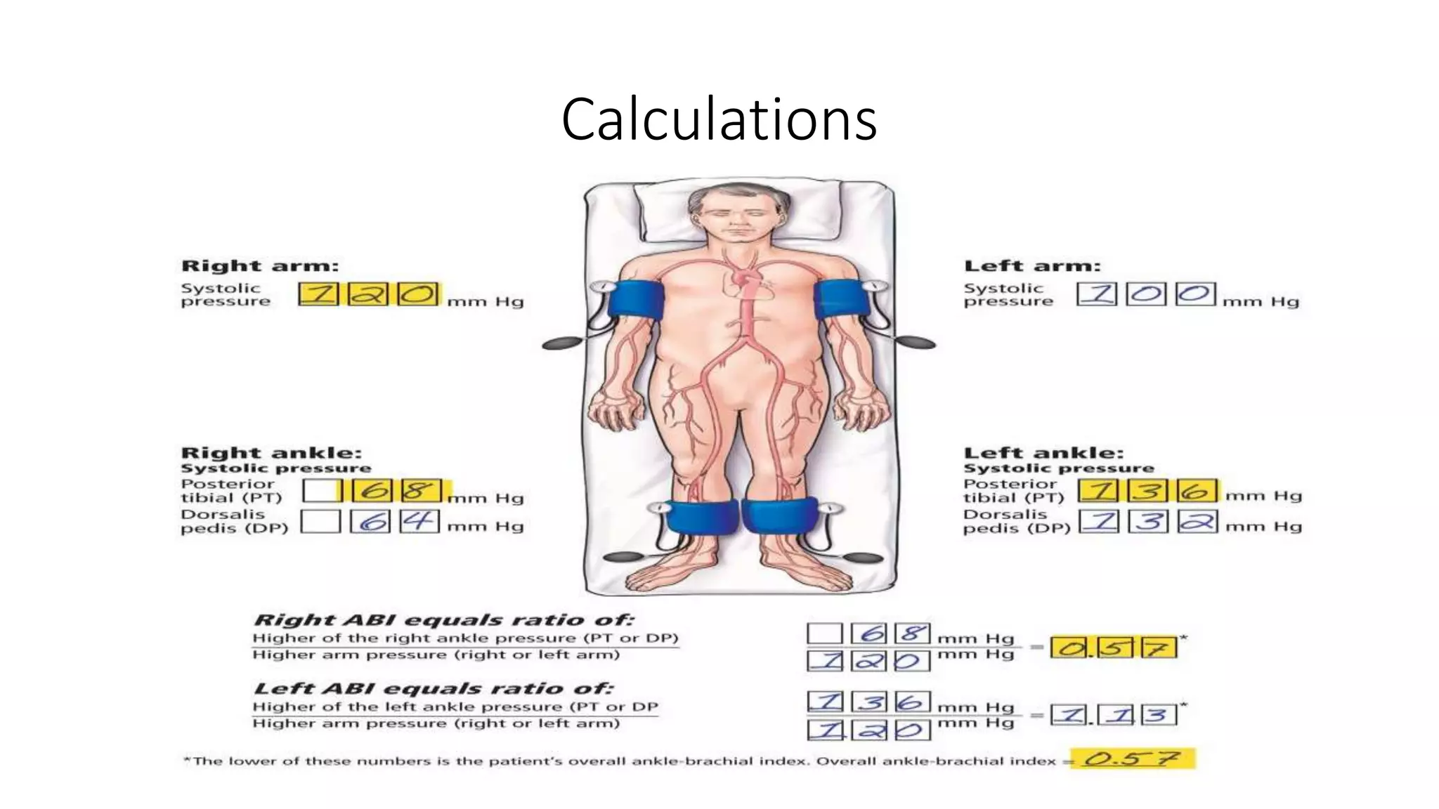
The ankle-brachial index (ABI) is a non-invasive diagnostic tool that measures the ratio of blood pressure in the lower legs to blood pressure in the arms. This measurement is primarily used to screen for peripheral artery disease (PAD), a condition where narrowed arteries reduce blood flow to the limbs. A value between 1.00 and 1.40 is considered normal, while lower values typically indicate varying degrees of arterial blockage.
Understanding the Ankle Brachial Index (ABI)
The ankle-brachial index is one of the most reliable and cost-effective methods for assessing the vascular health of the lower extremities. In the medical landscape of 2026, it remains a cornerstone of cardiovascular diagnostics because of its high sensitivity and specificity in detecting peripheral artery disease (PAD). PAD occurs when atherosclerosis—the buildup of fatty deposits or plaques—narrows the arteries that supply blood to the legs and feet.
By comparing the systolic blood pressure measured at the ankle with the systolic blood pressure measured at the arm (brachial artery), clinicians can calculate a ratio that reflects how well blood is reaching the distal portions of the body. If the pressure in the ankle is significantly lower than the pressure in the arm, it suggests that there is an obstruction or narrowing somewhere in the arterial pathway between the heart and the feet.
This test is not merely a "leg check." Because atherosclerosis is a systemic condition, a low ABI value often serves as a powerful predictor of cardiovascular events elsewhere in the body, such as myocardial infarction (heart attack) or stroke. Consequently, interpreting these numbers correctly is vital for both immediate limb preservation and long-term cardiovascular risk management.
The Mechanics of the ABI Procedure
To understand the numbers, one must understand how they are derived. The procedure involves the use of a standard blood pressure cuff and a handheld Doppler ultrasound device. The Doppler probe is essential because it allows the technician to hear the blood flow in specific arteries—the posterior tibial artery and the dorsalis pedis artery in the foot—even when the pulse is too weak to be felt by hand.
During the test, the patient lies flat (supine). Blood pressure is taken in both arms using the brachial artery. Then, blood pressure is measured at both ankles. The ABI is calculated for each leg individually by dividing the highest systolic pressure measured at that ankle by the highest systolic pressure measured in either arm.
In 2026, automated ABI systems have become more prevalent in primary care settings, using multiple cuffs that inflate simultaneously to provide a rapid reading. However, the manual Doppler method remains the gold standard for accuracy, particularly in patients with complex vascular presentations.
Interpreting the Numbers: A Categorical Breakdown
The result of an ABI test is a decimal value. Each range of values corresponds to a specific clinical interpretation, guiding physicians on the severity of arterial disease and the necessary interventions.
Normal Range (1.00 to 1.40)
A value within this range indicates that the blood flow to the lower extremities is adequate. It suggests that the arteries are open and blood pressure is being maintained throughout the limb. In a healthy individual, the pressure at the ankle should be equal to or slightly higher than the pressure in the arm. Values near 1.1 or 1.2 are frequently seen in healthy adults.
If a patient presents with leg pain but has a normal ABI at rest, clinicians may recommend an exercise ABI test. Some arterial narrowings only become significant when oxygen demand increases during walking, causing the pressure to drop only after physical exertion.
Borderline Range (0.91 to 0.99)
This range is often considered a "gray area." While not definitively diagnostic of PAD, values in this range are associated with an increased risk of cardiovascular events compared to those in the normal range. Patients with borderline ABI results may not experience classic symptoms like intermittent claudication (cramping pain during exercise), but they require close monitoring and aggressive management of risk factors like hypertension, high cholesterol, and smoking.
Mild to Moderate PAD (0.41 to 0.90)
Values in this category confirm the presence of peripheral artery disease.
- 0.71 to 0.90 (Mild): Patients often begin to experience intermittent claudication. This is characterized by muscle pain or cramping in the legs triggered by activity and relieved by rest.
- 0.41 to 0.70 (Moderate): At this stage, the narrowing is significant. Walking distances are usually limited by pain, and the patient may begin to show signs of skin changes or reduced hair growth on the lower legs.
Treatment at this stage focuses on lifestyle modifications, antiplatelet therapy, and specialized exercise programs designed to improve collateral circulation.
Severe PAD (0.40 and Below)
An ABI of 0.40 or lower is a medical red flag indicating severe arterial obstruction. At this level, blood flow may be insufficient to maintain the health of the tissue even at rest. This is often associated with critical limb ischemia (CLI).
Patients in this range may experience "rest pain"—a burning or aching sensation in the feet or toes that is often worse at night when the legs are elevated. There is a high risk of developing non-healing ulcers or gangrene. Urgent vascular consultation is mandatory for these patients to evaluate the possibility of revascularization through surgery or endovascular procedures (such as stenting or angioplasty).
High Values and Non-Compressible Vessels (>1.40)
A value higher than 1.40 does not mean "extra-healthy" blood flow. Instead, it usually indicates that the arterial walls in the ankle have become stiff and calcified. When the blood pressure cuff inflates, these rigid arteries do not compress normally, leading to a falsely elevated pressure reading.
This condition, known as medial arterial calcification, is most common in elderly patients, individuals with long-standing diabetes, and those with end-stage renal disease. Because an ABI over 1.40 is unreliable for diagnosing PAD, clinicians must use alternative tests, such as the Toe-Brachial Index (TBI), which measures pressure in the small arteries of the toes that are less prone to calcification.
Comparison of ABI Value Categories
The following table summarizes the standard interpretations for ABI results as recognized by major vascular societies in 2026.
| ABI Value Range | Interpretation | Clinical Significance & Action |
|---|---|---|
| Above 1.40 | Non-compressible | Likely arterial calcification; requires TBI or ultrasound. |
| 1.00 – 1.40 | Normal | Healthy blood flow; no immediate vascular action needed. |
| 0.91 – 0.99 | Borderline | Increased cardiovascular risk; monitor risk factors. |
| 0.71 – 0.90 | Mild PAD | Likely claudication; lifestyle changes and medication. |
| 0.41 – 0.70 | Moderate PAD | Significant impairment; referral to vascular specialist. |
| 0.00 – 0.40 | Severe PAD | High risk of limb loss; urgent surgical evaluation. |
Why These Numbers Matter: Risk Factors and Complications
The interpretation of ABI values is not just about leg pain; it is a window into the patient’s systemic health. Peripheral artery disease shares the same risk factors as coronary artery disease. If the ABI indicates significant blockage in the legs, there is a statistically high probability that similar blockages exist in the carotid arteries (leading to the brain) or the coronary arteries (supplying the heart).
According to data maintained by the American Heart Association, individuals with a low ABI have a significantly higher risk of mortality from cardiovascular causes. The primary risk factors that lead to abnormal ABI numbers include:
- Smoking: The single most significant modifiable risk factor. Tobacco use damages the endothelium (the lining of the blood vessels) and accelerates plaque formation.
- Diabetes Mellitus: Chronic high blood sugar damages vessel walls and promotes both atherosclerosis and calcification.
- Age: The prevalence of PAD increases sharply after age 65.
- Hypertension: High blood pressure places constant mechanical stress on arterial walls.
- Hyperlipidemia: High levels of LDL cholesterol provide the raw material for arterial plaques.
Early detection through ABI testing allows for intervention before a patient reaches the stage of severe PAD or suffers a major cardiac event.
Factors That May Influence ABI Accuracy
While the ABI is highly effective, certain physiological and technical factors can influence the results, requiring careful interpretation by a medical professional.
- Medial Calcification: As mentioned, diabetes and renal failure can cause falsely high readings. In these cases, the ABI cannot be the sole diagnostic tool.
- Subclavian Artery Stenosis: If a patient has a blockage in the artery supplying the arm (subclavian artery), the brachial blood pressure will be abnormally low. Since this is the denominator in the ABI equation, it can result in a falsely "normal" or "high" ABI even if leg disease is present. This is why pressure is always taken in both arms.
- Aortic Coarctation or Severe Proximal Disease: Blockages very high up in the arterial tree (like the aorta) can lower the pressure in both legs simultaneously, which might be missed if the clinician is only looking for asymmetry between the left and right legs.
- Patient Positioning: If the patient is not in a fully supine (flat) position, gravity will affect the pressure readings. The legs must be at the same level as the heart for the ratio to be valid.
Next Steps After Receiving Your Results
If you have undergone an ABI test and received your numbers, the next steps depend entirely on which category your results fall into.
For those in the normal range, the focus remains on prevention. Maintaining a healthy diet, regular exercise, and avoiding tobacco are essential to keep these numbers stable as you age.
For those in the borderline or mild PAD range, the primary goal is "risk factor modification." This typically involves:
- Starting a physician-supervised walking program to improve circulation.
- Prescription medications such as statins to lower cholesterol or ACE inhibitors to manage blood pressure.
- Antiplatelet therapy (like aspirin or clopidogrel) to prevent blood clots from forming on existing plaques.
For those with moderate to severe PAD, more advanced diagnostics are usually the next step. A vascular surgeon may order a Duplex Ultrasound, a CT Angiography (CTA), or an MR Angiography (MRA) to create a detailed map of the blockages. These images help the surgeon decide if a procedure is needed to bypass the blockage or open it from the inside using a balloon and stent.
In the current medical environment of 2026, many of these procedures are minimally invasive and can be performed on an outpatient basis, significantly reducing the risk of the most feared complication of PAD: amputation.
Frequently Asked Questions
What does it mean if my ABI is different in each leg?
It is common for PAD to progress at different rates in different limbs. The ABI is calculated for each leg separately. The lower of the two scores is typically used to categorize the patient's overall severity of PAD, but the specific score for each leg guides localized treatment, such as which leg might require a surgical intervention.
Can an ABI test be done at home?
While there are consumer-grade blood pressure monitors, a true ABI requires a Doppler ultrasound probe or a specialized multi-cuff automated system to get an accurate reading of the ankle arteries. Measurements taken with standard oscillometric cuffs (the kind that wrap around the arm and "beep") at the ankle are often inaccurate and are not recommended for diagnostic purposes.
How often should I have my ABI checked?
If you have risk factors such as smoking, diabetes, or are over the age of 65, the Centers for Disease Control and Prevention (CDC) and other health organizations suggest discussing a baseline screening with your doctor. If you have been diagnosed with PAD, your doctor may check your ABI every 6 to 12 months to monitor the effectiveness of your treatment and the progression of the disease.
Is the test painful?
No, the test is non-invasive and painless. It feels exactly like having your blood pressure taken at the doctor's office, though it takes slightly longer because the technician needs to locate the specific pulses in your feet using the Doppler device.
Can exercise change my ABI results?
In healthy individuals, exercise should cause the ABI to stay the same or increase slightly. In people with PAD, exercise usually causes the ABI to drop significantly because the narrowed arteries cannot keep up with the muscles' increased demand for blood. This "Exercise ABI" is a specific diagnostic tool used when resting results are inconclusive.
Practical Recommendation for Patients
If you are experiencing unexplained leg pain while walking or have been told you are at risk for vascular disease, request an ankle-brachial index test from your primary care provider. It is a simple, five-minute procedure that provides critical information about your circulatory health.
When you receive your numbers, ensure you ask your provider for the specific decimal value, not just a "normal" or "abnormal" designation. Knowing whether you are at 0.92 (borderline) versus 0.50 (moderate PAD) is essential for tailoring your lifestyle and medical interventions. In 2026, the emphasis in vascular medicine is on early detection; catching a declining ABI early can prevent the progression to critical limb ischemia and significantly lower your overall risk of heart attack and stroke.