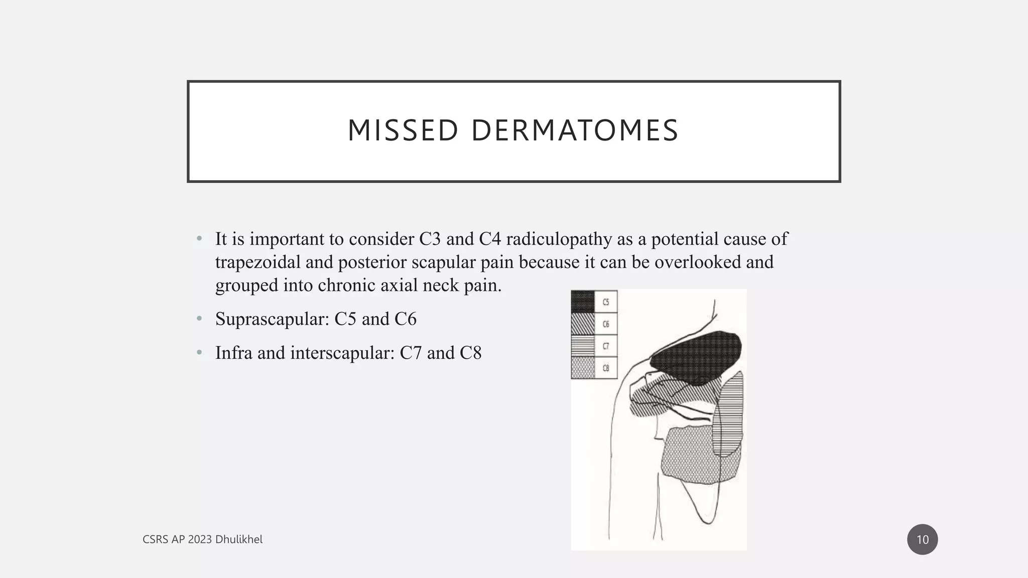
Cervical radiculopathy at the C6 level occurs when the C6 nerve root is compressed or irritated as it exits the spinal column, typically at the C5-C6 vertebral segment. This condition manifests as radiating pain, numbness, and muscle weakness that follows a specific path from the neck down to the thumb and index finger. Effective management requires a structured approach involving clinical diagnosis, diagnostic imaging, and a tiered treatment plan ranging from physical therapy to surgical intervention.
Step 1: Identifying the Sensory and Pain Distribution Patterns
The first step in gaining insight into C6 radiculopathy is recognizing the "geographic" signature of the pain. Unlike generalized neck pain, radiculopathy follows a dermatomal pattern. A dermatome is an area of skin supplied by a single spinal nerve. For the C6 nerve root, this pathway is remarkably consistent across most patients.
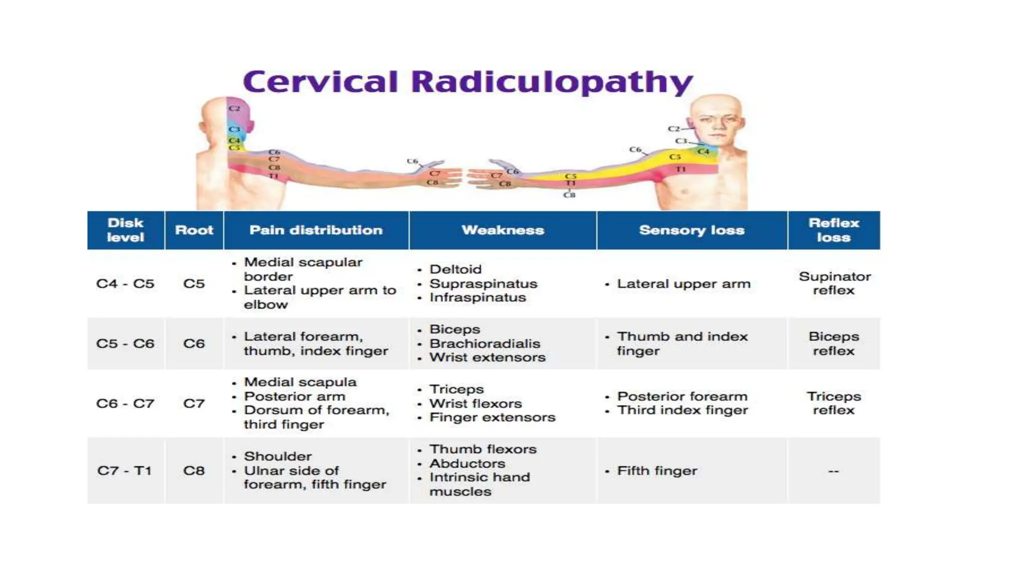
Pain or "paresthesia" (burning, tingling, or pins-and-needles) usually originates in the cervical spine or the top of the shoulder (suprascapular area). From there, the sensation travels down the lateral (outer) aspect of the arm and into the forearm. The hallmark of C6 involvement is the extension of these symptoms into the thumb and the radial side of the index finger.
In the clinical landscape of 2026, specialists distinguish between "referred pain" and "radicular pain." Referred pain is often a dull, aching sensation caused by muscle strain or joint irritation. In contrast, C6 radicular pain is often described as electric, sharp, or lancinating. Patients may also experience a "heavy" feeling in the arm, which can be exacerbated by certain head positions, such as tilting the head back or turning it toward the side of the pain (the Spurling maneuver). Understanding this specific sensory map is the primary prerequisite for differentiating C6 issues from other common conditions like carpal tunnel syndrome or C5 radiculopathy.
Step 2: Understanding the Pathophysiology of the C5-C6 Motion Segment
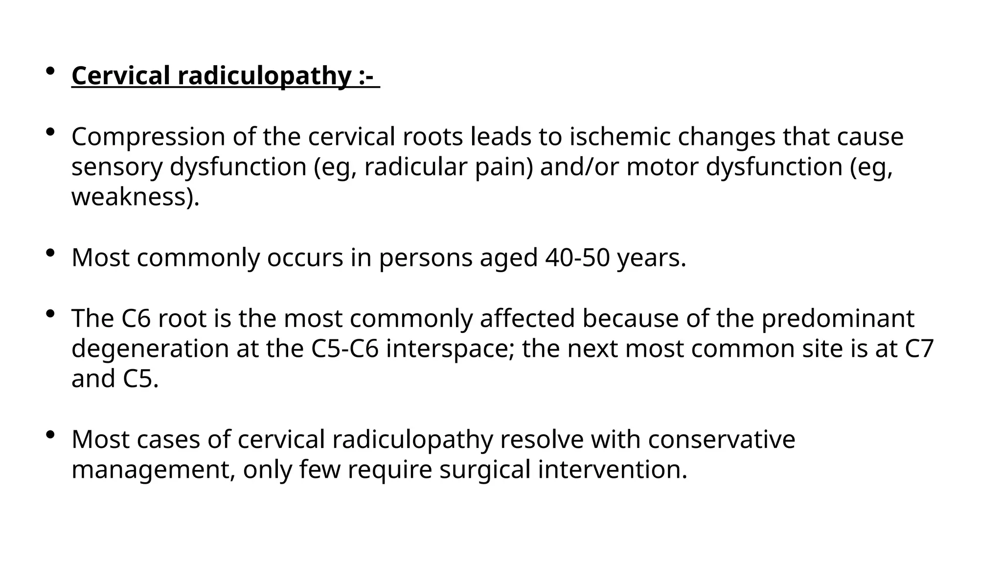
To understand why the C6 nerve root is compromised, one must look at the C5-C6 intervertebral space. This is one of the most mobile segments of the cervical spine, making it highly susceptible to wear and tear. There are two primary mechanisms of compression:
- Disc Herniation: The intervertebral disc acts as a shock absorber. If the tough outer layer (annulus fibrosus) tears, the soft inner material (nucleus pulposus) can bulge or extrude. If this occurs posterolaterally, it directly impinges upon the C6 nerve root. This is more common in younger patients and often follows acute trauma or sudden strain.
- Cervical Spondylosis (Degeneration): As we age, the discs lose water content and height. This leads to the development of osteophytes (bone spurs) around the neuroforamen—the bony window through which the nerve exits. In 2026, longitudinal studies continue to show that chronic C6 radiculopathy is frequently the result of these "hard" bony changes rather than "soft" disc herniations.
Beyond mechanical compression, there is a significant biochemical component. When a disc herniates, it releases inflammatory mediators like tumor necrosis factor-alpha (TNF-α). These chemicals can irritate the nerve root even if the physical pressure is relatively mild. This explains why some patients have severe pain despite "minor" findings on an MRI, while others with large herniations remain asymptomatic.
Step 3: Assessing Motor Deficits and Reflex Changes (Myotomes)
The third step involves a neurological exam to identify functional loss. Each nerve root powers specific muscles, known as a myotome. For C6, the primary muscle groups affected are the biceps brachii and the extensor carpi radialis (the muscles that allow you to "cock" your wrist back).
Clinical weakness in C6 radiculopathy often manifests in two ways:
- Elbow Flexion: The patient may find it difficult to bring their hand toward their shoulder against resistance.
- Wrist Extension: This is a classic C6 indicator. If a patient cannot maintain a strong, extended wrist when pushed downward, C6 involvement is highly suspected.
Furthermore, clinicians check the brachioradialis reflex. A healthy C6 nerve root facilitates a predictable "jerk" response when the tendon near the wrist is tapped with a reflex hammer. A diminished or absent reflex in this area, when compared to the opposite arm, provides objective evidence of nerve root dysfunction. By 2026, standardized neurological scoring systems have become even more rigorous, allowing therapists to track small changes in muscle power (on a 0–5 scale) to determine if a patient is improving or if the nerve damage is progressing toward a permanent state.
Step 4: The Diagnostic Pathway—Imaging and Electrophysiology
Once clinical suspicion is high, diagnostic tools are used to confirm the location and severity of the compression. In 2026, the diagnostic protocol typically follows a specific sequence:
- Magnetic Resonance Imaging (MRI): The gold standard. MRI provides high-resolution views of soft tissues, allowing doctors to see exactly how much room the C6 nerve has within the foramen. It can distinguish between a disc bulge, a full herniation, and foraminal stenosis caused by bone spurs.
- Electromyography (EMG) and Nerve Conduction Studies (NCS): While an MRI shows the anatomy, an EMG shows the physiology. It measures the electrical activity of muscles. If the C6 nerve is dying or severely compressed, the muscles it supplies will show "denervation" potentials. This is crucial for distinguishing C6 radiculopathy from peripheral nerve issues like a brachial plexus injury.
- Upright or Dynamic X-rays: These are used to assess stability. If the C5 vertebrae is sliding over the C6 (spondylolisthesis), it can "pinch" the nerve root during movement, a detail sometimes missed on a static, lying-down MRI.
It is important to note that many asymptomatic people have disc bulges on an MRI. Therefore, the imaging must correlate precisely with the patient's physical symptoms before a diagnosis of C6 radiculopathy is finalized.
Step 5: Conservative Management and Rehabilitation
The vast majority of C6 radiculopathy cases—approximately 75% to 90%—resolve with non-surgical treatment within 6 to 12 weeks. The goal of conservative care is to reduce inflammation and mechanical pressure.
Pharmacological Interventions
Initial treatment usually involves Non-Steroidal Anti-Inflammatory Drugs (NSAIDs) like ibuprofen or naproxen to quiet the chemical "storm" around the nerve. In 2026, physicians may also prescribe gabapentinoids for neuropathic pain or a short course of oral corticosteroids to rapidly reduce nerve edema.
Physical Therapy (PT)
PT is the cornerstone of recovery. A typical C6 program includes:
- Cervical Traction: Gently stretching the neck to manually open the neuroforamen and provide temporary relief.
- Postural Re-education: Addressing "forward head posture," which significantly increases the load on the C5-C6 segment.
- Nerve Gliding: Specific exercises designed to pull the nerve gently through its pathway, preventing adhesions and improving blood flow to the nerve fibers.
- The McKenzie Method: A system of repeated movements designed to "centralize" the pain—moving it out of the thumb and back toward the neck, which is a sign of healing.
Epidural Steroid Injections (ESIs)
If oral medications and PT are insufficient, a transforaminal epidural injection may be performed. Under X-ray guidance (fluoroscopy), a specialist injects a concentrated corticosteroid directly next to the C6 nerve root. This provides a potent anti-inflammatory effect that can last several months, often providing a "window of opportunity" for the patient to engage more effectively in physical therapy.
Step 6: Surgical Considerations and Long-term Prognosis
Surgery is reserved for patients who fail conservative care after 6–8 weeks, or those who exhibit "Red Flag" symptoms such as progressive muscle wasting or "drop wrist." The objective of surgery is to decompress the C6 nerve root.
Common Surgical Procedures
- Anterior Cervical Discectomy and Fusion (ACDF): The most common procedure. The surgeon removes the damaged disc from the front of the neck, clears the bone spurs, and fuses the C5 and C6 vertebrae together using a graft and a small metal plate.
- Cervical Disc Replacement (CDR): In 2026, CDR has become a preferred alternative for younger patients with single-level C6 radiculopathy. Instead of fusing the bones, an artificial, moving disc is inserted. This preserves motion and reduces the risk of "adjacent segment disease," where the discs above or below a fusion wear out prematurely.
- Posterior Foraminotomy: A "keyhole" surgery performed from the back of the neck. The surgeon shaves away a small portion of the bone (the lamina) to create more room for the nerve root without needing a fusion.
The long-term prognosis for C6 radiculopathy is generally excellent. Most patients return to full activity, although those who underwent fusion may need to modify certain high-impact sports.
Comparative Analysis: C5 vs. C6 vs. C7 Radiculopathy
| Feature | C5 Radiculopathy | C6 Radiculopathy | C7 Radiculopathy |
|---|---|---|---|
| Pain/Numbness Area | Lateral upper arm (Deltoid) | Lateral forearm, Thumb, Index Finger | Middle finger, back of hand |
| Primary Weakness | Shoulder abduction (Deltoid) | Wrist extension, Biceps | Elbow extension (Triceps) |
| Reflex Affected | Biceps reflex | Brachioradialis reflex | Triceps reflex |
| Common Disc Level | C4-C5 | C5-C6 | C6-C7 |
| Functional Impact | Difficulty lifting arm | Difficulty gripping/writing | Difficulty pushing doors |
Frequently Asked Questions
How long does it take for a C6 nerve root to heal?
Nerve tissue heals much more slowly than muscle or skin. While pain often improves within 2 to 4 weeks of starting treatment, full neurological recovery (getting strength and full sensation back) can take 3 to 6 months. In cases where the nerve was severely compressed for a long time, some mild numbness may persist indefinitely.
What is the best sleeping position for C6 radiculopathy?
The goal is to keep the cervical spine in a "neutral" position. Sleeping on your back with a contoured pillow that supports the natural curve of the neck is generally best. If you sleep on your side, ensure the pillow is high enough to keep your head aligned with your shoulders, and avoid tucking your chin toward your chest, as this can increase pressure on the exit points of the C6 nerve.
Can C6 radiculopathy cause chest pain?
Yes, this is known as cervical angina. Because the C6 nerve root supplies parts of the shoulder and upper chest wall, compression can mimic the pain of a heart condition. However, cervical angina is usually triggered by neck movement and is not accompanied by the shortness of breath or diaphoresis (sweating) typical of a cardiac event. Always consult a doctor to rule out heart issues first.
When is C6 radiculopathy considered an emergency?
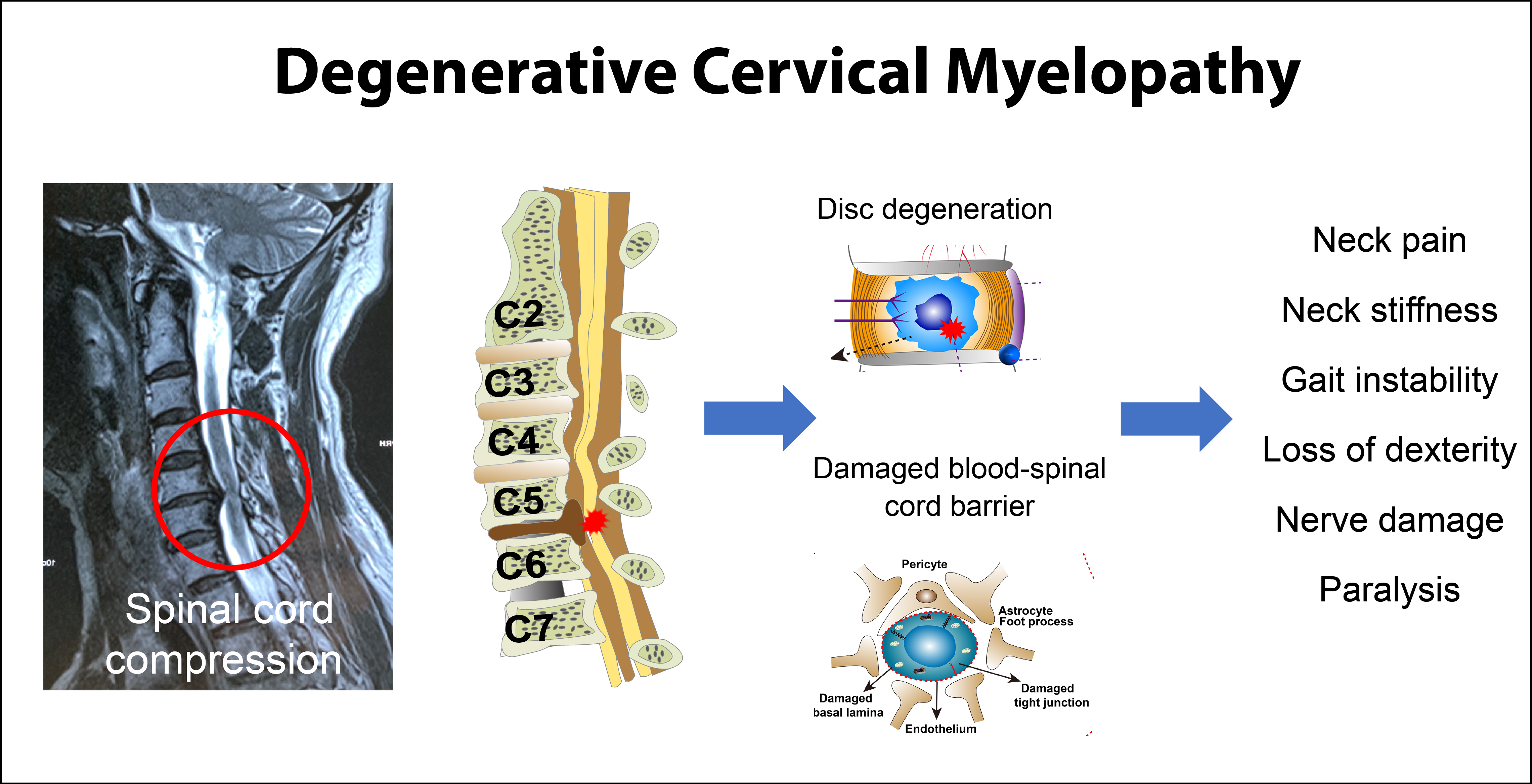
While most cases are managed electively, you should seek immediate care if you experience "saddle anesthesia" (numbness in the groin), sudden loss of bowel or bladder control, or significant weakness in both arms simultaneously. These may be signs of cervical myelopathy, where the spinal cord itself—not just a single nerve root—is being compressed.
Is carpal tunnel syndrome often confused with C6 radiculopathy?
Yes, they are frequently misdiagnosed because both involve the thumb and index finger. However, carpal tunnel syndrome usually only causes symptoms in the hand and wrist, whereas C6 radiculopathy almost always involves pain or stiffness in the neck and upper arm. An EMG/NCS study is the most effective way to distinguish between the two.
Practical Recommendations for 2026
If you suspect you are dealing with C6 radiculopathy, your first action should be activity modification. Avoid overhead lifting and prolonged "tech neck" (looking down at mobile devices), which increases the pressure on the C5-C6 segment. Early intervention with a physical therapist specializing in mechanical diagnosis and therapy (MDT) is statistically shown to provide the best outcomes. If weakness is present, do not "wait and see"; objective muscle loss is a signal that the nerve is under significant distress and requires immediate medical evaluation to prevent permanent atrophy.