The apical segment of the lung is the uppermost functional sub-division of the lung's upper lobe, situated at the peak (apex) of the thoracic cavity. Positioned just above the level of the first rib, this anatomical region plays a vital role in pulmonary ventilation and is clinically significant due to its susceptibility to specific conditions like tuberculosis, Pancoast tumors, and spontaneous pneumothorax.
Understanding Bronchopulmonary Segments
To understand the apical segment, one must first grasp how the human lungs are structurally divided. The respiratory system does not operate as two simple, uniform balloons. Instead, each lung is divided into lobes by anatomical fissures, and each lobe is further subdivided into self-contained functional units called bronchopulmonary segments.
A bronchopulmonary segment is an anatomically distinct portion of lung tissue supplied by its own segmental bronchus (a branch of the main tracheobronchial tree), a corresponding segmental artery, and dedicated autonomic nerves. Surrounding connective tissue septa isolate each segment from its neighbors. This compartmentalization offers two significant biological advantages:
- Disease Localization: Localized infections or localized tissue damage can remain confined to a single segment without immediately spreading throughout the entire lobe.
- Surgical Precision: Thoracic surgeons can perform a segmentectomy—removing a single diseased bronchopulmonary segment—while preserving the remaining healthy lung tissue and preserving overall pulmonary function.
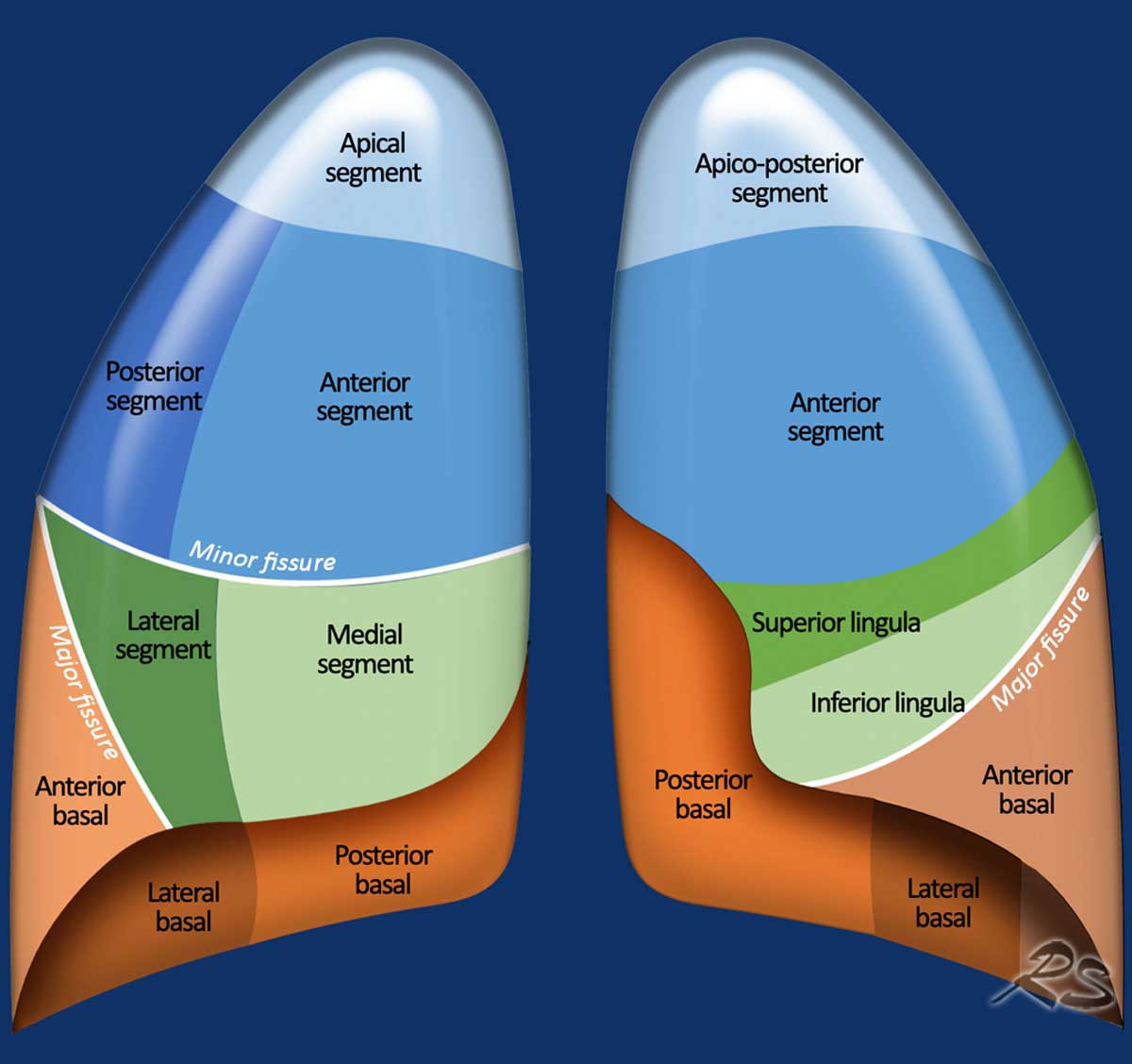
The right lung contains ten standard bronchopulmonary segments distributed across three lobes (superior, middle, and inferior). The left lung typically contains eight to ten segments across two lobes (superior and inferior). The apical segment occupies the apex—the very top—of the upper lobe in both lungs, though its exact anatomical arrangement differs slightly between the right and left sides.
Anatomical Breakdown of the Apical Segment
The apex of each lung extends superiorly into the root of the neck, reaching approximately 2.5 to 4 centimeters above the level of the sternal end of the first rib. Because of this high anatomical placement, the apical segment occupies a unique anatomical boundary between the chest cavity and the neck.
- Root of the Neck / Subclavian Vessels
- APICAL SEGMENT (S1) (Above 1st Rib)
- Rest of Upper Lobe
- Posterior Segment (S2)
- Anterior Segment (S3)
- Rest of Upper Lobe
- APICAL SEGMENT (S1) (Above 1st Rib)
Right Lung vs. Left Lung Architecture
In international anatomical nomenclature (Terminologia Anatomica), bronchopulmonary segments are assigned specific numbers.
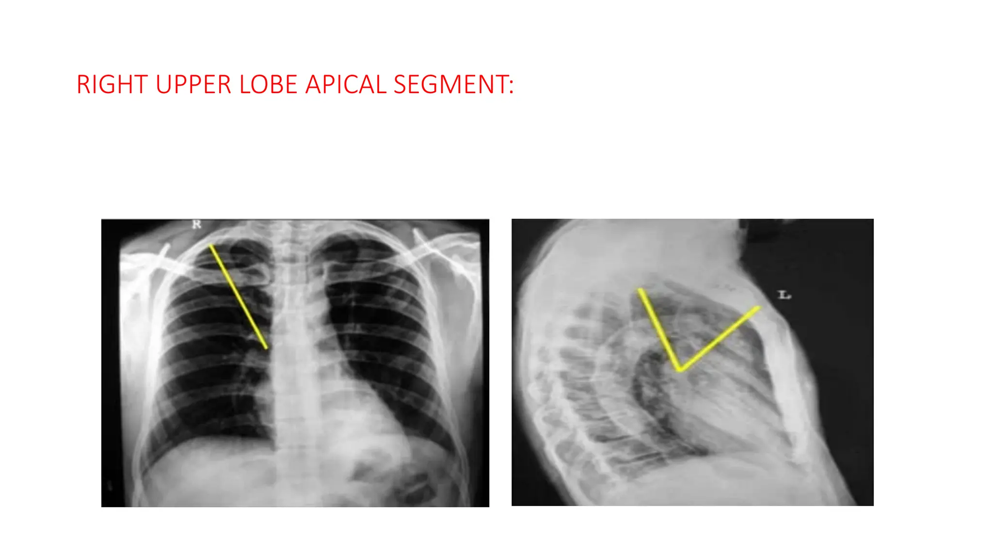
- Right Lung (Apical Segment - S1): In the right upper lobe, the apical segment is designated as Segment 1 (S1). It forms a distinct pyramidal shape with its apex pointing upward into the cervical region and its base resting upon the anterior and posterior segments of the right upper lobe.
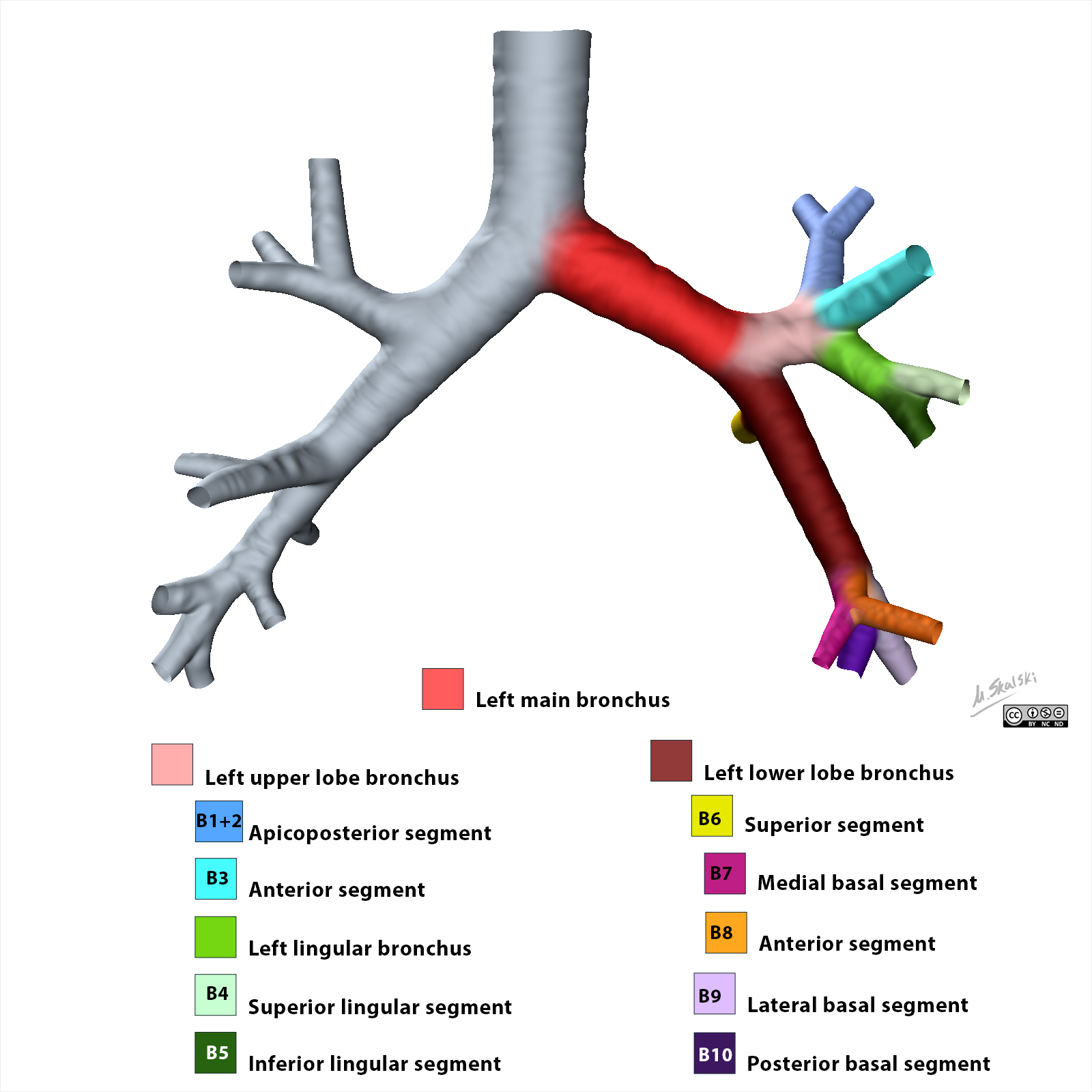
- Left Lung (Apicoposterior Segment - S1+S2): In the left lung, embryonic development causes the bronchial branches of the apical and posterior regions to merge. Consequently, classic human anatomy classifies the top of the left upper lobe as the apicoposterior segment (S1+S2). While some clinicians discuss the left apical portion separately, it usually shares a combined apicoposterior segmental bronchus.
Adjacent Anatomical Boundaries
Because the apical segment projects into the root of the neck, it shares intimate anatomical boundaries with major neural, vascular, and musculoskeletal structures:
- Subclavian Artery and Vein: Pass anterior and slightly inferior to the pulmonary apex, leaving a distinct groove on the surface of the apical pleura.
- Brachial Plexus: The lower roots of the brachial plexus (specifically the C8 and T1 nerve roots) run directly adjacent to the apical segment.
- Cervical Sympathetic Chain: The stellate ganglion (cervicothoracic ganglion) sits posterior to the lung apex near the neck of the first rib.
- Cervical Pleura (Suprapleural Membrane / Sibson’s Fascia): A fibrous membrane that covers the apical pleura, anchoring the lung apex to the inner border of the first rib and the transverse process of the C7 vertebra.
Airway and Vascular Supply
The apical segment receives dedicated tracheobronchial and vascular branches:
- Apical Segmental Bronchus: Originates from the right upper lobe bronchus (or the apicoposterior trunk on the left) and directs inhaled air into the segment.
- Apical Segmental Artery: A direct branch of the pulmonary artery supplying deoxygenated blood from the right ventricle of the heart into the apical capillary beds for oxygenation.
- Segmental Pulmonary Veins: Collect oxygenated blood from the apical capillaries and run in the intersegmental septa, draining ultimately into the superior pulmonary vein toward the left atrium.
Key Functions and Physiological Dynamics
Like all bronchopulmonary segments, the primary function of the apical segment is gas exchange—absorbing oxygen into the bloodstream and eliminating carbon dioxide. However, because of its anatomical position at the highest point of the vertical chest axis, the apical segment experiences unique physiological dynamics governed by gravity.
Gravity and the Ventilation-Perfusion (V/Q) Ratio
When a person stands or sits upright, gravity exerts a profound effect on pulmonary blood flow and intrapleural pressure:
- Blood Flow (Perfusion, Q): Hydrostatic pressure causes blood to pool toward the bases of the lungs. As a result, blood flow at the apical segment is significantly lower than at the lung bases.
- Airflow (Ventilation, V): Because the weight of the lung pulls downward on the pleural cavity, intrapleural pressure at the apex is more negative than at the base. This keeps the alveoli in the apical segment expanded even at the end of a normal exhalation. However, because these apical alveoli are already nearly fully stretched, they undergo less volume change during normal tidal breathing compared to basal alveoli.
These dynamics result in a high Ventilation-Perfusion ratio ($\text{V/Q} > 1.0$) in the apical segment. Because ventilation exceeds blood perfusion at the apex, carbon dioxide is washed out rapidly, and the local alveolar partial pressure of oxygen ($\text{P}_{\text{O}_2}$) is noticeably higher in the apical segment than in any other part of the lung.
Clinical Significance and Pathologies
The physiological and anatomical characteristics of the apical segment make it a frequent focal point in pulmonology, radiology, and thoracic surgery. Several major medical conditions display a strong predilection for the apical segment.
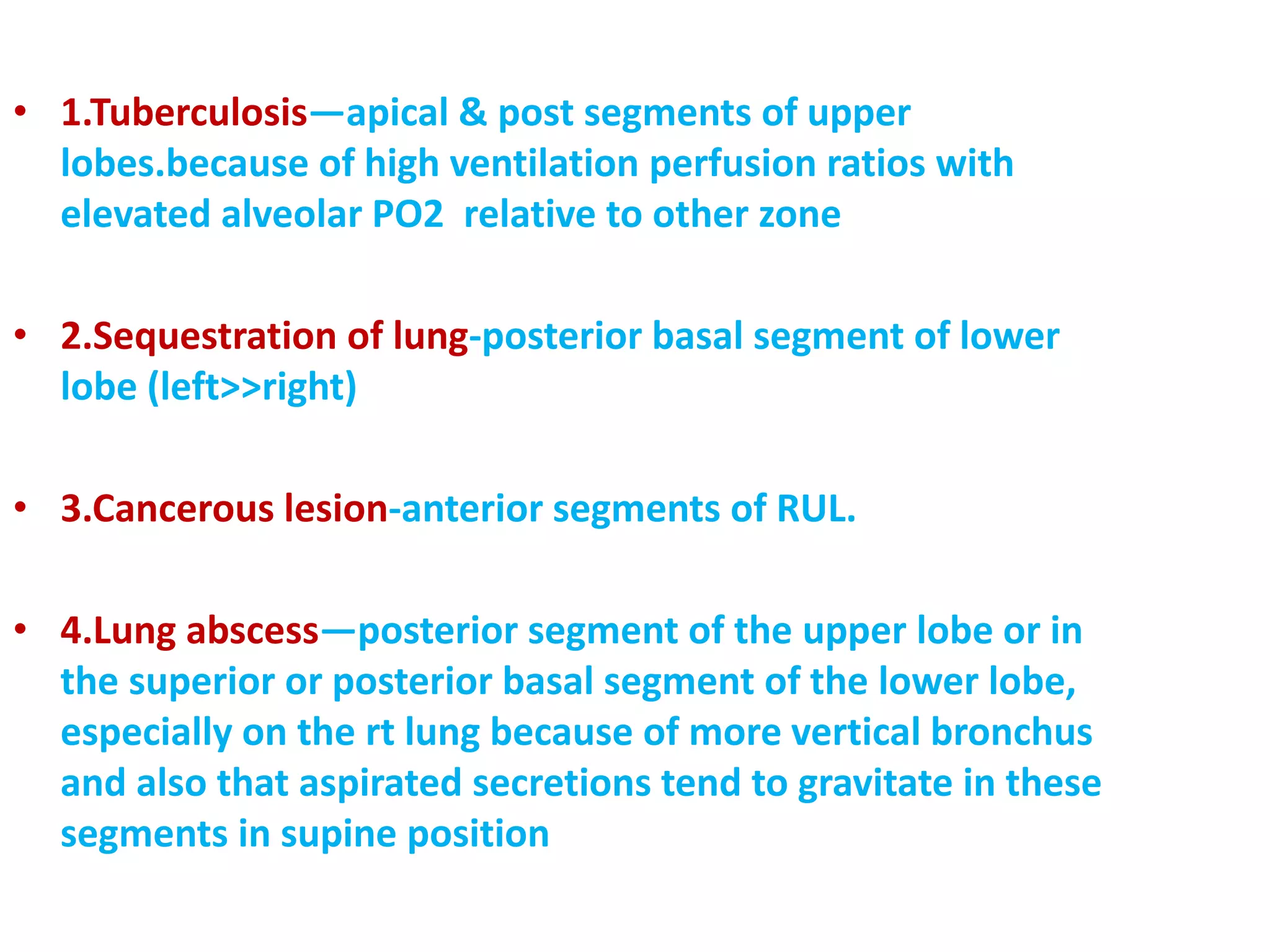
1. Reactivation Pulmonary Tuberculosis (TB)
Mycobacterium tuberculosis, the bacterium responsible for tuberculosis, is a strict obligate aerobe that thrives in oxygen-rich environments.
When a person develops secondary (reactivation) tuberculosis—often years after an initial latent infection—the bacteria overwhelmingly favor the apical and posterior segments of the upper lobes. The high alveolar oxygen tension ($\text{P}_{\text{O}_2}$) in the apical segment provides the optimal metabolic environment for bacterial replication.
Reactivation TB in the apical segment typically presents on imaging as:
- Patchy consolidation or infiltrates at the lung apex.
- Cavitary lesions: Tissue necrosis created as the immune system fights the organism, forming hollowed-out spaces that can harbor millions of multiplying bacteria.
- Fibrotic scarring and calcified granulomas (Ghon complexes) as long-term sequelae.
Detailed clinical protocols for diagnosing and managing pulmonary tuberculosis are available through the Centers for Disease Control and Prevention (CDC) TB Guidance.
2. Pancoast Tumors (Superior Sulcus Tumors)
A Pancoast tumor, also referred to as a superior sulcus tumor, is a form of lung cancer—typically non-small cell lung carcinoma (NSCLC)—that arises in the extreme apex of the lung within the apical segment.
Because of the apical segment's proximity to the root of the neck, growing apical tumors can invade adjacent structures before causing typical respiratory symptoms such as coughing or shortness of breath. This produces a classic clinical constellation known as Pancoast Syndrome:
- Severe Shoulder and Arm Pain: Caused by tumor invasion into the lower trunks of the brachial plexus (C8, T1), often radiating down the inner arm to the fourth and fifth fingers.
- Horner's Syndrome: Resulting from invasion and compression of the cervical sympathetic chain (stellate ganglion). Horner's syndrome is characterized by a triad of symptoms on the affected side of the face:
- Ptosis: Drooping of the upper eyelid.
- Miosis: Constriction of the pupil.
- Anhidrosis: Loss of sweating on the affected side of the face.
- Subclavian Vessel Compression: Can cause arm swelling or weakened arterial pulses on the ipsilateral side.
Diagnostic standards and staging guidelines for Pancoast tumors can be reviewed via the National Cancer Institute (NCI) Physician Data Query.
3. Primary Spontaneous Pneumothorax and Apical Blebs
A pneumothorax occurs when air enters the pleural space between the lung and the chest wall, causing the lung to collapse. Primary spontaneous pneumothorax (PSP) occurs in individuals without underlying chronic lung disease, most commonly tall, thin young adults (typically males aged 15 to 30).
The primary driver of PSP is the rupture of small, air-filled sacs called subpleural blebs or bullae, which form predominantly along the apex of the apical segment.
- Normal Apical Pleura
- Lung Tissue
- Intact Pleura
- Subpleural Bleb Formation
- Weakened Bleb
- Lung Tissue
- Rupture Risk leading to Pneumothorax
Why do blebs form at the apical segment?
- Transpulmonary Stress: In tall individuals, the vertical distance from the base to the apex is greater, increasing the gravitational traction hanging the lung from its apex.
- Apical Ischemia: Rapid height growth during adolescence can outpace blood vessel development at the extreme apex, causing localized tissue weakness and subpleural alveolar enlargement.
When an apical bleb ruptures, air leaks directly into the pleural cavity. Treatment ranges from simple observation or needle aspiration for small leaks to video-assisted thoracoscopic surgery (VATS) with apical bullectomy (removing the bleb-bearing apex) and pleurodesis for recurrent cases.
4. Radiation-Induced Lung Injury
Patients undergoing radiation therapy for cancers of the head, neck, breast, or esophagus may have upper thoracic radiation fields that encompass the apical segment. Because apical pulmonary tissue is sensitive to ionizing radiation, patients can develop:
- Acute Radiation Pneumonitis: Inflammatory exudate and alveolar edema appearing 1 to 6 months post-treatment.
- Chronic Apical Radiation Fibrosis: Permanent scarring and retraction of apical lung tissue occurring months to years later, often appearing on CT scans as dense, geometric opacities corresponding exactly to the radiation beam portal.
5. Surgical Segmentectomy vs. Lobectomy
In modern thoracic surgery, preserving healthy lung tissue is paramount, especially for patients with borderline pulmonary function. When early-stage lung cancer (such as a small ground-glass nodule) or localized bronchiectasis is isolated strictly to the apical segment, thoracic surgeons perform an anatomical apical segmentectomy (S1 resection).
This procedure requires individual dissection and ligation of the apical segmental bronchus, apical segmental artery, and segmental veins while preserving the adjacent anterior and posterior segments. Surgical guidance from the Society of Thoracic Surgeons (STS) outlines standardized surgical pathways for segmentectomy versus formal lobectomy.
Diagnostic Imaging of the Apical Segment
Evaluating the apical segment requires specialized radiologic techniques because the overlying bones of the shoulder girdle—including the clavicles, first ribs, and scapulae—can obscure soft tissue details on standard imaging.
| Imaging Modality | Diagnostic Utility in the Apical Segment |
|---|---|
| Posteroanterior (PA) Chest X-ray | Standard screening view; clavicles often overlap the apical segment, potentially hiding small lesions. |
| Apical Lordotic Radiograph | Patient leans backward; projects clavicles upward, unmasking the apical segment for clear inspection. |
| High-Resolution CT (HRCT) Scan | Gold standard; provides cross-sectional 1mm slices to evaluate blebs, nodules, cavitary TB, and invasion. |
| Contrast MRI (Chest / Neck) | Excellent for evaluating suspected Pancoast tumors to map brachial plexus and subclavian vessel involvement. |
The Apical Lordotic View
On a standard posteroanterior (PA) chest X-ray, the clavicle and first rib cross directly over the apical segment. To overcome this limitation, radiologists historically utilized an apical lordotic radiograph. In this positioning, the patient stands angled backward. This projects the clavicles above the pulmonary apex on the image, unmasking the apical segment so small tuberculous cavities, apical scarring, or subtle opacities can be identified.
High-Resolution Computed Tomography (HRCT)
Today, High-Resolution CT scanning is the primary tool for detailed evaluation of apical anatomy. Thin-slice CT images allow radiologists to:
- Detect subpleural blebs as small as 1 to 2 millimeters.
- Differentiate between active tuberculous consolidation and chronic fibrotic scarring.
- Measure solitary pulmonary nodules (SPNs) located within Segment 1.
- Assess chest wall or neurovascular invasion in apical malignancies.
Comparative Matrix: Right S1 vs. Left S1+S2
Understanding how the apical segment differs between the right and left lungs helps contextualize anatomical reports and surgical planning.
| Parameter | Right Upper Lobe: Apical Segment (S1) | Left Upper Lobe: Apicoposterior Segment (S1+S2) |
|---|---|---|
| Anatomical Designation | Segment 1 (S1) | Segment 1 + Segment 2 combined (S1+S2) |
| Bronchial Tree Branch | Apical segmental bronchus (direct branch of Right Upper Lobe bronchus) | Apicoposterior segmental bronchus (bifurcates into apical and posterior sub-branches) |
| Vascular Architecture | Apical artery branch from main right pulmonary artery | Apicoposterior arterial trunks from left pulmonary artery |
| Surrounding Boundaries | First rib, right subclavian artery/vein, right vagus and phrenic nerves | First rib, left subclavian artery, aortic arch, left recurrent laryngeal nerve |
| Common Pathologies | Reactivation Tuberculosis, Pancoast tumor, Primary Spontaneous Pneumothorax | Reactivation Tuberculosis, Left-sided Pancoast tumor, Radiation fibrosis |
| Surgical Resection Type | Isolated Right S1 Segmentectomy | Combined Left S1+S2 Segmentectomy (less frequently isolated S1) |
Practical Guidance: Understanding Radiology Findings
If you are reviewing a medical report, chest X-ray, or CT scan result that mentions the "apical segment" or "lung apex," here is how to interpret common radiologic terms in consultation with your physician:
Common Terms Found in Imaging Reports
- "Apical Scarring" or "Apical Pleuroparenchymal Thickening": Often benign, represents healed prior inflammation, past minor infections, or age-related pleuropleural thickening. Common in older adults or former smokers.
- "Apical Bulla / Subpleural Bleb": Thin-walled, air-filled space at the edge of the apical lung. Requires evaluation if the patient has a history of sudden chest pain or shortness of breath.
- "Apical Opacity or Infiltrate": Indicates tissue density in the apex where air should be. May represent localized pneumonia, active infection (such as TB), or focal atelectasis (lung collapse).
- "Apical Nodule": A round, discrete lesion. Most nodules are benign (such as old granulomas), but new, growing, or spiculated nodules require follow-up CT surveillance or biopsy to rule out malignancy.
Next Steps for Patients
- Review with Your Clinician: Never interpret an imaging report in isolation. A finding must be correlated with symptoms (e.g., fever, night sweats, cough, weight loss, shoulder pain).
- Compare Prior Scans: Comparing new CT or X-ray images with older scans ("prior films") is crucial. A stability of findings over two or more years strongly suggests a benign condition.
- Pulmonary Function Testing (PFTs): If surgical intervention in the apical segment is considered, PFTs measure baseline spirometry and diffusion capacity ($\text{DLCO}$) to ensure safe pulmonary reserve.
- Specialist Consultation: Persistent, unexplained apical abnormalities should be evaluated by a board-certified pulmonologist or thoracic surgeon.
Frequently Asked Questions
Why is tuberculosis most commonly found in the apical segment of the lung?
Mycobacterium tuberculosis is an obligate aerobic bacterium that requires high concentrations of oxygen to multiply. In an upright human, gravity causes lower blood perfusion to the lung tops, resulting in a high ventilation-perfusion ratio ($\text{V/Q} > 1.0$) and elevated oxygen partial pressure ($\text{P}_{\text{O}_2}$) in the apical segments. This oxygen-rich environment allows reactivated TB bacteria to thrive and cause tissue cavitation.
What is the difference between Segment 1 on the right lung and the apex of the left lung?
The right upper lobe has a distinct, dedicated Apical Segment designated as Segment 1 (S1). In the left upper lobe, the apical bronchial branch typically shares a common trunk with the posterior bronchial branch, combining them into an Apicoposterior Segment (S1+S2). Functionally and clinically, both apex regions share similar disease susceptibilities.
How does an apical segment lesion cause arm or eyelid symptoms?
Because the apical segment extends above the first rib into the root of the neck, mass lesions such as Pancoast tumors can grow directly into neighboring nerve structures. Compression of the lower brachial plexus (C8-T1) causes shoulder and inner arm pain. Compression of the adjacent cervical sympathetic chain (stellate ganglion) disrupts sympathetic innervation to the face, leading to Horner's syndrome (drooping eyelid, pupil constriction, and lack of facial sweating).
What is an apical bleb, and how dangerous is it?
An apical bleb is a small, air-filled blister on the outer surface of the apical lung pleura, often formed due to mechanical stretching forces at the apex. While an intact bleb causes no symptoms, if it ruptures, air leaks into the chest cavity, causing a primary spontaneous pneumothorax (collapsed lung). Unruptured blebs discovered incidentally on CT scans do not always require surgery, but recurring collapses are treated surgically via thoracoscopic bleb removal.
Can a person live normally after having their apical lung segment removed?
Yes. An apical segmentectomy (surgical removal of Segment 1) removes only a small fraction of total lung volume. Because neighboring healthy segments expand to fill the space and the remaining lung tissue continues normal gas exchange, most patients maintain excellent overall lung function and quality of life after recovering from surgery.
Professional Medical Disclaimer
This article is provided for educational and informational purposes only and does not constitute formal medical advice, diagnosis, or treatment recommendations. Clinical decisions involving pulmonary conditions, radiologic findings, or thoracic surgical procedures must always be made in direct consultation with a qualified healthcare provider or pulmonologist.