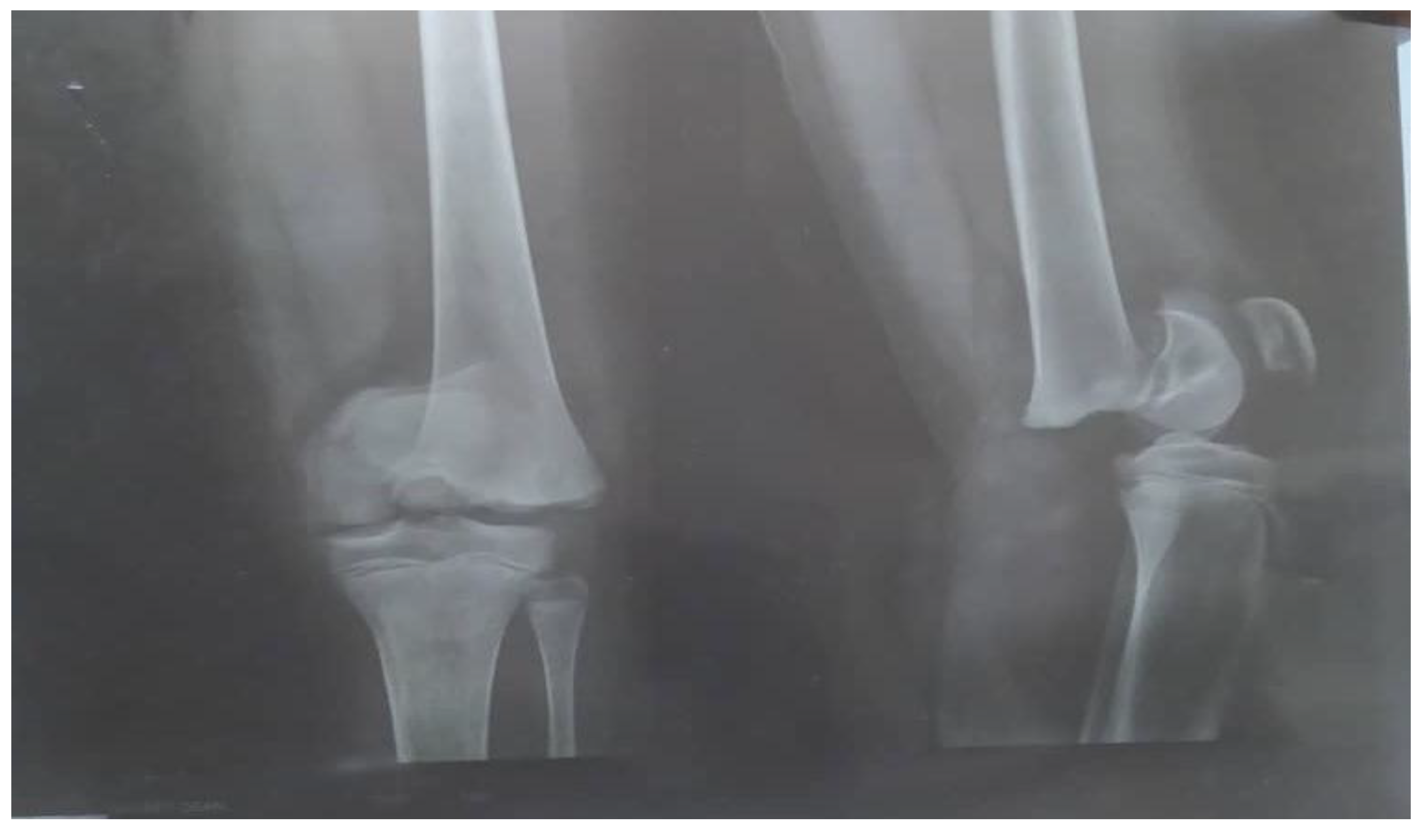
A Salter-Harris Type 1 fracture is a specific injury involving the growth plate, or physis, of a bone in children and adolescents. It occurs when the bone separates through the growth plate without breaking the bone itself. While typically stable and highly treatable, these injuries require precise medical evaluation to ensure proper bone development and prevent long-term growth complications.
Anatomy and Mechanics of the Growth Plate
To understand a Salter-Harris Type 1 injury, one must first understand the anatomy of a growing bone. In children, bones are not yet fully ossified. Between the ends of the bone (the epiphysis) and the shaft (the diaphysis) lies the physis, or growth plate. This layer of actively dividing cartilage is responsible for the longitudinal growth of the bone.
Because this cartilage is structurally weaker than the surrounding bone or the fibrous ligaments that hold joints together, it is a common site of injury during childhood. In a Type 1 fracture, the shearing or compressive force is applied directly across this growth plate. The epiphysis remains intact, and the metaphysis (the wide portion of the bone near the joint) remains intact; the injury occurs strictly through the plane of the growth plate itself.
The Salter-Harris Classification System
The Salter-Harris system, first described in 1963, remains the gold standard for classifying pediatric fractures involving the physis. The classification is essential because the pattern of the fracture dictates the risk of growth arrest and determines the appropriate management strategy.
- Type 1 (S): Straight across. The fracture line goes directly through the growth plate.
- Type 2 (A): Above. The fracture line goes through the growth plate and upward into the metaphysis.
- Type 3 (L): Lower. The fracture line goes through the growth plate and downward through the epiphysis.
- Type 4 (T): Through. The fracture line passes through the metaphysis, physis, and epiphysis.
- Type 5 (ER): Erasure or Compression. The growth plate is crushed by a compressive force.
Type 1 fractures are often characterized by a clinical presentation that may look like a ligament sprain, as there is often no obvious radiographic evidence of a break if the bone has not been significantly displaced.
Clinical Presentation and Diagnosis
Diagnosing a Salter-Harris Type 1 fracture can be challenging. Because children’s growth plates are radiolucent (transparent to X-rays), a non-displaced Type 1 fracture may appear perfectly normal on standard radiographs.
Physical Assessment
Physicians rely heavily on physical examination findings. Tenderness located directly over the physis—often described as point tenderness—is the primary clinical indicator. If a child presents with a history of trauma, joint swelling, and significant tenderness specifically localized to the growth plate area, medical professionals generally treat the injury as a Salter-Harris Type 1 fracture, regardless of whether the X-ray confirms it.
Diagnostic Imaging
- Standard Radiography: Frontal and lateral views are the initial step. While the fracture line may not be visible, secondary signs such as soft tissue swelling or joint effusion may suggest an underlying injury.
- Comparison Views: In some clinical settings, doctors may order X-rays of the uninjured limb for comparison, although this practice is decreasing in favor of clinical diagnosis.
- Advanced Imaging: If the diagnosis is unclear and the impact on treatment is significant, MRI or ultrasound may be utilized. In 2026, ultrasound is increasingly recognized for its ability to visualize cartilaginous structures in real-time, offering a non-ionizing alternative to CT scans.
Comparative Characteristics of Salter-Harris Fractures
The management of these injuries varies significantly based on their type. The following table highlights why Type 1 is distinct in its presentation and prognosis.
| Type | Fracture Path | Prognosis for Growth | Common Management |
|---|---|---|---|
| Type 1 | Across the Physis | Excellent | Casting/Splinting |
| Type 2 | Physis and Metaphysis | Excellent | Reduction/Casting |
| Type 3 | Physis and Epiphysis | Guarded | Often Surgical |
| Type 4 | All three components | Poor | Surgical/ORIF |
| Type 5 | Crushed Physis | Very Poor | Surgical/Monitoring |
Management and Treatment Strategies
Because the growth plate in a Type 1 fracture is typically undisplaced or minimally displaced, the treatment is generally conservative. The primary objective is to immobilize the limb to allow the cartilage and surrounding periosteum to heal without interruption.
Immobilization
The standard of care is the application of a cast or splint. The duration of immobilization is determined by the age of the child and the specific bone involved, typically ranging from three to six weeks. During this time, weight-bearing or activity involving the injured limb is restricted to prevent displacement of the fracture.
Reduction
If a Type 1 fracture is significantly displaced, a physician may need to perform a closed reduction. This involves realigning the bone fragments under local or general anesthesia. Once reduced, the bone is held in place with a cast. It is rare for a Type 1 fracture to require open reduction and internal fixation (surgery), unless the injury cannot be managed through closed methods.
Follow-Up
Periodic follow-up is mandatory. Because there is a theoretical risk of premature closure of the growth plate (physeal arrest), serial X-rays may be performed over several months to years to ensure the bone continues to grow symmetrically.
Common Risks and Complications
While Salter-Harris Type 1 fractures have the best prognosis of all physeal injuries, they are not immune to complications.
- Growth Arrest: If the germinal layer of the growth plate is damaged at the time of the initial trauma, the bone may stop growing prematurely. This can lead to limb length discrepancy or angular deformities.
- Malunion: If the bone heals in an abnormal position due to improper immobilization, the structural integrity of the limb may be compromised.
- Stiffness: Prolonged immobilization can lead to temporary joint stiffness. Physiotherapy is often recommended once the cast is removed to restore range of motion and muscle strength.
Addressing Parental Concerns
When a child suffers a bone injury, parents are understandably concerned about the long-term impact on the child's development. It is important to emphasize that Type 1 fractures are generally "benign" in the context of growth potential compared to Types 3, 4, and 5. Most children achieve full recovery with no lasting impact on their stature or limb alignment.
However, adherence to the medical team’s instructions is paramount. Allowing a child to return to sports or high-impact activities too early can convert a stable Type 1 injury into a more complex, displaced injury, which carries a significantly higher risk of complications.
Frequently Asked Questions
1. How long does a Salter-Harris Type 1 fracture take to heal? Healing time depends on the bone involved and the patient's age. Generally, the fracture requires 4 to 6 weeks of immobilization, followed by a period of rehabilitation to restore full function.
2. Is a Salter-Harris Type 1 fracture the same as a sprain? Clinically, they may feel similar. However, a sprain is an injury to the ligaments, whereas a Type 1 fracture is an injury to the growth plate cartilage. Because the growth plate is a site of future bone development, it requires more careful monitoring than a standard ligament sprain.
3. Will this injury affect my child's height? In the vast majority of cases, a Type 1 injury does not cause significant growth disturbance. Growth arrest is much more common in high-grade fractures (Types 4 and 5) that cross the germinal zones of the growth plate.
4. Why was the fracture not seen on the first X-ray? The growth plate is made of cartilage, which does not show up on X-rays like bone does. If the fracture did not shift the bone, the X-ray will look normal. Doctors often diagnose these based on physical examination and the location of the pain.
5. What is the most common location for this injury? Type 1 fractures are frequently seen in the distal fibula (the outer bone of the ankle) and the proximal humerus (the shoulder area), often resulting from falls or sports-related trauma.
Recommendations for Next Steps
If you suspect your child has a Salter-Harris Type 1 fracture, seek an evaluation by a pediatric orthopedist or a qualified emergency room provider immediately. Do not attempt to move or straighten the limb yourself. Apply ice to manage swelling and use a makeshift sling or splint to stabilize the limb until professional medical imaging and examination can be performed. Ensure you strictly follow the follow-up schedule provided by your clinician, as long-term monitoring is the best way to safeguard your child’s bone health.
Disclaimer: This article is for informational purposes only and does not constitute professional medical advice, diagnosis, or treatment. Always seek the advice of your physician or other qualified health provider with any questions regarding a medical condition.