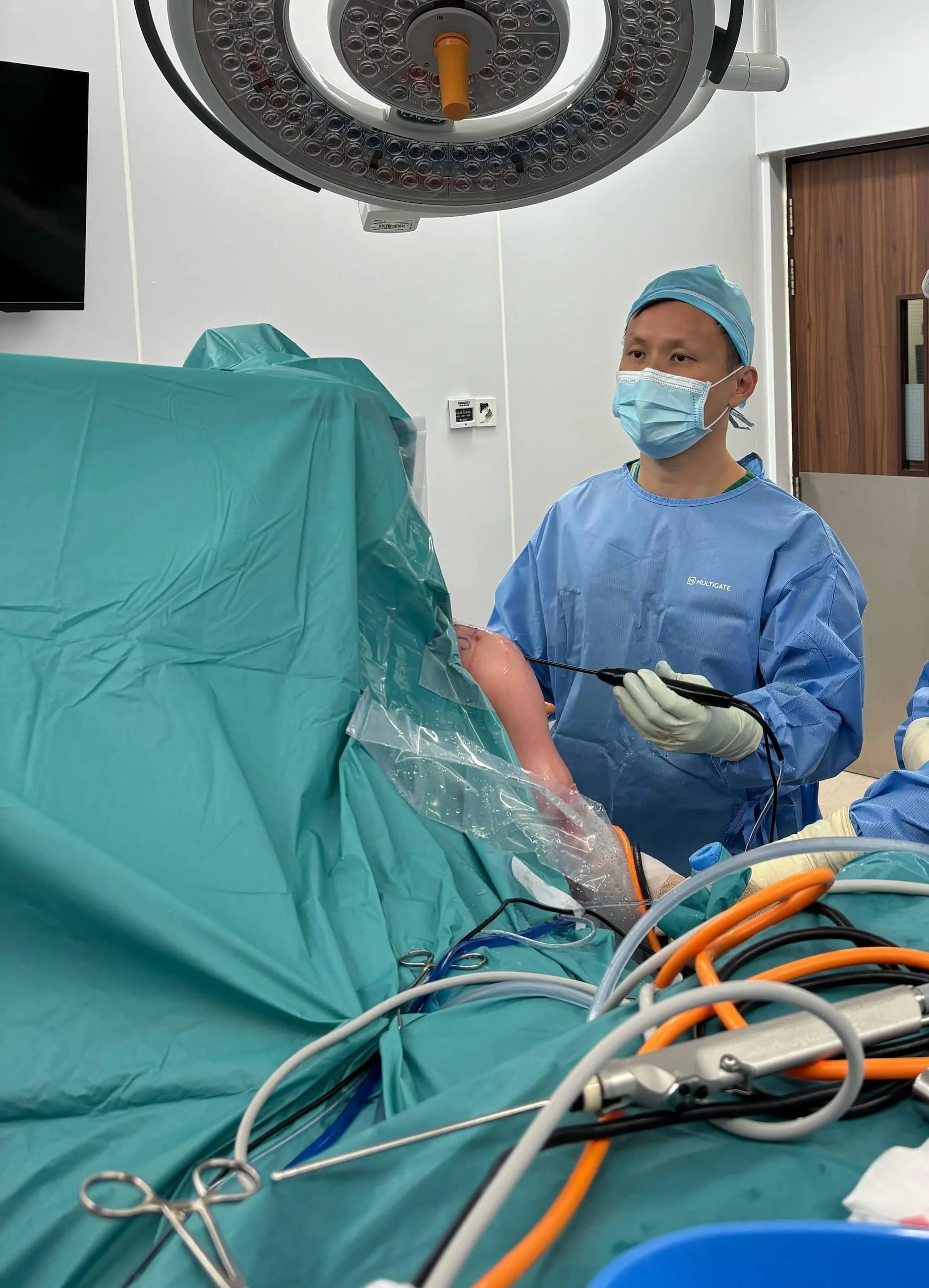
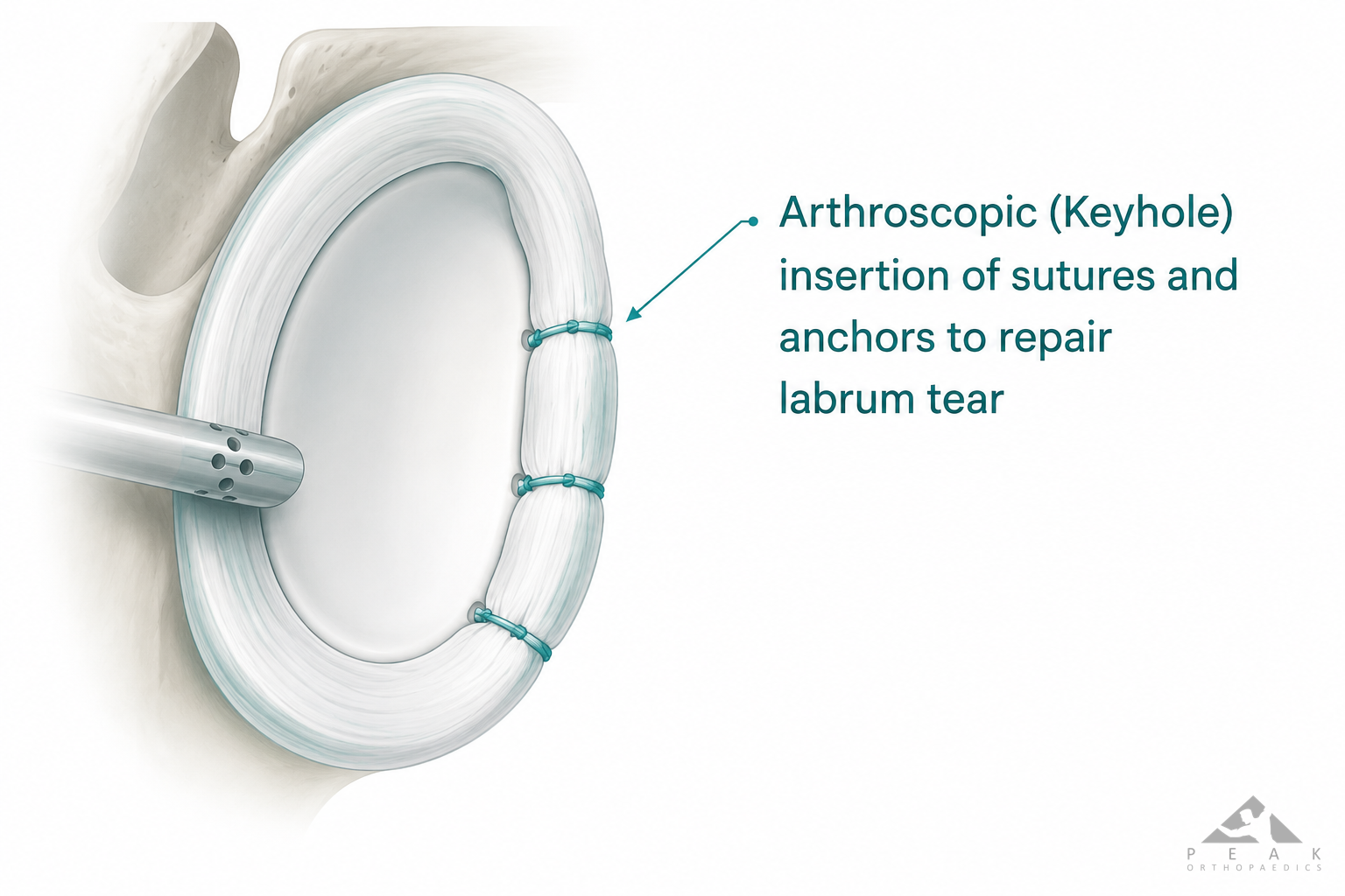
Arthroscopic labral repair is a minimally invasive surgical procedure performed to reattach a torn or detached labrum—the ring of cartilage that stabilizes the shoulder joint—to the glenoid bone. By utilizing specialized instruments and small incisions, surgeons restore shoulder stability, alleviate pain, and enable a return to functional activities for patients suffering from persistent instability or labral tears.
Understanding the Shoulder Labrum and Pathology
The glenoid labrum is a fibrocartilaginous structure that encircles the glenoid fossa (the socket of the shoulder joint). Its primary role is to deepen the socket and provide a secure attachment point for the joint capsule and ligaments, effectively acting as a bumper to prevent the humeral head from sliding out of place.
When this tissue is damaged, usually due to trauma, repetitive overhead activity, or chronic wear, patients often experience mechanical symptoms. Common pathologies include Bankart lesions (tears in the anterior-inferior labrum, often resulting from dislocation) and SLAP tears (Superior Labrum from Anterior to Posterior), which involve the top part of the labrum where the biceps tendon attaches. Arthroscopic repair is indicated when conservative measures—such as physical therapy, activity modification, and anti-inflammatory medications—fail to address the pain or the mechanical instability of the joint.
Clinical Evaluation and Prerequisites
Before scheduling an arthroscopic labral repair, a orthopedic surgeon must confirm the diagnosis through a combination of clinical assessment and diagnostic imaging.
- Physical Examination: The surgeon performs provocative tests to isolate the labrum. For instance, the O’Brien’s test is often used to assess SLAP lesions, while the Apprehension-Relocation test evaluates anterior instability.
- Imaging: Magnetic Resonance Imaging (MRI) or MR Arthrography (MRA)—where contrast dye is injected into the joint prior to the scan—remains the gold standard for visualizing soft tissue tears.
- Medical Clearance: Because this procedure typically involves anesthesia, patients must undergo preoperative clearance to ensure they are fit for surgery. This includes blood work, electrocardiograms for older patients, and a review of current medications—specifically blood thinners—that may need to be paused prior to the procedure.
The Surgical Procedure: What to Expect
Arthroscopic labral repair is typically performed in an outpatient setting, meaning the patient returns home the same day. The process follows a structured sequence:
- Anesthesia: General anesthesia is most common, sometimes combined with a regional nerve block to provide extended pain relief post-operatively.
- Portals: The surgeon makes several small (roughly 5mm to 10mm) incisions around the shoulder. Through these portals, a camera (arthroscope) and specialized surgical tools are introduced.
- Debridement and Preparation: The joint is cleared of loose tissue or scar tissue. The glenoid rim is abraded (gently roughened) to encourage a robust biological healing response (bone-to-soft-tissue adhesion).
- Anchor Placement: Suture anchors—small, bio-absorbable, or metal implants—are inserted into the bone at the rim of the glenoid.
- Repair: Using the sutures attached to these anchors, the labrum is pulled back into its anatomical position and secured to the bone, effectively "stitching" the tissue down to promote healing.
- Closure: Once the repair is verified for tension and placement, the instruments are removed, and the small portals are closed with sutures or adhesive strips.
Comparison of Labral Repair Techniques
| Feature | Suture Anchor Repair | Knotless Anchor Systems |
|---|---|---|
| Fixation Strength | High, traditional gold standard | High, lower profile |
| Complexity | Involves tying complex knots | Simplifies tensioning |
| Material | PEEK, Metal, or Bio-composite | PEEK or Bio-composite |
| Post-op Risk | Potential for "knot prominence" | Reduced risk of hardware irritation |
Post-Operative Rehabilitation and Recovery
The success of arthroscopic labral repair is heavily dependent on the post-operative rehabilitation protocol. Recovery is a marathon, not a sprint, generally spanning four to six months for full return to pre-injury activity.
- Phase 1 (Weeks 0–6): Protection is paramount. The patient must wear an immobilizing sling to prevent movement that could stress the repair. Passive range-of-motion exercises may be introduced under the guidance of a physical therapist to prevent "frozen shoulder" (adhesive capsulitis).
- Phase 2 (Weeks 6–12): The sling is discarded, and active range-of-motion begins. Strengthening focuses on the rotator cuff and periscapular muscles.
- Phase 3 (Months 3–6): Progression to sports-specific or work-specific loading. Plyometrics and dynamic stability exercises are integrated as the tissue regains sufficient biological strength.
Patients should be aware that "return to play" timelines vary by individual and the specific demands of their activities. Returning too early risks re-tearing the repair.
Managing Expectations and Potential Complications
While arthroscopic techniques have high success rates, no surgery is without risk. Complications are rare but can include:
- Infection: Managed through sterile technique and prophylactic antibiotics.
- Nerve Injury: Temporary neuropraxia can occur from traction during surgery.
- Recurrent Instability: In some patients, the tissue may not heal with enough tensile strength, or a new injury may occur, leading to persistent instability.
- Stiffness: The most common post-operative challenge, typically resolved with physical therapy.
Frequently Asked Questions
1. Is arthroscopic labral repair painful? Post-operative pain is expected but is typically managed effectively with a combination of nerve blocks, oral analgesics, and cold therapy. Most patients find that the sharp, "catching" pain associated with the tear is replaced by manageable surgical soreness.
2. How long does the surgery take? Most arthroscopic labral repairs take between 60 to 90 minutes, depending on the complexity of the tear and whether other issues, such as loose bodies or biceps tendon pathology, are addressed simultaneously.
3. Will I need physical therapy after the surgery? Yes. Physical therapy is not optional; it is a critical component of the surgery. Without structured rehabilitation to regain strength and coordination, the shoulder may remain stiff or fail to regain its full functional stability.
4. Can I return to contact sports after this surgery? Generally, yes, but only after a clearance process that includes testing for full strength, pain-free motion, and psychological readiness. This typically occurs between the 6th and 9th month post-operation.
5. How much does an arthroscopic labral repair cost? Costs vary significantly based on geographic location, the surgical center (hospital vs. ambulatory surgery center), and the specifics of your health insurance plan. In 2026, patients should consult their provider to understand their specific deductibles, coinsurance, and any pre-authorization requirements.
Recommendation for Next Actions
If you suspect you have a labral tear, the first step is to schedule an appointment with an orthopedic specialist who focuses on sports medicine or shoulder surgery. Request an evaluation that includes a physical exam and, if deemed necessary by the physician, an MRA scan. If surgery is recommended, ask your surgeon about their specific volume of this procedure—high-volume surgeons generally report fewer complications. Prior to your surgery, prepare your home for a period of one-armed recovery, arranging for assistance with daily tasks like meal preparation and dressing, as your operative arm will be immobilized in a sling for the first six weeks.