Ubiquitin detection is a cornerstone of proteomic research, essential for mapping the ubiquitination landscape and understanding protein degradation pathways, cell signaling, and DNA repair. The primary challenge in this field lies in the transient, heterogeneous nature of polyubiquitin chains, which necessitates robust, highly specific antibodies to distinguish between different linkages and modification states within complex biological lysates.
The Complexity of Ubiquitin Detection
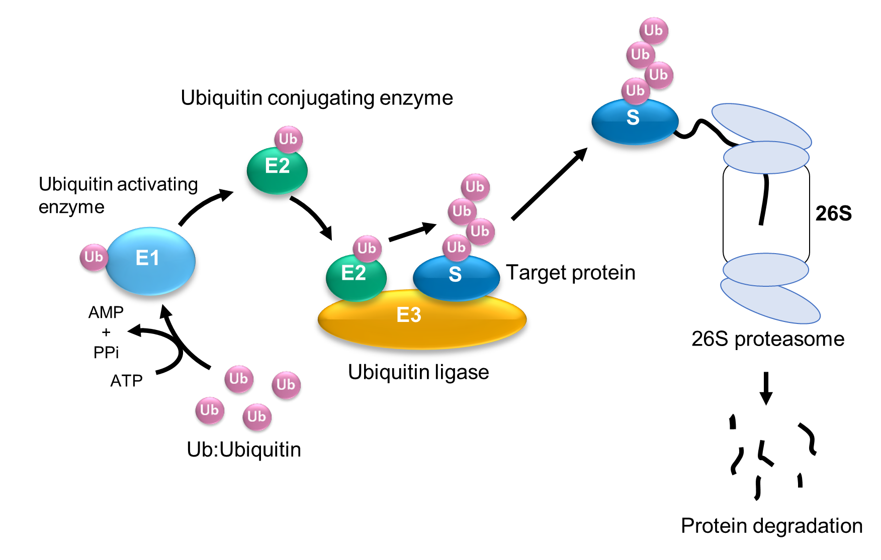
Ubiquitin is a small, highly conserved regulatory protein that attaches to substrate proteins—most commonly via a lysine residue—to form chains. These chains dictate the fate of the protein, ranging from proteasomal degradation (K48-linked) to signaling cascades (K63-linked) and DNA damage response. Detecting these modifications requires an analytical approach that can resolve both the identity of the ubiquitinated protein and the architecture of the ubiquitin chain itself.
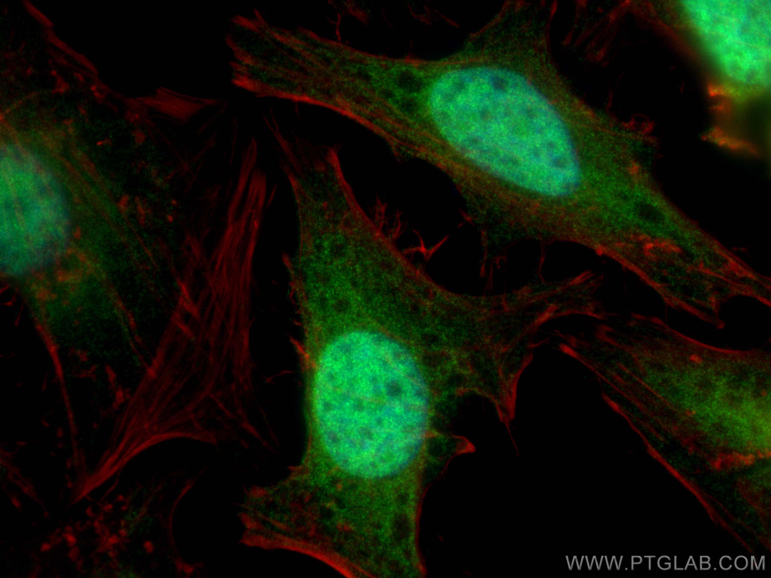
Researchers primarily utilize immunoblotting (Western blot), immunoprecipitation (IP), and immunofluorescence (IF) to visualize these modifications. However, because ubiquitin is abundant in cells, non-specific background signals often obscure the detection of specific, low-abundance ubiquitinated substrates. Furthermore, the rapid activity of deubiquitinating enzymes (DUBs) during sample preparation can lead to the artificial loss of ubiquitin signals, rendering detection inaccurate.
Prerequisites for Successful Detection
Before initiating an experiment, the researcher must account for the preservation of the ubiquitinated state. Ubiquitination is a dynamic process; without inhibition, endogenous DUBs will strip ubiquitin from proteins as soon as cells are lysed.
- DUB Inhibitors: Always supplement lysis buffers with potent DUB inhibitors, such as N-ethylmaleimide (NEM) or specialized protease inhibitor cocktails that specifically block thiol-dependent DUBs.
- Buffer Optimization: High-salt buffers or detergents that effectively solubilize membrane proteins are often required, but they must be balanced against the binding affinity of the chosen antibody.
- Denaturing Conditions: If the goal is to isolate a specific protein and detect its ubiquitin modification, perform a denaturing IP. This involves boiling the sample in SDS before diluting it to non-denaturing concentrations, ensuring that the detected ubiquitin is covalently attached to the target protein rather than being part of a non-covalent protein complex.
Selection and Characterization of Ubiquitin Antibodies
The efficacy of your research depends heavily on the specificity of the antibody. Antibodies for ubiquitin detection generally fall into two categories: anti-ubiquitin antibodies and linkage-specific antibodies.
- Pan-Ubiquitin Antibodies: These recognize the ubiquitin moiety regardless of its linkage. They are ideal for confirming the presence of ubiquitination on a target but do not provide information regarding the topology of the chain.
- Linkage-Specific Antibodies: These are engineered to recognize specific junctions between ubiquitin molecules (e.g., K48, K63, M1). These are critical for determining the biological consequence of the modification. When using these, it is imperative to verify the manufacturer’s validation data, specifically regarding the antibody’s performance in the intended application (e.g., WB vs. IP).
| Antibody Type | Best Application | Key Consideration |
|---|---|---|
| Pan-Ubiquitin | Western Blot / IF | High sensitivity; detects total ubiquitination. |
| K48-Linked | Western Blot / IP | Signals proteasomal degradation pathways. |
| K63-Linked | Western Blot / IP | Often associated with signal transduction. |
| Monoclonal | IP / Specificity | Higher lot-to-lot consistency; reduced cross-reactivity. |
| Polyclonal | General Screening | Higher signal strength; potential for non-specific binding. |
Methodological Workflow: From Lysate to Visualization
To achieve reliable results, follow a structured workflow that accounts for the potential interference of free ubiquitin and the structural instability of the target proteins.
1. Sample Preparation and Stabilization
Begin with rapid cell lysis at 4°C. As mentioned, the inclusion of DUB inhibitors is non-negotiable. If investigating a specific protein, perform a targeted immunoprecipitation. Use magnetic beads to improve the purity of the pull-down and reduce non-specific binding that is common with agarose-based resins.
2. Electrophoresis and Transfer
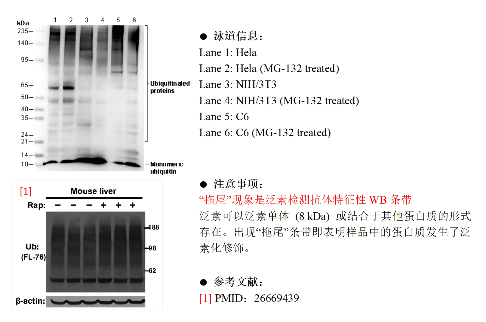
Ubiquitinated proteins appear as a "smear" on a Western blot because they exist in a heterogeneous distribution of chain lengths and attachment sites. Ensure your gel percentage allows for the visualization of high-molecular-weight species. Transferring to a PVDF membrane is generally preferred over nitrocellulose for its superior mechanical strength, especially when dealing with the high-molecular-weight complexes typical of multi-ubiquitinated proteins.
3. Blocking and Antibody Incubation
Non-specific background is the enemy of ubiquitin blots. Avoid using milk-based blocking agents if possible, as they contain high levels of endogenous biotin and can interfere with certain detection reagents. Use bovine serum albumin (BSA) or specialized animal-free blocking buffers. Incubating the primary antibody at 4°C overnight often improves the signal-to-noise ratio compared to short, room-temperature incubations.
Common Troubleshooting and Optimization
If you are failing to detect ubiquitin, or if your signal is buried in background noise, investigate the following areas:
- The "Smear" Problem: If you see no signal, your proteins may be degrading or the DUBs are too active. Re-verify the concentration of your NEM or alternative DUB inhibitors.
- Non-Specific Binding: If the entire lane is dark, try reducing the concentration of the primary antibody. Ubiquitin antibodies often exhibit high avidity; starting with a higher dilution (e.g., 1:2000 instead of 1:500) can often clean up the blot.
- Detection Thresholds: Ubiquitinated forms of a protein often constitute only a small fraction of the total protein pool. If you cannot see the ubiquitinated species, you may need to enrich for ubiquitinated proteins using tandem ubiquitin-binding entities (TUBEs) before performing Western blotting.
Regulatory and Safety Considerations
Work involving the manipulation of cellular proteostasis involves standard Biosafety Level 2 (BSL-2) practices. When using chemical inhibitors like NEM, exercise caution; it is a potent alkylating agent and can be toxic if inhaled or absorbed through the skin. Always handle these reagents in a chemical fume hood and dispose of waste according to institutional guidelines.
Frequently Asked Questions
Why does my Western blot for ubiquitin look like a vertical smear rather than a distinct band? Ubiquitin modifications are inherently heterogeneous. Because a single protein can be modified with chains of varying lengths and linkages, the target protein will migrate at various molecular weights, creating a smear. This is a normal, expected result in ubiquitin research and confirms the presence of polyubiquitination.
Are DUB inhibitors necessary if I am performing denaturing lysis? Yes. Even with denaturing detergents like SDS, DUB activity can occur during the window between cell disruption and boiling. To ensure absolute preservation, include inhibitors in your lysis buffer prior to the application of heat.
What is the advantage of using TUBEs over conventional antibodies? TUBEs (Tandem Ubiquitin-Binding Entities) bind to polyubiquitin chains with much higher affinity than conventional antibodies. They are particularly useful for pulling down polyubiquitinated proteins from lysates where the target protein is present in very low concentrations, essentially acting as an enrichment tool before detection.
Can I reuse a membrane for multiple ubiquitin antibodies? Yes, stripping and reprobing is possible. However, ensure that the stripping buffer is not so harsh that it damages the underlying protein or removes the target protein from the membrane entirely. Always validate that your signal is gone after stripping before applying a new antibody.
How do I distinguish between K48 and K63 linkages on the same blot? You must use linkage-specific antibodies. Because these antibodies are designed to recognize the specific geometry of the isopeptide bond between two ubiquitin molecules, you can run parallel lanes or strip and reprobe the membrane to compare the intensity of K48 versus K63 signals, provided the antibodies are well-validated for Western blot applications.
Moving Forward
Effective ubiquitin detection in 2026 relies on a rigorous combination of sample stabilization, high-affinity reagents, and a clear understanding of the heterogeneous nature of ubiquitin chains. For your next experiment, begin by characterizing the ubiquitination status of your protein of interest using a pan-ubiquitin antibody to establish a baseline. If you find the signal is too weak, transition to TUBE-based enrichment. If the biological context requires knowing the chain topology, proceed to linkage-specific antibodies, ensuring that each reagent is validated for your specific sample type and application.