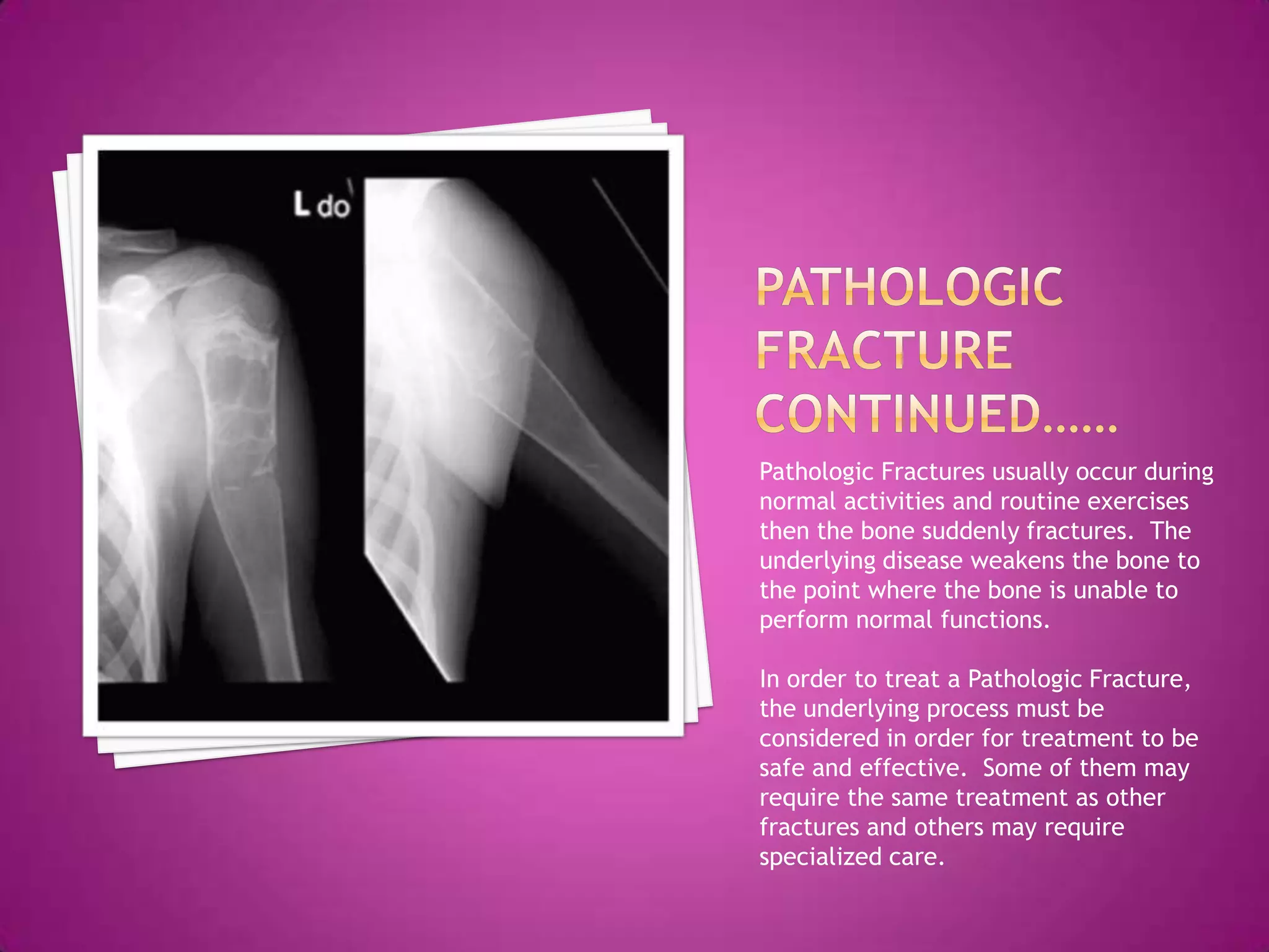
Yes, a bone fracture and a bone break are exactly the same thing. In medical terminology, a "fracture" is the formal word used by healthcare professionals to describe any loss of continuity in a bone, ranging from a microscopic hairline crack to a bone that has shattered into multiple fragments.
The confusion between the two terms often stems from colloquial usage. Many people mistakenly believe that a "break" implies a complete separation of the bone into two or more pieces, while a "fracture" suggests something less severe, such as a "crack." In reality, an orthopedic surgeon will use the word fracture to describe a shattered femur just as readily as they would use it for a minor stress fracture in a toe. Whether a doctor tells you that your arm is broken or that you have a radial fracture, they are describing the same underlying physical trauma.
The Medical Definition of a Bone Fracture vs. a Break
To understand why these terms are synonymous, we must look at the linguistics of medicine. The word fracture originates from the Latin fractura, meaning "a breach or a break." In a clinical setting, physicians prefer precise, standardized language to ensure clear communication across different specialties and regions. "Break" is considered a layperson’s term; it is descriptive and accurate, but it lacks the clinical specificity required for medical coding, surgical planning, and academic research.
From a physiological standpoint, a fracture occurs when the physical force applied to a bone exceeds its structural integrity. Bones are remarkably resilient, designed to withstand significant compression and tension, but they have a breaking point. When that point is reached, the mineralized matrix of the bone snaps or cracks. Because "fracture" is the technical term, you will find it on every X-ray report, insurance claim, and discharge summary. Using the word "break" in those contexts would be akin to a cardiologist using the word "heart throb" instead of "arrhythmia."
Common Misconceptions: Is a Fracture Less Serious?
One of the most persistent myths in first aid and emergency medicine is that a fracture is "just a crack" and therefore does not require the same level of attention as a "break." This misconception can be dangerous, as it often leads patients to delay seeking professional medical evaluation.
The severity of a bone injury is determined by its location, pattern, and displacement, not by which word is used to describe it. For example, a "stress fracture" in the hip can be life-altering and may require surgical intervention to prevent a total collapse of the femoral head. Conversely, a "complete break" of a small distal phalanx (the tip of a pinky toe) might simply be treated with "buddy taping" and rest.
In 2026, medical professionals continue to emphasize that any suspected injury to the skeletal system should be treated as a fracture until imaging proves otherwise. The distinction between a "hairline fracture" and a "clean break" is a matter of degree, not a matter of kind. Both represent a failure of the bone tissue that requires a biological healing process to repair.
The Anatomy of a Bone Fracture: How It Happens
Bones are living tissues, constantly undergoing a process of "remodeling" where old bone is broken down by cells called osteoclasts and new bone is built by osteoblasts. This dynamic nature allows bones to be strong yet flexible. However, several factors contribute to how and why a bone eventually fractures:
- Traumatic Impact: High-force events such as car accidents, falls from heights, or sports-related collisions provide more energy than the bone can dissipate.
- Repetitive Stress: Also known as overuse injuries, these occur when repeated sub-maximal loading (like long-distance running) causes micro-cracks that the body cannot repair fast enough.
- Pathological Factors: Conditions like osteoporosis, bone cysts, or certain cancers weaken the internal architecture of the bone, allowing it to fracture under "normal" loads that would not typically cause an injury.
When the bone breaks, the damage extends beyond the hard mineralized tissue. It involves the periosteum (the highly sensitive outer membrane), the endosteum (the inner lining), and the bone marrow. The rupture of blood vessels within these structures leads to internal bleeding and the formation of a hematoma, which is the first step in the body’s complex inflammatory response.
Types of Bone Fractures and Their Classifications
Because "fracture" is a broad term, orthopedists categorize them into specific types to guide treatment. Understanding these classifications is more important than the "break vs. fracture" debate.
Displacement and Alignment
- Non-displaced Fracture: The bone cracks or breaks, but the pieces remain aligned in their normal anatomical position.
- Displaced Fracture: The bone snaps into two or more parts, and the ends are no longer lined up. These often require "reduction" (realigning the bone) before they can be casted.
Interaction with the Environment
- Closed (Simple) Fracture: The bone breaks, but the skin remains intact. While less prone to infection, these can still involve significant internal soft-tissue damage.
- Open (Compound) Fracture: The broken bone pierces the skin or there is a deep wound that exposes the bone. This is a medical emergency due to the high risk of osteomyelitis (bone infection).
Fracture Patterns
- Transverse: The break is a straight horizontal line across the bone shaft.
- Oblique: The break occurs at an angle.
- Spiral: The break spirals around the bone; this is usually caused by a twisting or rotational force.
- Comminuted: The bone is shattered into three or more pieces.
- Greenstick: An incomplete fracture where the bone bends and cracks but doesn't break completely. This is most common in children, whose bones are more flexible and "springy" than adult bones.
Symptoms and Immediate Warning Signs
Determining if a bone is fractured can sometimes be difficult without an X-ray, as symptoms can overlap with severe sprains or ligament tears. However, certain "red flags" strongly suggest a fracture:
- Deformity: If the limb or joint looks misshapen or is at an unnatural angle.
- Crepitus: A grating or grinding sensation or sound caused by the broken bone ends rubbing together.
- Inability to Bear Weight: If you cannot put any weight on a leg or use an arm without excruciating pain.
- Rapid Swelling and Bruising: While sprains cause swelling, a fracture often results in immediate, intense discoloration as blood leaks from the bone's internal supply.
- Point Tenderness: Pain that is localized precisely over the bone rather than the surrounding soft tissue.
The Diagnostic Process in 2026
When you arrive at a clinic or emergency room with a suspected break, the diagnostic process follows a structured path to ensure no secondary injuries (like nerve or vascular damage) are missed.
- Physical Examination: The clinician will check for distal pulses (blood flow) and sensation (nerve function) beyond the site of the injury.
- X-ray (Radiography): The gold standard for initial diagnosis. At least two views (usually frontal and lateral) are required because a fracture might be invisible from one angle but obvious from another.
- CT Scan (Computed Tomography): If the fracture involves a complex joint like the ankle, wrist, or spine, a CT scan provides a 3D view of the bone fragments.
- MRI (Magnetic Resonance Imaging): Often used for stress fractures or injuries where the bone itself is intact but there is "bone bruising" or occult damage that doesn't show up on a standard X-ray.
| Fracture Type | Common Cause | Typical Severity | Estimated Healing Time |
|---|---|---|---|
| Stress | Overuse/Running | Low to Moderate | 6–8 Weeks |
| Transverse | Direct Blow/Impact | Moderate | 8–12 Weeks |
| Spiral | Twisting/Skiing | Moderate to High | 10–16 Weeks |
| Comminuted | High-speed Crash | Very High | 4–6 Months+ |
| Compound | Severe Trauma | Extreme (Surgical) | 6 Months+ |
Treatment Modalities: How Fractures are Fixed
The primary goal of treating any fracture is immobilization. For a bone to heal, the two ends must be held still so that the body can bridge the gap with new tissue. Depending on the severity, several methods may be employed:
Non-Surgical Management
Most simple fractures are treated with a cast or a functional brace. In 2026, "smart casts" and 3D-printed lightweight splints have become more common, allowing for better airflow and even integrated ultrasound sensors that monitor bone healing. These devices ensure the bone remains in the correct position (reduction) while the initial callus forms.
Surgical Intervention
If the fracture is unstable, displaced, or involves a joint surface, surgery is often necessary.
- Internal Fixation: This involves "Open Reduction and Internal Fixation" (ORIF). The surgeon repositions the bone fragments and secures them with metal plates, screws, or intramedullary nails (rods placed down the center of the bone).
- External Fixation: Used primarily for severe compound fractures or when the surrounding skin is too damaged for immediate internal surgery. Metal pins are placed into the bone above and below the fracture site and connected to a rigid frame outside the body.
The Biological Healing Process: How Bone Knits Together
Whether you call it a break or a fracture, the body’s repair mechanism is a marvel of biological engineering. It occurs in four distinct, overlapping stages:
- Inflammation (Days 1–5): Immediately after the injury, the body creates a blood clot around the fracture. This "fracture hematoma" provides a framework for healing cells to migrate to the area.
- Soft Callus Formation (Week 1–3): Cells called fibroblasts begin producing collagen, and chondroblasts produce fibrocartilage. This creates a "bridge" across the break. At this stage, the connection is soft and cannot support weight, but it begins to stabilize the bone.
- Hard Callus Formation (Week 2–10): Osteoblasts begin to replace the soft cartilage with hard, mineralized bone. This "bony callus" is often visible on X-rays as a cloudy mass around the break site.
- Bone Remodeling (Months to Years): This is the longest phase. The body slowly reshapes the hard callus, removing excess bone and restoring the bone to its original shape and strength. Eventually, the bone may become so well-repaired that the original fracture line is nearly invisible on future imaging.
Complications and Long-term Management
While most fractures heal without incident, certain complications can arise. A "non-union" occurs when the bone fails to heal, often due to poor blood supply or excessive movement. A "mal-union" occurs when the bone heals in a crooked or misaligned position, which can lead to chronic pain or early-onset arthritis in nearby joints.
For older adults, a fracture—particularly of the hip—carries significant risks. According to the Centers for Disease Control and Prevention (CDC), hip fractures are associated with a high rate of secondary complications like pneumonia or blood clots due to prolonged immobility. This is why immediate mobilization and physical therapy are core components of fracture management in modern medicine.
Prevention and Bone Health
Maintaining skeletal integrity is the best way to prevent fractures. This involves a combination of nutrition and lifestyle choices:
- Calcium and Vitamin D: Essential for maintaining bone mineral density.
- Weight-Bearing Exercise: Activities like walking, weightlifting, and tennis signal the body to strengthen bone tissue.
- Fall Prevention: Especially critical for those with osteoporosis. This includes removing trip hazards in the home and improving balance through exercises like Tai Chi.
- Avoiding Tobacco: Smoking significantly impairs bone healing by constricting blood vessels and slowing the action of osteoblasts.
Frequently Asked Questions
Can a fracture be worse than a break?
Since they are the same thing, one cannot be "worse" than the other based on terminology alone. However, a comminuted fracture (shattered bone) is much more severe than a simple break (clean snap). The severity depends on the pattern of the injury, not whether you call it a break or a fracture.
Why do people say 'hairline' fracture?
A hairline fracture is a real clinical term used to describe a non-displaced, thin crack in the bone. It is often used to reassure patients that the bone hasn't shifted, but it is still a "break" that requires proper care to prevent it from becoming a full-blown displaced fracture.
Do fractures always show up on X-rays?
Not always. Stress fractures or very small cracks may not be visible on an X-ray for 10 to 14 days, until the healing process begins and the bone starts to change. In these cases, a doctor might treat the injury as a fracture and re-image it later, or order an MRI for immediate confirmation.
Is it true that a broken bone heals stronger than it was before?
This is a common myth. While the "hard callus" formed during the healing process is very strong, the bone eventually undergoes remodeling to return to its original strength and shape. The site of a former fracture is generally no more or less likely to break again than any other part of the bone.
What is the difference between a fracture and a sprain?
A fracture is a break in the bone, whereas a sprain is a stretching or tearing of ligaments (the tissues that connect bones to each other). Both can be equally painful and may present with similar symptoms like swelling and bruising.
Summary of Next Actions
If you suspect you have sustained a bone injury, the first step is immobilization. Do not attempt to "walk it off" or realign the limb yourself. Apply ice to reduce swelling and seek a professional evaluation at an urgent care center or emergency room. Regardless of whether the final diagnosis uses the word "break" or "fracture," follow the prescribed rehabilitation plan closely. Proper rest and physical therapy are the only ways to ensure the bone returns to its full functional capacity without long-term complications.