Second-degree heart block is an interruption in the electrical signals traveling from the upper chambers (atria) of the heart to the lower chambers (ventricles), causing some electrical impulses to be dropped rather than conducted normally. This conduction delay can lead to a slower or irregular heartbeat, which may require monitoring, medication adjustments, or a pacemaker depending on the specific subtype and patient symptoms.
Understanding Cardiac Conduction and Heart Block
The human heart relies on a sophisticated electrical system to maintain a steady and coordinated rhythm. Electrical impulses originate in the sinus node, located in the right atrium, and travel through the atrioventricular (AV) node down into the ventricles. This pathway ensures that the atria contract first to fill the ventricles with blood, followed immediately by ventricular contraction to pump that blood out to the lungs and the rest of the body. When a heart block occurs, the transmission of these electrical signals through the AV node or the His-Purkinje system is delayed, partially obstructed, or completely interrupted.
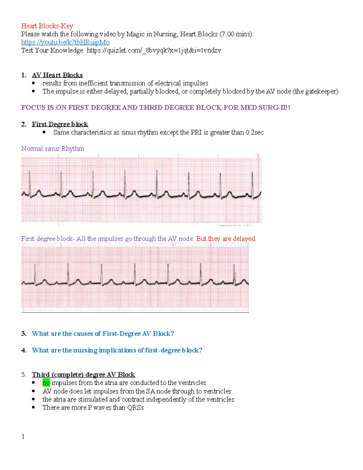
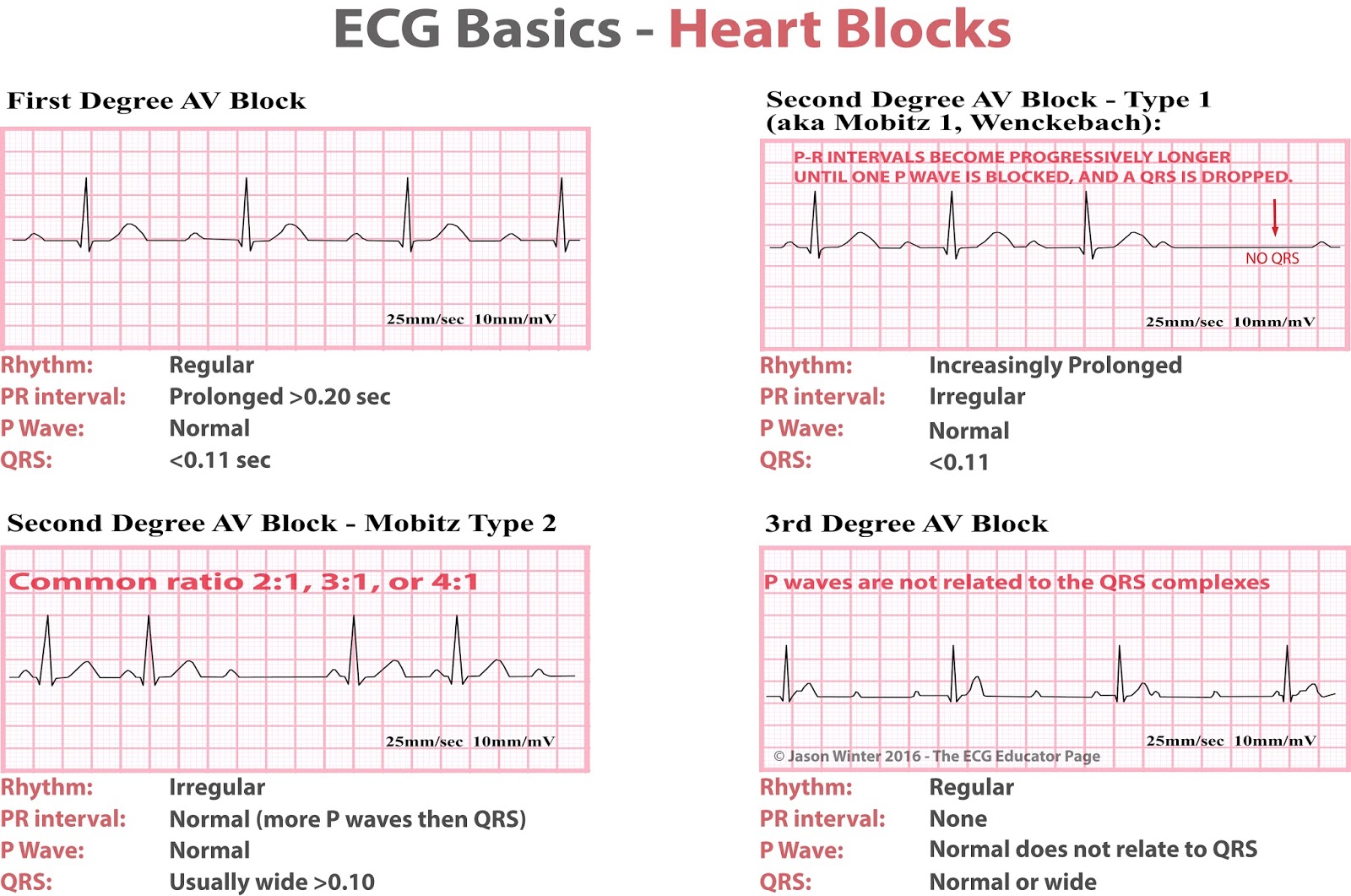
Physicians categorize AV blocks into three primary degrees based on severity. First-degree heart block involves a simple delay where every signal gets through, albeit slowly. Third-degree (complete) heart block means no electrical signals pass from the atria to the ventricles at all, forcing the lower chambers to rely on an unreliable backup rhythm. Second-degree heart block sits in the middle: some impulses successfully reach the ventricles, but others are blocked entirely. This intermittent failure creates dropped beats, which disrupt the heart’s normal pumping efficiency and can result in symptoms ranging from mild fatigue to sudden syncope (fainting).
Healthcare professionals evaluate these conduction abnormalities primarily through a 12-lead electrocardiogram (ECG), which records the heart's electrical activity over a brief window. For intermittent symptoms, patients may wear ambulatory monitors, such as a Holter monitor for 24 to 48 hours or an event recorder for weeks, to capture episodes of dropped beats during daily activities. Understanding the precise mechanics of the block is critical because the two main subtypes of second-degree heart block carry vastly different prognoses and require distinct management strategies.
Subtypes of Second-Degree Heart Block: Mobitz Type I vs. Mobitz Type II
Distinguishing between the two main types of second-degree heart block—Mobitz Type I and Mobitz Type II—is the most crucial step in clinical management. These subtypes differ significantly in their anatomical location, underlying causes, response to physiological stress, and long-term risk of progressing to complete heart block.
Mobitz Type I (Wenckebach)
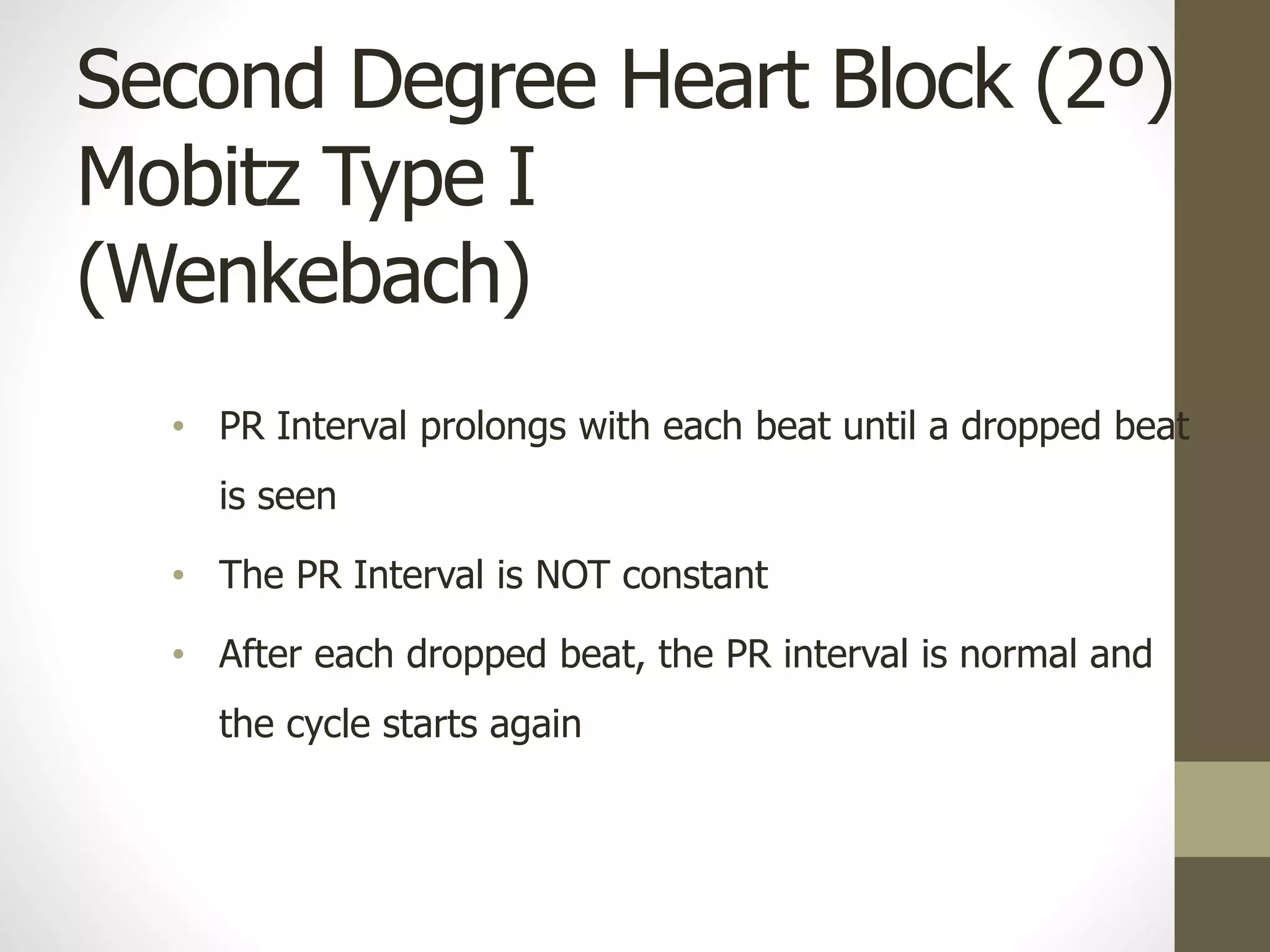
Mobitz Type I, commonly known as Wenckebach block, is characterized by a progressive lengthening of the PR interval on an ECG strip with each successive heartbeat until an atrial impulse is completely blocked and a QRS complex is dropped. Following the dropped beat, the conduction system resets, and the cycle repeats.
- Anatomical Location: The delay typically occurs directly within the AV node.
- Prognosis: Generally benign. It is often asymptomatic and can occur normally in healthy individuals, young athletes with high vagal tone, or during sleep.
- Causes: Can be induced by medications that slow AV node conduction (such as beta-blockers, calcium channel blockers, digoxin) or transient ischemia (reduced blood flow) affecting the right coronary artery.
- Management Focus: Often requires no intervention beyond monitoring, unless the patient experiences significant symptoms like dizziness or severe bradycardia, or if a reversible drug cause is identified and discontinued.
Mobitz Type II
Mobitz Type II heart block features intermittent dropped ventricular beats without the progressive lengthening of the PR interval seen in Wenckebach. When the electrical impulse does get through, the PR interval remains constant.
- Anatomical Location: The block almost always occurs below the AV node, within the bundle of His or the bundle branches (infra-Hisian disease).
- Prognosis: Considered significantly more dangerous. It has a high propensity to progress unpredictably to third-degree heart block, which can cause sudden cardiac arrest or Stokes-Adams attacks (sudden fainting spells due to inadequate cerebral perfusion).
- Causes: Frequently associated with structural heart disease, extensive coronary artery disease, anterior myocardial infarction, fibrosis of the conduction system (Lenègre's or Lev's disease), or infiltrative heart conditions.
- Management Focus: Generally requires permanent pacemaker implantation due to the high risk of sudden progression to complete heart block, regardless of whether the patient is currently experiencing severe symptoms.
| Feature | Mobitz Type I (Wenckebach) | Mobitz Type II (Classic) |
|---|---|---|
| PR Interval | Progressively lengthens until a beat is dropped | Constant (fixed) before and after dropped beats |
| Anatomical Site | Atrioventricular (AV) Node | Below AV Node (Bundle of His / Purkinje system) |
| Risk of Progression | Low; usually benign course | High; often progresses to complete heart block |
| Association with CAD | Less common; often right coronary artery | Frequently associated with extensive anterior wall disease |
| Primary Treatment | Observation; discontinue causative drugs if needed | Permanent pacemaker implantation |
Clinical Presentation and Diagnostic Evaluation
Recognizing the clinical manifestations of second-degree heart block allows for timely intervention before complications arise. While some individuals with Mobitz Type I remain entirely asymptomatic, others experience noticeable alterations in their cardiac output.
Common symptoms associated with symptomatic heart block include fatigue, exercise intolerance, lightheadedness, dizziness, and shortness of breath. When the heart drops beats frequently, the overall heart rate decreases (bradycardia), reducing the volume of blood pumped to the brain and body per minute. In more severe cases—particularly with Mobitz Type II—patients may experience presyncope, sudden fainting spells, or chest pain if the underlying cause involves coronary ischemia.
The diagnostic workup extends beyond a standard resting ECG. Because conduction blocks can be paroxysmal (occurring intermittently), clinicians rely on extended rhythm monitoring.
Patient Presents with Symptoms or Incidental Bradycardia
12-Lead Electrocardiogram (ECG) Evaluation
- Mobitz Type I (Wenckebach) identified
- Asymptomatic: Monitor & Review Meds
- Symptomatic: Investigate Reversible Causes / Consider Pacing
- Mobitz Type II identified
- High Risk: Electrophysiology Study / Permanent Pacemaker Evaluation
- Mobitz Type I (Wenckebach) identified
Holter Monitors: Wearable devices recording continuous ECG data for 24 to 48 hours to correlate symptoms with rhythm abnormalities.
Event Monitors and Mobile Cardiac Telemetry (MCT): Devices worn for weeks that allow patients to record symptoms manually or transmit data automatically when arrhythmias occur.
Echocardiogram: An ultrasound of the heart to assess structural integrity, ejection fraction, valve function, and rule out structural heart disease or infiltrative cardiomyopathies.
Electrophysiology Study (EPS): An invasive procedure where catheter electrodes are placed inside the heart to map the electrical conduction system precisely, determining the exact level of the block when non-invasive tests are inconclusive.
Laboratory Testing: Blood work to check for electrolyte imbalances (such as severe hyperkalemia), thyroid dysfunction, or cardiac biomarkers indicating myocardial injury.
Management Strategies and Therapeutic Approaches
Management of second-degree heart block depends heavily on the subtype, the presence of symptoms, and the identification of any reversible secondary causes. Clinicians stratify patients carefully to determine whether conservative observation, medical therapy, or invasive intervention is appropriate.
Identifying and Treating Reversible Causes
Before committing to permanent hardware, physicians investigate and address temporary or reversible triggers. Common reversible causes include acute ischemia or myocardial infarction, electrolyte derangements, and negative chronotropic medications.
If a patient is taking AV nodal blocking agents—such as beta-blockers, non-dihydropyridine calcium channel blockers (verapamil or diltiazem), or digoxin—for hypertension or heart failure, adjusting or stopping these medications under medical supervision may resolve a Mobitz Type I block. However, medications should never be altered independently without consulting a cardiologist or primary care physician.
Pharmacological Management in Acute Settings
In emergency settings where a patient is hemodynamically unstable (experiencing hypotension, acute heart failure, or altered mental status) due to symptomatic bradycardia from heart block, temporary medical measures may be utilized while preparing for pacing:
- Atropine: An anticholinergic drug that can enhance AV nodal conduction. It is typically effective for Mobitz Type I (nodal block) but is often ineffective or potentially paradoxical in Mobitz Type II (infra-nodal block).
- Temporary Pacing: Transcutaneous pacing (via external pads) or transvenous pacing (via a catheter inserted into a central vein) provides immediate electrical stability for unstable patients with Mobitz Type II or refractory Mobitz Type I.
Permanent Pacemaker Implantation
For patients with persistent, symptomatic second-degree heart block, or those with asymptomatic Mobitz Type II, a permanent pacemaker is the gold standard of therapy. Modern pacemakers are sophisticated, programmable devices implanted subcutaneously, typically in the upper chest, with leads threaded through veins directly into the chambers of the heart.
- Dual-Chamber Pacemakers: These devices use two leads—one in the right atrium and one in the right ventricle—to preserve the natural synchronous timing between the upper and lower chambers.
- Rate-Responsive Features: Contemporary pacemakers can detect physical motion or changes in breathing parameters to increase pacing rates during exercise, improving quality of life and functional capacity.
Patients with permanent pacemakers require regular follow-up appointments and remote monitoring transmissions to check battery life, lead impedance, and device diagnostics. While modern pacemakers are highly reliable and resistant to most common electromagnetic interference, patients must still inform medical staff before undergoing procedures such as magnetic resonance imaging (MRI), though many newer devices are MR-conditional.
Frequently Asked Questions
What is the main difference between Mobitz Type I and Mobitz Type II heart block?
Mobitz Type I (Wenckebach) involves a gradual delay of electrical signals through the AV node before a beat is dropped, and it is usually benign. Mobitz Type II features sudden, unpredictable dropped beats originating below the AV node and carries a high risk of progressing to complete heart block.
Can second-degree heart block go away on its own?
Yes, if the conduction block is caused by a temporary trigger such as a medication side effect, Lyme disease, or mild acute inflammation, it may resolve completely once the underlying cause is treated or the offending drug is cleared from the system. Structural blocks, however, typically require long-term management.
Is a pacemaker always required for second-degree heart block?
A pacemaker is not always required for Mobitz Type I if the patient has no symptoms and no dangerous structural heart disease. However, a permanent pacemaker is almost always indicated for Mobitz Type II due to the unpredictable risk of sudden progression to complete heart block.
What lifestyle modifications should someone with heart block adopt?
Patients should follow their cardiologist's guidance regarding physical activity, avoid unprescribed medications that slow heart rate, attend all routine device checks or cardiac monitoring appointments, and immediately report symptoms such as dizziness, shortness of breath, or fainting.
Can stress or anxiety cause heart block?
Stress and anxiety do not directly cause structural heart block, but they can influence autonomic tone. High vagal tone can occasionally unmask or worsen Mobitz Type I blocks, though persistent conduction abnormalities warrant formal cardiological evaluation rather than stress management alone.
Conclusion and Next Actions
Navigating a diagnosis of second-degree heart block requires a structured approach centered on accurate subtype classification and risk stratification. Patients experiencing unexplained dizziness, fatigue, or fainting should consult a primary care physician or a cardiologist promptly for a comprehensive 12-lead ECG and appropriate rhythm monitoring. Adhering to prescribed medication reviews, maintaining scheduled cardiac evaluations, and understanding the warning signs of clinical progression ensure the safest long-term outcomes for those living with conduction disorders.