Infections caused by Escherichia coli O157:H7 demand specialized supportive care primarily because standard antibiotic treatments can trigger the release of Shiga toxins, significantly increasing the risk of life-threatening complications such as hemolytic uremic syndrome.
Introduction to E. coli O157:H7 and Pathophysiology
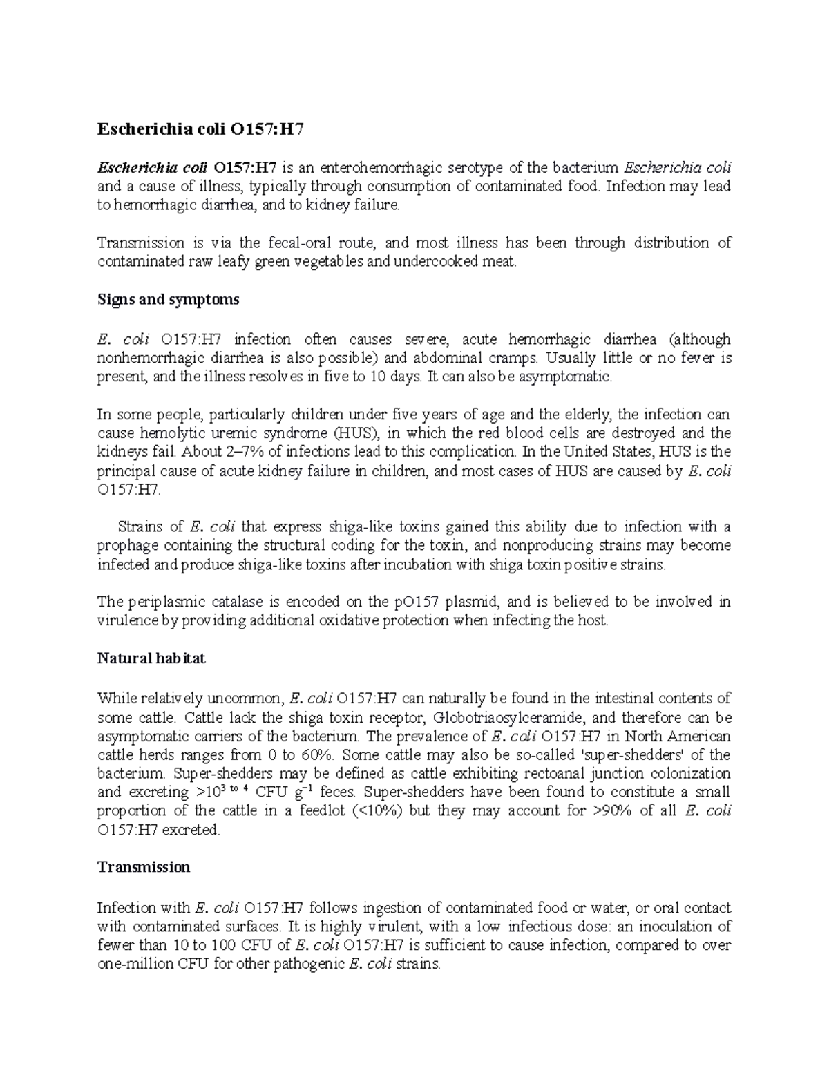
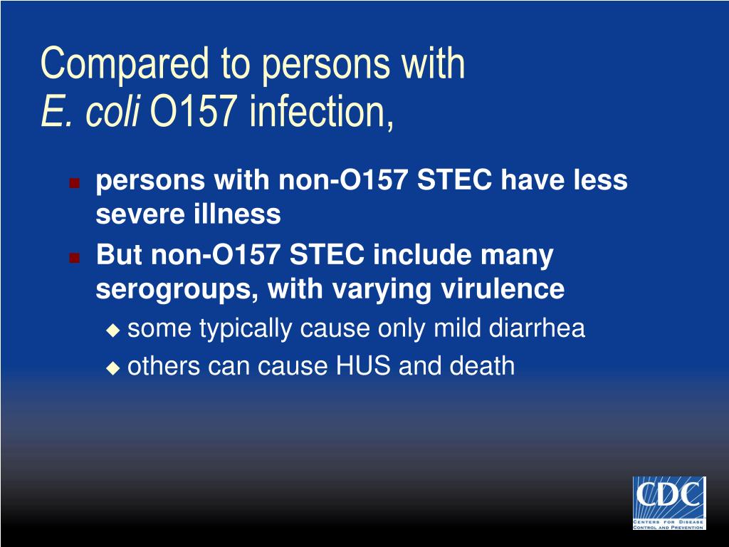
Escherichia coli O157:H7 is a prominent member of the Shiga toxin-producing E. coli (STEC) group, frequently categorized alongside other enterohemorrhagic strains. Understanding this bacterium requires examining its unique pathogenic mechanisms, which distinguish it from non-pathogenic enteric flora and even other diarrheagenic bacterial strains. The defining characteristic of E. coli O157:H7 is its capacity to produce potent cytotoxins known as Shiga toxins (Stx1 and Stx2), which are structurally and functionally similar to the toxins produced by Shigella dysenteriae.
Once ingested—typically through contaminated water, undercooked ground beef, unpasteurized dairy or juices, or direct contact with farm animals—the bacteria survive the acidic environment of the stomach and colonize the large intestine. Adherence to the intestinal epithelium is mediated by a chromosomal pathogenicity island known as the locus of enterocyte effacement (LEE). This genetic machinery allows the bacterium to form characteristic attaching and effacing (A/E) lesions, destroying the microvilli of enterocytes and disrupting normal absorptive functions.
The clinical danger, however, lies beyond local intestinal damage. The secreted Shiga toxins cross the intestinal epithelial barrier and enter the bloodstream. These toxins bind specifically to globotriaosylceramide ($Gb_3$) receptors, which are densely expressed on endothelial cells of the renal microvasculature and the central nervous system. Upon binding, the toxin enters the cell, inhibits protein synthesis, and induces cellular apoptosis. This triggers a cascade of microvascular endothelial injury, platelet activation, and microthrombi formation, setting the stage for systemic complications.
| Feature / Parameter | E. coli O157:H7 Infection Characteristics |
|---|---|
| Primary Reservoirs | Ruminants, primarily cattle and sheep |
| Infectious Dose | Extremely low (fewer than 100 to 1,000 organisms) |
| Incubation Period | Typically 3 to 4 days (range: 1 to 10 days) |
| Key Virulence Factors | Shiga toxins (Stx1, Stx2), LEE pathogenicity island |
| Hallmark Symptom | Sudden onset of severe, painful abdominal cramps and bloody diarrhea |
| Primary Risk Factor for HUS | Administration of antibiotics to children and high-risk patients |
Clinical Presentation and Diagnostic Evaluation
Recognizing an E. coli O157:H7 infection promptly is vital for preventing inappropriate therapeutic interventions and monitoring high-risk patients for systemic complications. The clinical presentation typically follows a distinct chronological progression, beginning days after exposure.
Symptom Progression
The illness often starts with non-bloody diarrhea and moderate abdominal discomfort within 3 to 4 days of ingestion. Within 24 to 48 hours, the clinical picture evolves dramatically. Abdominal cramps become intensely severe, mimicking acute surgical conditions like appendicitis, and the diarrhea transitions from watery to frankly bloody (hemorrhagic colitis). Low-grade fever or no fever at all is characteristic, helping clinicians differentiate STEC infections from invasive bacterial pathogens such as Salmonella, Shigella, or Campylobacter, which typically present with high fevers.
Diagnostic Testing Protocols
Because clinical symptoms alone cannot definitively distinguish E. coli O157:H7 from other causes of bloody diarrhea, laboratory confirmation is essential.
- Stool Culture: Stool samples must be plated on selective media, specifically Sorbitol MacConkey (SMAC) agar. Unlike most other E. coli strains, E. coli O157:H7 fails to ferment sorbitol within 24 hours, appearing as colorless colonies on SMAC plates.
- Toxin Assays: Enzyme immunoassay (EIA) or rapid membrane enzyme immunoassays can detect Shiga toxins 1 and 2 directly in stool samples or broth cultures.
- Molecular Testing: Polymerase chain reaction (PCR) assays are increasingly utilized to rapidly identify the genes encoding Stx1 and Stx2, offering high sensitivity and specificity.
- Laboratory Safety: Clinical laboratories must follow strict biosafety protocols when handling suspected STEC specimens to protect laboratory personnel from accidental transmission.
The Core Dilemma: Antibiotic Contraindications
One of the most critical tenets in the management of E. coli O157:H7 is the strict avoidance of antibiotic therapy. While physicians instinctively reach for antimicrobial agents when managing bacterial infections, prescribing antibiotics for Shiga toxin-producing E. coli is contraindicated.
Mechanism of Antibiotic-Induced Harm
Bacteriostatic and bactericidal antibiotics—such as fluoroquinolones, trimethoprim-sulfamethoxazole, and beta-lactams—stress the bacterial cell wall and DNA. In response to this genotoxic stress, the bacteria upregulate the SOS response pathways of their genome. Because the genes encoding Shiga toxins (specifically Stx2) are often integrated into prophages residing within the bacterial chromosome, the activation of the bacterial SOS response triggers the lytic cycle of the bacteriophage.
This causes massive bacterial lysis, releasing preformed Shiga toxins and dramatically increasing the concentration of circulating toxin within the gastrointestinal tract. The surge in free toxin absorption accelerates systemic dissemination, directly elevating the patient's risk of developing hemolytic uremic syndrome.
Clinical Exceptions and Nuances
While antibiotic therapy is withheld during active acute infection, clinical debate persists regarding very specific scenarios, such as extra-intestinal manifestations or overwhelming sepsis. However, for uncomplicated intestinal E. coli O157:H7 infection, guideline-concordant care mandates abstaining from antimicrobials. Anti-motility agents (such as loperamide or diphenoxylate) are similarly contraindicated. By slowing intestinal motility, these agents prolong mucosal contact time with Shiga toxins, increase systemic toxin absorption, and heighten the likelihood of severe systemic disease.
Supportive Management and Hydration Strategies
Without targeted antimicrobial options, the cornerstone of E. coli O157:H7 treatment is aggressive, meticulous supportive care aimed at maintaining intravascular volume, correcting electrolyte disturbances, and managing pain.
Fluid Resuscitation and Electrolyte Balance
Severe diarrhea and vomiting can rapidly lead to significant volume depletion and acute kidney injury, particularly in vulnerable populations like young children and older adults.
- Oral Rehydration Therapy (ORT): For patients able to tolerate oral intake, balanced oral rehydration solutions containing appropriate ratios of sodium, potassium, and glucose are initiated immediately.
- Intravenous (IV) Hydration: Patients exhibiting signs of severe dehydration, persistent vomiting, or hemodynamic instability require prompt admission for isotonic IV crystalloid administration (such as normal saline or lactated Ringer's solution).
- Monitoring: Serial measurements of serum electrolytes, blood urea nitrogen (BUN), creatinine, and complete blood counts (CBC) are mandatory to track renal function and watch for early hematologic signs of hemolysis.
Nutritional Support and Pain Management
Nutritional management during acute hemorrhagic colitis requires balancing the patient's caloric needs with gastrointestinal rest.
- Dietary Progression: Once acute abdominal pain and vomiting subside, early introduction of bland, easily digestible foods (such as bananas, rice, applesauce, and toast) is encouraged.
- Pain Control: Managing severe abdominal cramping can be challenging. Opiate-based analgesics must be used with extreme caution or avoided entirely due to their potential antimotility effects, which mirror the risks associated with anti-motility drugs. Non-opioid supportive pain management should be coordinated carefully under inpatient supervision.
Hemolytic Uremic Syndrome (HUS): Recognition and Critical Care
Hemolytic uremic syndrome is the most feared and severe complication of E. coli O157:H7 infection, occurring in approximately 5% to 15% of infected children and a smaller percentage of adults. Early recognition of HUS criteria is lifesaving.
Clinical Triad of HUS
The classic clinical presentation of HUS is defined by three hallmark findings:
- Microangiopathic Hemolytic Anemia: Characterized by rapid hemoglobin decline, elevated serum lactate dehydrogenase (LDH), undetectable haptoglobin, and the presence of fragmented red blood cells (schistocytes) on peripheral blood smear.
- Thrombocytopenia: Consumption of platelets within damaged microvessels leads to low platelet counts, sometimes accompanied by petechiae or purpura.
- Acute Kidney Injury (AKI): Manifests as elevated serum creatinine, oliguria, or complete anuria, resulting from extensive thrombotic microangiopathy within the renal glomeruli.
Advanced Medical Management
When HUS develops, management transitions from basic supportive care to intensive multidisciplinary intervention in an inpatient or pediatric intensive care unit (PICU) setting.
- Renal Replacement Therapy: A significant proportion of patients with HUS require dialysis (either hemodialysis or continuous renal replacement therapy) to manage fluid overload, severe hyperkalemia, and uremia while the kidneys recover.
- Blood Component Support: Transfusions of packed red blood cells are administered carefully for severe anemia. Platelet transfusions are generally avoided unless active, life-threatening bleeding occurs, as infused platelets can potentially fuel microvascular thrombus formation.
- Complement-Targeted Therapy: In rare, atypical presentations or severe cases where complement-mediated thrombotic microangiopathy cannot be ruled out, terminal complement inhibitors (such as eculizumab) may be considered, though their efficacy in pure STEC-HUS remains an area of ongoing clinical investigation.
Prevention, Public Health, and Safe Handling Practices
Given the challenges of treating established E. coli O157:H7 infections and the severity of HUS, primary prevention remains the most effective strategy for reducing disease burden. Public health agencies focus heavily on food safety, agricultural practices, and consumer education.
Consumer Food Safety Guidelines
Households and food handlers can dramatically lower infection risks by adhering to standardized hygiene and cooking protocols:
- Thorough Cooking: Ground beef must be cooked to an internal temperature of at least 160°F (71°C). Using a digital meat thermometer is the only reliable way to verify safe internal temperatures.
- Cross-Contamination Prevention: Raw meats should be kept separate from produce, ready-to-eat foods, and utensils. Cutting boards, knives, and countertops must be washed thoroughly with hot soapy water after handling raw meat.
- Produce Hygiene: Fresh fruits and vegetables eaten raw should be washed thoroughly under running water.
- Beverage Safety: Consumers should avoid raw milk, unpasteurized dairy products, and unpasteurized ciders or juices.
Occupational and Environmental Safety
Because E. coli O157:H7 has a remarkably low infectious dose, human-to-human transmission in settings like daycare centers, nursing homes, and households is common. Rigorous handwashing with soap and water after handling animals, visiting agricultural fairs, using the restroom, or changing diapers is essential. Public health authorities mandate that infected food handlers and child-care workers be excluded from their occupations until stool tests confirm they are no longer shedding the organism.
Note: This article is for informational and educational purposes only and does not substitute for professional medical evaluation, diagnosis, or treatment. If you or someone in your care exhibits symptoms of bloody diarrhea or severe abdominal pain, seek immediate medical attention.