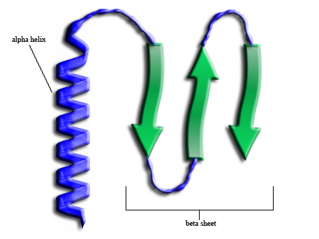
Alpha helices and beta sheets are the two primary patterns of secondary protein structure, stabilized by hydrogen bonds between the peptide backbone's atoms. While alpha helices form tightly coiled, rod-like spirals, beta sheets consist of extended strands linked laterally, providing essential mechanical stability and functional scaffolding for all known proteins.
Proteins are the workhorses of the biological world, performing functions ranging from catalytic enzyme activity to structural support in tissues. The functionality of a protein is dictated entirely by its three-dimensional shape, which is achieved through a hierarchical folding process. At the heart of this process lies the secondary structure—the local spatial arrangement of the polypeptide backbone. In 2026, our ability to predict these structures using advanced computational models has reached near-atomic precision, yet the fundamental physics governing the alpha helix and beta sheet remain the cornerstone of molecular biology.
The Chemical Foundation: The Peptide Bond and Backbone
To understand how alpha helices and beta sheets form, one must first examine the nature of the polypeptide chain. Proteins are polymers of amino acids linked by peptide bonds. A peptide bond is formed through a dehydration reaction between the carboxyl group of one amino acid and the amino group of the next.
The peptide bond possesses a partial double-bond character due to resonance. This restricts rotation around the C-N bond, keeping the peptide unit planar. However, the bonds on either side of the alpha carbon ($\text{C}_\alpha$)—the Phi ($\phi$) and Psi ($\psi$) angles—are technically free to rotate. These rotations are not infinite; steric hindrance between side chains and the backbone atoms limits the allowable angles.
In the early 1950s, Linus Pauling and Robert Corey identified that specific combinations of these angles allow the backbone to form repetitive, stable patterns held together by hydrogen bonds. These patterns are the alpha helix and the beta sheet. By 2026, structural biologists use these geometric constraints to validate experimental data from cryo-electron microscopy and X-ray crystallography, ensuring that proposed models adhere to the energetic "sweet spots" of protein folding.
The Alpha Helix: The Molecular Spiral
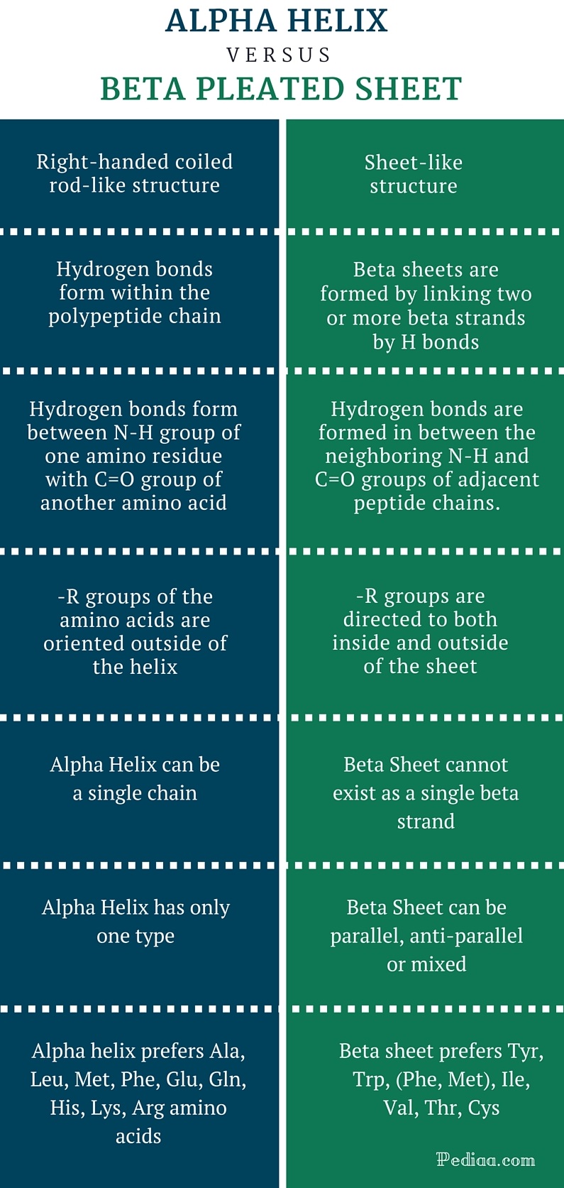
The alpha helix ($\alpha$-helix) is perhaps the most recognizable element of protein structure. It is a right-handed coiled conformation in which every backbone N-H group donates a hydrogen bond to the backbone C=O group of the amino acid located four residues earlier in the protein sequence.
Geometric Specifications
The geometry of the alpha helix is remarkably consistent across different proteins. A standard alpha helix has:
- 3.6 residues per turn: This means the helix completes a full 360-degree rotation every 3.6 amino acids.
- A rise of 1.5 Å per residue: This is the translation along the helical axis for each amino acid.
- A pitch of 5.4 Å: This is the total vertical distance covered in one full turn of the helix.
The hydrogen bonds in an alpha helix are parallel to the helical axis, providing significant longitudinal stability. Because the bonds are internal to the coil, the helix acts as a rigid, rod-like structure.
Side Chain Orientation
One of the most critical features of the alpha helix is the orientation of the amino acid side chains (R-groups). In an alpha helix, the R-groups point outward and slightly downward from the helical axis. This arrangement prevents steric interference between the side chains and the backbone. This outward orientation allows the alpha helix to interact with its environment. For example, amphipathic helices have hydrophobic side chains on one side and hydrophilic ones on the other, allowing them to sit at the interface of membranes or to bundle together in the protein core.
Constraints and Variations
While the 3.6${13}$ helix (the standard alpha helix) is the most common, variations exist, such as the tighter 3${10}$ helix or the wider $\pi$-helix. Furthermore, certain amino acids act as "helix breakers." Proline, for instance, has a cyclic side chain that prevents the N-H group from participating in hydrogen bonding and creates a structural kink. Conversely, Alanine is highly favored in helices due to its small, non-bulky side chain that fits perfectly into the coiled geometry.
The Beta Sheet: The Pleated Scaffold
The beta sheet ($\beta$-sheet) is the second major form of secondary structure. Unlike the compact alpha helix, the beta sheet is composed of beta strands that are almost fully extended. These strands align side-by-side to form a sheet-like structure held together by lateral hydrogen bonds.
Parallel vs. Antiparallel Orientations
Beta sheets are categorized based on the relative directionality of the constituent strands:
- Antiparallel Beta Sheets: The strands run in opposite N-to-C directions. This is the most stable configuration because the hydrogen bonds are linear (180 degrees), which is the strongest orientation for a hydrogen bond.
- Parallel Beta Sheets: The strands run in the same N-to-C direction. The hydrogen bonds in this configuration are slightly distorted or slanted, making the structure marginally less stable than the antiparallel version.
- Mixed Beta Sheets: Some proteins contain sheets that feature both parallel and antiparallel strand arrangements.
The "Pleat" and Side Chain Alternation
The term "beta pleated sheet" refers to the zig-zag or pleated appearance of the backbone. Because the carbon atoms are not in a straight line, the sheet has a rippled texture. This pleating is functionally significant because it causes the R-groups of adjacent amino acids to point in alternating directions—one above the plane of the sheet, the next below it.
This alternation is a vital design principle in proteins. It allows one side of a beta sheet to be entirely hydrophobic (facing the protein interior) while the other side is hydrophilic (facing the aqueous environment).
Comparative Analysis of Secondary Structures
The choice between an alpha helix or a beta sheet at any given point in a protein sequence is determined by the local amino acid sequence and the global energetic landscape of the folding protein.
| Feature | Alpha Helix | Beta Sheet |
|---|---|---|
| Basic Form | Single strand coiled into a spiral | Multiple strands aligned laterally |
| Hydrogen Bond Pattern | Intrachain ($i$ to $i+4$) | Interstrand (lateral between strands) |
| Side Chain Direction | Projected outward from the spiral | Alternating above and below the sheet |
| Distance per Residue | ~1.5 Å (compact) | ~3.2 to 3.5 Å (extended) |
| Typical Stability | High longitudinal rigidity | High lateral/mechanical tensile strength |
| Common Amino Acids | Ala, Glu, Leu, Met | Val, Ile, Tyr, Trp |
| Structure Breakers | Proline, Glycine | Proline |
The Ramachandran Plot: Mapping the Geometry
To understand how these structures are verified in 2026, we look at the Ramachandran Plot. This is a 2D visualization of the $\phi$ and $\psi$ torsion angles of the polypeptide backbone.
Because of steric clashes, only specific regions of the plot are "allowed." Alpha helices and beta sheets occupy distinct, well-defined clusters on this map:
- Alpha Helices: Cluster in the lower-left quadrant (negative $\phi$, negative $\psi$).
- Beta Sheets: Cluster in the upper-left quadrant (negative $\phi$, positive $\psi$).
When researchers solve a new protein structure today, the Ramachandran plot serves as a primary quality control metric. If residues fall into "disallowed" regions without a clear functional reason (like a strained active site), the model is likely incorrect.
Supersecondary Structures and Motifs
Rarely do alpha helices and beta sheets exist in isolation. They combine into recognizable patterns called supersecondary structures or motifs. These motifs serve as the building blocks for protein domains.
- Beta-Alpha-Beta Motif: A common way to connect two parallel beta strands using an alpha helix.
- Hairpin Loop: A simple connection between two antiparallel beta strands.
- Greek Key: A complex fold involving four antiparallel beta strands folded over each other.
- Beta Barrel: A large beta sheet that twists and coils to form a closed cylinder. These are frequently found in porins (membrane-spanning proteins) where the interior of the barrel acts as a channel.
These motifs are highly conserved across species. In 2026, evolutionary biology relies heavily on identifying these motifs to determine the function of newly sequenced genomes, as structure is often more conserved than the raw amino acid sequence itself.
Influence of Amino Acid Propensity
Not all amino acids are equally likely to be found in a helix or a sheet. This "propensity" is based on the chemical nature of the side chains:
- Branching at the Beta-Carbon: Amino acids like Valine, Threonine, and Isoleucine have branches close to the backbone. These branches cause steric interference in an alpha helix, making these residues more likely to be found in beta sheets.
- Small Side Chains: Glycine has only a hydrogen atom as its side chain. It is so flexible that it often acts as a "helix breaker" because it favors too many other conformations, although it is essential for the tight turns often found at the ends of beta strands.
- Electrostatics: If a sequence has many similarly charged residues (e.g., a string of Lysines), the electrostatic repulsion will destabilize an alpha helix.
Clinical Relevance: Misfolding and Secondary Structure
The balance between alpha helices and beta sheets is delicate. Many neurodegenerative diseases are characterized by a transition in secondary structure.
In Alzheimer’s disease and Prion diseases, proteins that are normally rich in alpha helices undergo a conformational change into a "cross-beta" structure. This transition turns soluble proteins into insoluble, protease-resistant amyloid fibrils. These fibrils are dominated by beta sheets that stack tightly together, creating the plaques observed in diseased brain tissue.
Understanding the transition from helix to sheet is a major focus of pharmacology in 2026. Current therapeutic strategies often involve "chaperone" molecules designed to stabilize the alpha-helical state or prevent the lateral aggregation of beta strands into toxic oligomers.
Practical Guidance for Structural Analysis
If you are a student or researcher analyzing protein structures, follow these steps to accurately identify and evaluate secondary structural elements:
- Examine the H-Bonding Pattern: Use visualization software (like PyMOL or ChimeraX) to display hydrogen bonds. Look for the $(i, i+4)$ pattern for helices and the inter-strand lateral bonds for sheets.
- Consult the Ramachandran Plot: Ensure the $\phi$ and $\psi$ angles align with the expected regions for the identified structure.
- Identify Amphipathicity: Look at the distribution of hydrophobic and hydrophilic residues. A helix with one hydrophobic face is likely a surface-exposed or protein-interaction helix.
- Check for Motifs: Identify if the helices and sheets form a known fold (e.g., a Rossmann fold or a Zinc finger). This often provides immediate clues to the protein's function.
FAQs
Why is the alpha helix right-handed instead of left-handed?
While left-handed alpha helices are theoretically possible, they are extremely rare in nature. The right-handed twist is energetically preferred because it results in significantly less steric interference between the side chains and the C=O groups of the backbone. Almost all naturally occurring L-amino acids form right-handed helices.
Can a protein exist without alpha helices or beta sheets?
Yes, though it is less common for functional globular proteins. Some proteins or regions of proteins are "intrinsically disordered." These regions lack a fixed secondary structure and instead exist as a dynamic ensemble of shapes. However, even these regions often adopt a helix or sheet structure upon binding to a target molecule.
How do beta turns differ from beta sheets?
A beta turn is a short sequence (usually four amino acids) that allows the polypeptide chain to reverse direction. While they often connect the strands of a beta sheet, they are a distinct structural element. They are stabilized by a single hydrogen bond between the first and fourth residue of the turn.
What is the role of AI in structural biology in 2026?
By 2026, AI tools like AlphaFold have evolved to predict not just static structures but also the "folding pathways." These models help researchers understand how a sequence of amino acids chooses between forming an alpha helix or a beta sheet in real-time, accounting for pH, temperature, and molecular crowding.
Why do beta sheets "pleat"?
The pleating is a result of the tetrahedral geometry of the alpha carbon ($\text{C}_\alpha$). To maintain the optimal bond angles and distances for the hydrogen bonds between strands, the backbone must adopt a non-linear, zig-zag path, which naturally creates the pleated appearance.
Next Actions
To deepen your understanding of protein secondary structure, utilize 2026-standard structural databases such as the Protein Data Bank (PDB). Select a well-known protein, such as Myoglobin (mostly alpha-helical) or an Immunoglobulin (mostly beta-sheet), and use a molecular viewer to isolate the hydrogen bonding patterns. Comparing the physical distance between residues in these two models will provide a clear, visual representation of the density differences between helical coils and extended sheets.