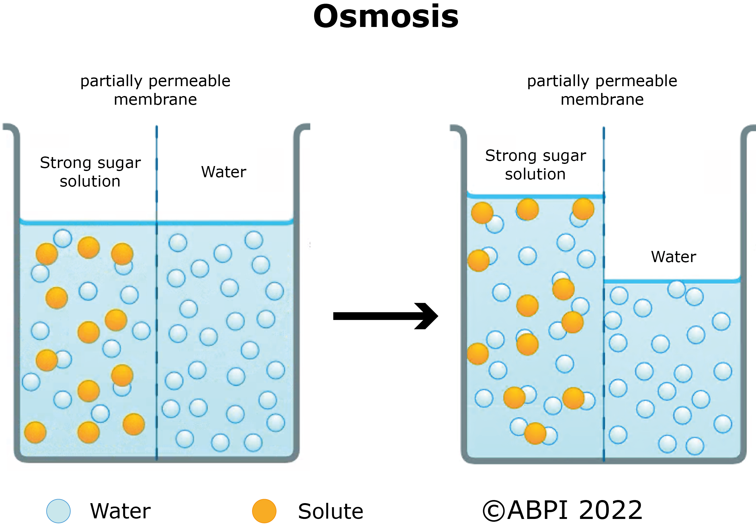
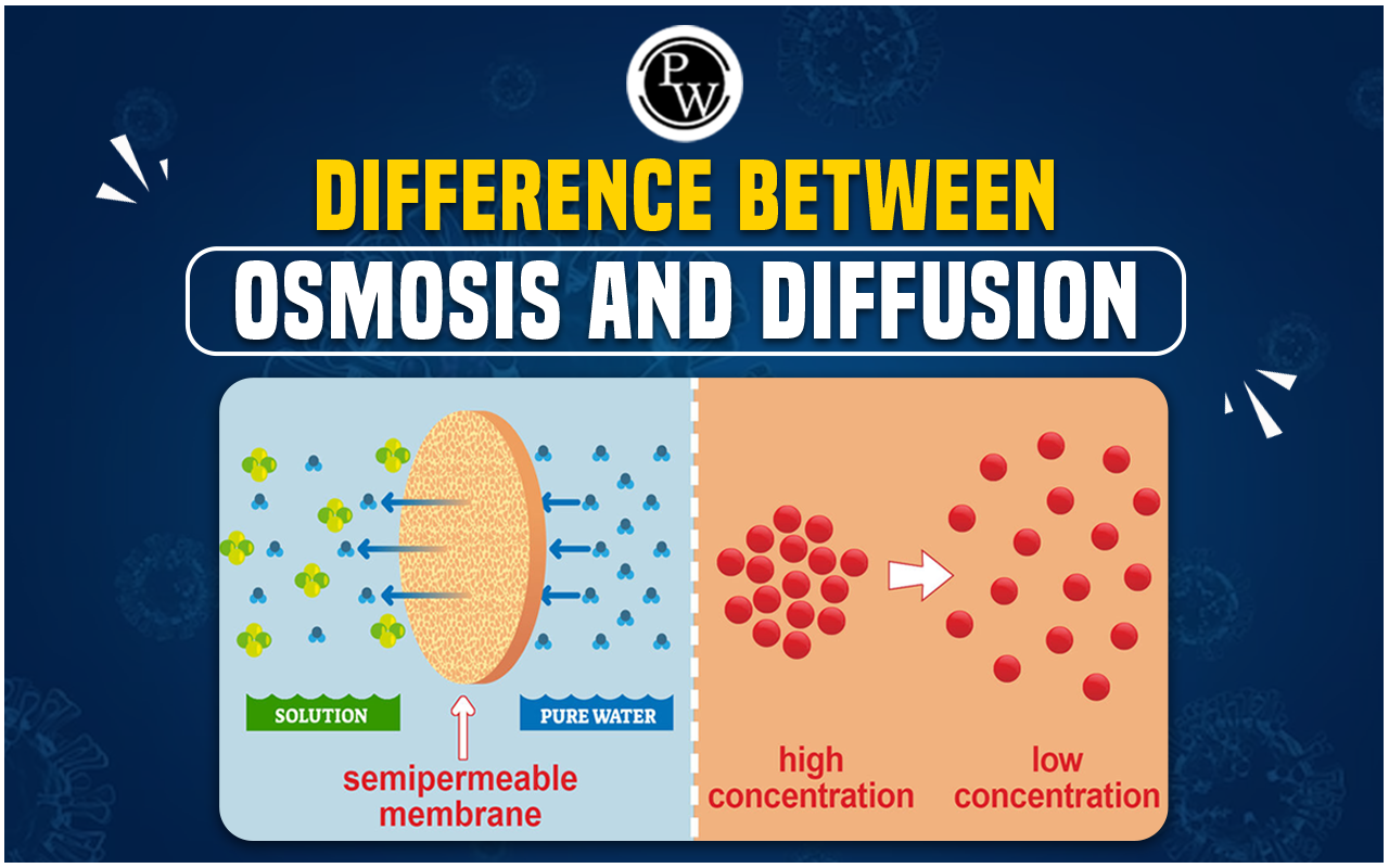
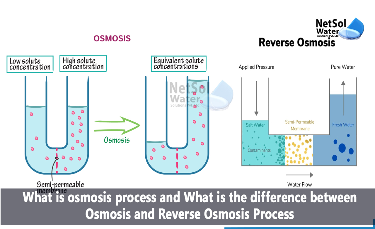
Osmosis is a vital physiological process defined as the net movement of water molecules across a selectively permeable membrane from an area of lower solute concentration to an area of higher solute concentration. In the human body, this passive transport mechanism maintains fluid balance, regulates blood pressure, enables kidney function, supports nutrient absorption in the gastrointestinal tract, and ensures proper cellular hydration.
Introduction to Osmosis in Biological Systems
Water constitutes approximately 60% of an adult human's total body weight. This massive volume of fluid is strictly compartmentalized. Intracellular fluid (ICF) exists inside trillions of individual cells, while extracellular fluid (ECF) surrounds them, further divided into interstitial fluid and blood plasma. Maintaining a stable environment across these compartments—a state known as homeostasis—depends heavily on osmotic pressures.
Unlike active transport, which requires cellular energy in the form of adenosine triphosphate (ATP), osmosis is driven entirely by thermodynamic gradients. Water molecules move spontaneously down their concentration gradient, or more accurately, down their water potential gradient, attempting to equalize the concentration of solutes (such as sodium, potassium, glucose, and proteins) on both sides of a membrane.
Understanding how this phenomenon operates in human physiology sheds light on everything from daily hydration to emergency critical care medicine. Below are the five key examples of osmosis at work in the human body.
1. Cellular Hydration and Maintenance of Shape
Every single cell in the human body is enclosed by a plasma membrane that acts as a selectively permeable barrier. This membrane allows water to pass freely via specialized channel proteins called aquaporins while restricting the movement of most dissolved solutes. The balance of water movement across this membrane dictates cell volume and structural integrity.
Depending on the solute concentration of the surrounding extracellular fluid, cells experience three distinct osmotic environments:
- Isotonic Environment: The solute concentration outside the cell matches the inside. Water moves in and out at equal rates, keeping the cell stable and functional.
- Hypotonic Environment: The extracellular fluid has a lower solute concentration than the cell interior. Water rushes into the cell via osmosis. In red blood cells, this can cause swelling and eventual bursting, a process called hemolysis.
- Hypertonic Environment: The extracellular fluid has a higher solute concentration than the cell interior. Water is drawn out of the cell. This causes red blood cells to shrink and shrivel, a process known as crenation.
In a healthy body, homeostatic mechanisms—primarily regulated by the brain, kidneys, and lungs—ensure that the extracellular fluid remains strictly isotonic relative to the intracellular fluid, protecting cells from structural failure.
2. Kidney Function and Urine Concentration
The kidneys are master regulators of water and electrolyte balance in the human body. Through an intricate network of microscopic filtering units called nephrons, the kidneys filter approximately 150 to 180 liters of blood daily, yet typically excrete only 1 to 2 liters of urine. The rest of the water is reabsorbed, and osmosis is the driving engine behind this reclamation process.
Within the loop of Henle and the collecting ducts of the nephron, the body establishes a steep osmotic gradient in the surrounding kidney tissue (the renal medulla). As the filtrate travels through the descending limb of the loop of Henle, the surrounding tissue becomes progressively more concentrated with solutes like sodium and urea.
Because the descending limb is freely permeable to water but impermeable to salts, water leaves the tubule via osmosis, moving out of the filtrate and back into the surrounding blood vessels (the vasa recta). This concentrates the remaining fluid inside the nephron. Later, under the influence of the antidiuretic hormone (ADH), the collecting ducts become more permeable to water, allowing even more water to be drawn out via osmosis if the body is dehydrated, thereby producing concentrated urine.
3. Water Absorption in the Gastrointestinal Tract
Every day, the gastrointestinal tract processes several liters of fluid, consisting of ingested liquids and secretions from salivary glands, the stomach, the pancreas, and the liver. By the time this digested material reaches the large intestine (colon), the vast majority of water must be recovered to prevent dehydration.
Nutrients such as glucose, amino acids, and sodium are actively transported out of the intestinal lumen and into the epithelial cells lining the gut. This active transport of solutes creates a high solute concentration inside the tissue cells and blood vessels relative to the watery contents of the intestinal lumen.
As a direct result, water follows these solutes by moving across the intestinal mucosal membrane via osmosis. This osmotic water reabsorption ensures that the body conserves vital fluids and transforms liquid chyme into solid feces for elimination. When this osmotic balance is disrupted—such as through bacterial toxins or malabsorbed sugars—water fails to be reabsorbed or is actually drawn into the gut, resulting in osmotic diarrhea.
4. Cerebrospinal Fluid Dynamics and Brain Protection
The central nervous system, comprising the brain and spinal cord, is suspended and cushioned within cerebrospinal fluid (CSF). This specialized fluid protects delicate neural tissue from mechanical shock, provides chemical stability, and supplies nutrients while removing metabolic waste products.
The production and absorption of CSF rely heavily on osmotic gradients. CSF is primarily secreted by the choroid plexuses within the brain's ventricles. Specialized ependymal cells actively transport ions, particularly sodium and chloride, into the ventricles. Water follows these ions passively through osmosis, generating the fluid pressure required to keep the brain buoyant.
Furthermore, imbalances in osmotic pressure within the cranial vault can have catastrophic medical consequences. If blood plasma becomes significantly hypotonic relative to brain tissue (as can occur in severe hyponatremia or water intoxication), water moves via osmosis into the brain cells, causing cerebral edema (brain swelling). Because the skull is a rigid, fixed container, this swelling increases intracranial pressure, leading to neurological deficits, seizures, or herniation.
5. Salivary and Exocrine Gland Secretion
Exocrine glands—including salivary glands, sweat glands, and the pancreas—produce secretions essential for digestion, thermoregulation, and metabolic support. The production of saliva is a classic textbook illustration of epithelial fluid transport driven by osmosis.
The process begins in the acinar cells of the salivary glands, which actively secrete sodium, chloride, and potassium ions into the lumen of the gland. This localized accumulation of solutes creates a hypertonic environment inside the duct. Because the epithelial lining of the salivary ducts is permeable to water, water is immediately drawn out of the surrounding interstitial fluid and into the duct lumen via osmosis, dissolving the secreted proteins (such as amylase) to form fluid saliva.
As the saliva travels down the striated ducts toward the oral cavity, specialized cells reabsorb sodium and chloride ions. However, because these duct walls are relatively impermeable to water, the final product becomes hypotonic relative to blood plasma, which is why saliva tastes flat and helps protect oral tissues from hyperosmolar damage.
Summary of Osmotic Mechanisms in Physiology
| Physiological System | Primary Location | Direction of Osmotic Water Movement | Biological Purpose |
|---|---|---|---|
| Cellular Hydration | Plasma Membrane | Inward or Outward (based on ECF tonicity) | Prevents cellular swelling (lysis) or shrinkage (crenation). |
| Renal Filtration | Loop of Henle / Collecting Ducts | Out of nephron tubule into renal medulla | Conserves body water and concentrates urine. |
| Gastrointestinal Tract | Intestinal Mucosa | From intestinal lumen into epithelial cells/blood | Recovers dietary and secreted water; prevents dehydration. |
| Central Nervous System | Ventricles / Blood-Brain Barrier | Into ventricles (secretion) or brain tissue (pathology) | Maintains buoyancy of the brain via CSF; prevents edema. |
| Exocrine Glands | Salivary and Sweat Glands | From interstitial fluid into gland lumens | Generates fluid secretions for digestion and cooling. |
Frequently Asked Questions
What is the primary difference between osmosis and diffusion?
Diffusion is the passive movement of any molecules (solutes or gases) from an area of higher concentration to an area of lower concentration. Osmosis is a specific type of diffusion: the movement of water molecules specifically across a selectively permeable membrane down their water potential gradient.
Why is intravenous (IV) saline usually 0.9% concentration?
A 0.9% sodium chloride solution is considered isotonic to human blood plasma (often referred to as normal saline). Because it matches the osmotic pressure of red blood cells, administering it intravenously prevents water from rushing into cells (causing them to burst) or drawing water out of cells (causing them to shrink).
Can drinking too much water disrupt osmosis in the body?
Yes. Consuming excessive water in a short period can dilute the sodium concentration in the blood plasma, leading to a dangerous condition called hyponatremia (water intoxication). This creates a hypotonic extracellular environment, causing water to rush via osmosis into brain cells and other tissues, resulting in life-threatening swelling.
How do aquaporins assist in osmosis?
Aquaporins are specialized transmembrane channel proteins that facilitate the rapid transport of water molecules across cell membranes. While water can cross lipid bilayers slowly on its own due to its small size, aquaporins dramatically increase membrane permeability, which is especially critical in organs with high water-transport demands like the kidneys.
Practical Clinical Takeaways and Next Actions
Recognizing how osmosis governs human physiology is critical for anyone studying biology, nursing, medicine, or allied health fields. Clinically, monitoring fluid and electrolyte balance prevents severe complications like dehydration, edema, and electrolyte imbalances. If you or someone you know is experiencing persistent symptoms of fluid imbalance—such as extreme thirst, severe swelling in the extremities, or sudden neurological changes—seek immediate professional medical evaluation, as these may indicate underlying systemic or renal dysfunction.