Aquaporins are specialized integral membrane proteins that function as biological water channels, facilitating the rapid and selective transport of water molecules across cellular membranes. By enabling water to bypass the hydrophobic lipid bilayer, these proteins maintain osmotic balance and facilitate essential physiological processes across all domains of life, including humans, plants, and microbes.
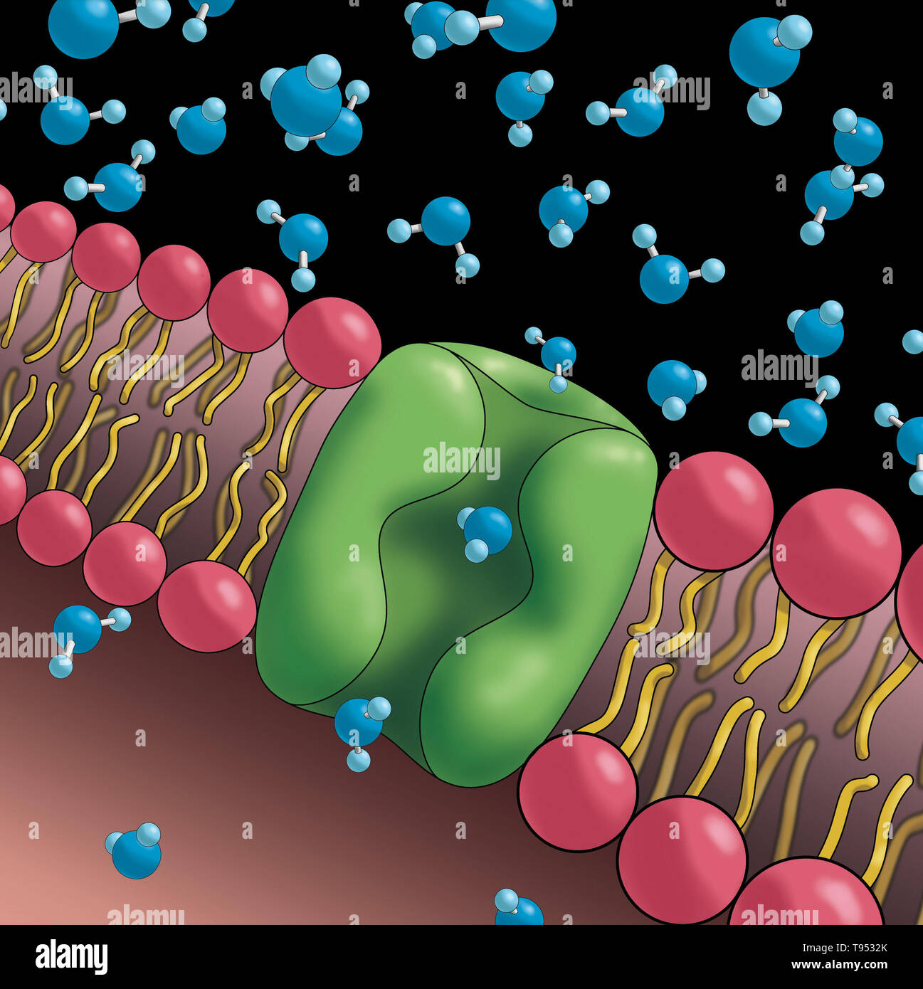
The fundamental challenge of cellular life involves moving water into and out of cells with high efficiency while maintaining the integrity of the cell membrane. Passive diffusion of water through the lipid bilayer is inherently slow due to the hydrophobic core of the plasma membrane, which repels polar molecules. Aquaporins—often referred to as "the plumbing system of the cell"—solve this by providing a highly specific, pore-like pathway. These proteins ensure that the osmotic gradient is utilized effectively, allowing cells to regulate their volume, internal pressure, and fluid distribution with extreme precision.
The Molecular Architecture of Aquaporins
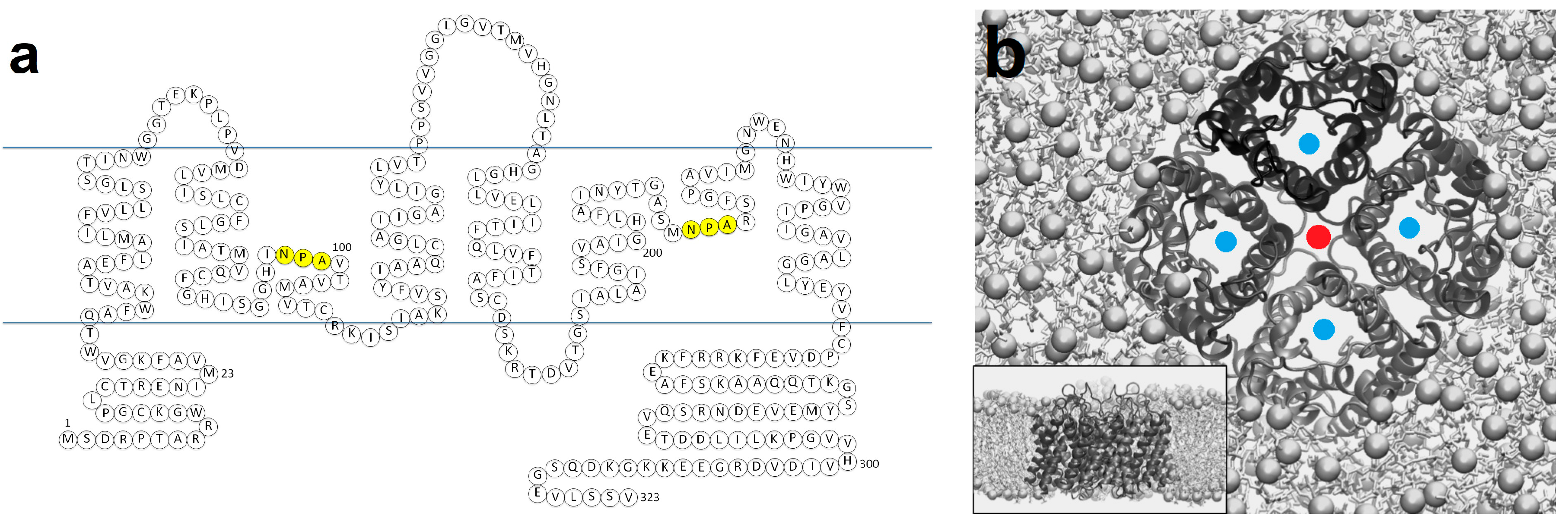
To understand how aquaporins function, one must look at their structural configuration. Each aquaporin typically exists as a tetramer within the cell membrane. While they function as a group of four, each individual monomer contains a distinct channel. This structure is conserved across species, reflecting its vital importance to biological stability.
Each monomer consists of six transmembrane alpha-helices that span the lipid bilayer. These helices organize to form a narrow, hourglass-shaped pore. The interior of this channel is lined with specific amino acids that dictate its selectivity. A critical feature of the aquaporin architecture is the presence of two highly conserved motifs known as NPA (asparagine-proline-alanine) boxes. These motifs fold into the center of the pore, creating a selectivity filter that prevents the passage of protons (hydrogen ions) and other solutes, ensuring that only water molecules move through the channel.
The exclusion of protons is a remarkable feat of molecular engineering. If protons were allowed to pass through the water channel, they would disrupt the electrochemical gradients that cells rely on for energy production and signaling. Aquaporins achieve this exclusion by using the dipoles of the water molecules themselves to reorient them as they pass through the narrowest part of the pore, effectively "breaking" the chain of hydrogen-bonded water molecules that would otherwise conduct electricity or ions.
Physiological Significance in the Human Body
In human physiology, the expression of different types of aquaporins—designated as AQP1 through AQP13—is tissue-specific. This specialization allows different organs to manage fluid dynamics according to their unique requirements.
- Kidney Function: The kidneys are perhaps the most prominent example of aquaporin utility. In the renal collecting ducts, AQP2 channels are regulated by the hormone vasopressin. When the body needs to conserve water, vasopressin triggers the translocation of AQP2 to the surface of the cell membrane, vastly increasing water reabsorption from the urine back into the bloodstream. Dysfunction in this system is linked to clinical conditions such as diabetes insipidus, where the body cannot adequately concentrate urine.
- Ocular Health: In the eye, AQP1 and AQP4 are vital for maintaining the clarity of the lens and the proper pressure within the intraocular space. Fluid movement through these channels ensures that the eye maintains the refractive index necessary for vision.
- Brain and Nervous System: AQP4 is highly concentrated in the end-feet of astrocytes within the brain. It plays a central role in the glymphatic system, a process that facilitates the clearance of metabolic waste products from the interstitial fluid during sleep. The discovery of this pathway has significantly updated our understanding of neurological health and neurodegenerative disease progression.
Comparison of Aquaporin Functional Classes
While all aquaporins facilitate the movement of water, some members of the family exhibit broader permeability. Scientists generally categorize these proteins based on their substrate specificity and structural nuances.
| Channel Type | Primary Substrate | Notable Distribution | Key Biological Role |
|---|---|---|---|
| Classical Aquaporins | Pure Water | Kidney, Red Blood Cells | Rapid osmotic water movement |
| Aquaglyceroporins | Water, Glycerol, Urea | Liver, Adipose Tissue | Metabolic regulation and fuel transport |
| S-aquaporins | Water, Gases, Ions | Intracellular organelles | Intracellular compartment homeostasis |
This functional diversity demonstrates that aquaporins are not merely passive holes in the membrane; they are active, regulated, and specialized transport systems that integrate cellular metabolism with fluid balance.
Plant Biology and Osmotic Stress
Aquaporins are equally critical in the botanical world. In plants, the movement of water from the roots to the leaves—often against the force of gravity—is driven by transpiration and osmotic pressure. Plant aquaporins (often called Major Intrinsic Proteins) are regulated by environmental signals such as drought, salinity, and cold.
When a plant experiences water stress, it may downregulate the expression or activity of its aquaporins to minimize water loss. Conversely, when water is abundant, these channels open to facilitate rapid nutrient uptake and cell expansion. This dynamic response allows plants to adapt to fluctuating soil conditions. Research into these channels has significant implications for agriculture in 2026, as scientists seek to engineer crops that are more resilient to the increasing frequency of arid conditions and soil salinization.
Investigative Techniques and Verification
Determining the presence and activity of aquaporins requires sophisticated laboratory techniques. Because these proteins are embedded within lipid membranes, they cannot be analyzed in isolation without maintaining their native folded state.
- X-Ray Crystallography: This method provided the first detailed, atomic-resolution images of the aquaporin tetramer. By crystallizing the protein and bombarding it with X-rays, researchers visualized the internal path through which water travels.
- Stopped-Flow Light Scattering: This is a kinetic approach used to measure the rate at which cells shrink or swell when exposed to osmotic gradients. By expressing specific aquaporins in Xenopus oocytes (frog eggs), researchers can verify the protein's water permeability by observing the speed of volume change.
- Immunofluorescence: This technique uses antibodies tagged with fluorescent markers to identify exactly where specific aquaporins are located within a tissue sample, allowing for the mapping of these proteins in complex biological structures like the kidney or brain.
Common Misconceptions and Troubleshooting
A common misunderstanding is the belief that aquaporins allow for the free, unregulated flow of all fluids. In reality, they are highly selective. They do not transport ions like sodium, potassium, or chloride, nor do they transport large molecules. If a substance is not specifically "gated" by the aquaporin's selectivity filter, it cannot pass through the channel.
Furthermore, students and researchers often confuse passive diffusion through the bilayer with facilitated transport through aquaporins. While both are "passive" in the sense that they do not require ATP directly, they differ in speed and regulation. Facilitated transport via aquaporins is orders of magnitude faster than simple diffusion through the lipid membrane and is subject to hormonal control, whereas simple diffusion is entirely dependent on the physical properties of the lipid bilayer and the concentration gradient.
When researching or troubleshooting laboratory experiments involving these proteins, consider the following:
- Protein Stability: Aquaporins are extremely sensitive to the detergent used in extraction. If the wrong detergent is used, the protein will denature, rendering it non-functional.
- Membrane Composition: The lipid environment surrounding the protein can influence its "gating." Some aquaporins require specific lipid interactions to open or close, meaning that purified protein studies may show different activity levels than the protein in its native cell membrane.
Clinical and Practical Implications
The study of aquaporins has moved from basic biochemistry into the realm of therapeutic development. By understanding the structure and function of these channels, medical researchers are exploring ways to modulate them to treat disease.
For instance, in cases of edema—the swelling of tissues due to fluid accumulation—the potential for small-molecule inhibitors that can temporarily block specific aquaporin channels is a subject of active research. Similarly, in oncology, certain cancers have been shown to overexpress aquaporins, which may contribute to the rapid growth and migration of tumor cells. Targeting these channels represents a promising, albeit complex, frontier in precision medicine.
As of 2026, the primary challenge remains the development of pharmacological agents that are sufficiently selective. Because there are so many different types of aquaporins, a drug designed to block an aquaporin in the brain must be precise enough not to interfere with the vital functions of aquaporins in the kidney or lungs.
Frequently Asked Questions
Are aquaporins present in all living cells? Yes, aquaporins are found in all three domains of life: Bacteria, Archaea, and Eukarya. Their ubiquitous presence across such a broad evolutionary spectrum underscores their essential role in life's basic requirement: water homeostasis.
Do aquaporins require energy (ATP) to move water? No, aquaporins do not consume ATP directly to transport water. The movement of water through these channels is driven by osmotic and hydrostatic pressure gradients. They are passive facilitators that simply provide a path of least resistance.
Can aquaporins transport anything other than water? Yes, some aquaporins, known as aquaglyceroporins, are capable of transporting small, uncharged molecules like glycerol, urea, and sometimes even carbon dioxide or ammonia, in addition to water. However, the classical aquaporins are strictly limited to water molecules.
What happens if a human lacks functional aquaporins? Complete absence of aquaporins is likely lethal due to the inability to maintain cellular volume and fluid balance. However, mutations that impair specific aquaporin function lead to well-documented medical conditions. For example, mutations in AQP2 result in nephrogenic diabetes insipidus, characterized by an inability to concentrate urine, leading to extreme dehydration.
How do aquaporins prevent the passage of ions? The channel features a narrow constriction site and an electrostatic filter. The presence of positively charged amino acid residues and the specific orientation of water molecules as they pass through the NPA motifs create a barrier that is energetically unfavorable for charged ions or protons, effectively blocking them.
Final Recommendation for Further Inquiry
To further your understanding of aquaporins, focus your study on the structural biology of the NPA motifs, as this explains the fundamental "how" of proton exclusion. If you are conducting laboratory work, prioritize the verification of your membrane environment, as the lipid-protein interface is the most common point of failure in experimental setups. For clinical context, the ongoing 2026 research into AQP4's role in the glymphatic system offers the most up-to-date insight into the relationship between fluid dynamics and neurological health. By integrating these mechanical, physiological, and clinical perspectives, you will gain a robust understanding of why these proteins are considered the essential conduits of life.