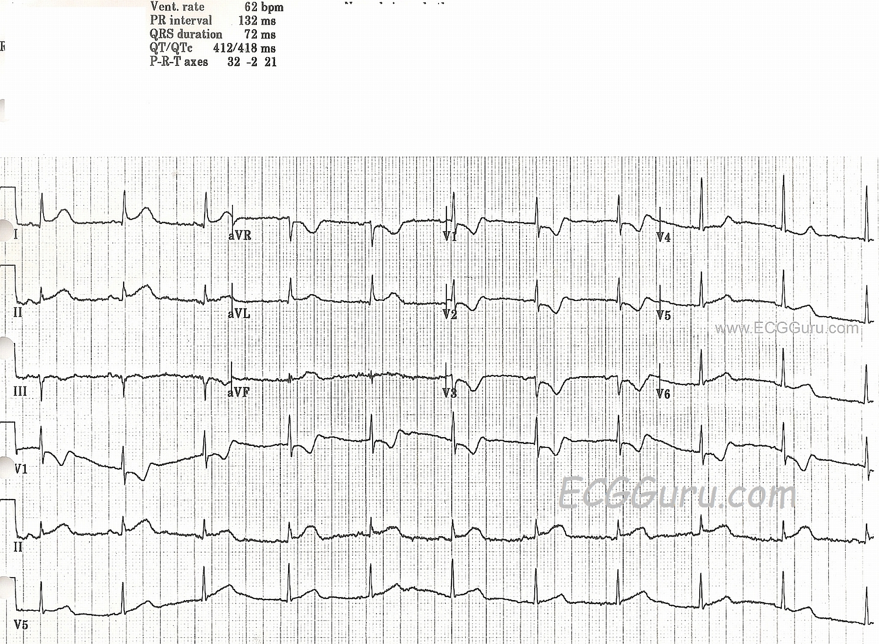
An anterior infarct, or anterior myocardial infarction, is a critical medical event evidenced on an electrocardiogram (ECG) by ST-segment elevation or significant Q-waves in the precordial leads V1 through V6. These patterns indicate significant damage or ischemia to the anterior wall of the heart, typically resulting from an occlusion of the left anterior descending (LAD) coronary artery. Early recognition through precise ECG lead analysis is vital for initiating reperfusion therapy and improving patient outcomes.
Understanding the Anatomy of an Anterior Infarct
To interpret an anterior infarct ECG, one must first understand the relationship between the coronary arteries and the heart’s electrical representation on paper. The heart is a three-dimensional organ, and the 12-lead ECG provides a snapshot of its electrical activity from twelve different "angles."
When a thrombus blocks the left anterior descending artery—often referred to as the "widow maker" due to the massive amount of myocardium it supplies—the anterior wall of the left ventricle becomes ischemic or necrotic. This region includes the front of the heart and the interventricular septum. Because the precordial leads (V1–V6) are positioned across the anterior chest wall, they are the primary leads that capture the electrical disturbances occurring in the anterior myocardium.
The Role of Coronary Perfusion
The LAD artery bifurcates from the left main coronary artery. It provides blood supply to the anterior wall, the anterior two-thirds of the interventricular septum, and the apex of the heart. Because of the vast territory this artery covers, an anterior infarct is often more hemodynamically significant than infarctions in smaller or more distal branches. An ECG showing changes in the precordial leads is the definitive diagnostic starting point for identifying the involvement of this critical vessel.
Recognizing Diagnostic ECG Features
Anterior myocardial infarction does not present as a single, uniform pattern. Instead, it evolves based on the degree of occlusion and the time elapsed since the event. A standard anterior infarct ECG will display specific findings that distinguish it from other cardiac events.
ST-Segment Elevation (STEMI)
The most urgent finding is ST-segment elevation. In an anterior STEMI, you will observe elevation in the leads corresponding to the anterior wall. Depending on the extent of the infarct, this may manifest as:
- Septal leads (V1–V2): Damage to the septum.
- Anterior leads (V3–V4): Damage to the front wall.
- Anterolateral leads (V5–V6): Damage extending toward the lateral wall.
When ST elevation is present in all these leads (V1–V6), it is often termed an "extensive anterior MI," suggesting an occlusion of the LAD near its origin, before it branches extensively.
Reciprocal Changes
One of the most useful diagnostic tools in ECG interpretation is the concept of "reciprocal changes." While the anterior leads (V1–V6) show elevation, the inferior leads (II, III, and aVF) often show ST-segment depression. These reciprocal depressions are not merely artifacts; they are the electrical "mirror image" of the injury occurring on the opposite side of the heart. Observing reciprocal changes significantly increases the clinical confidence that the ST elevation in the anterior leads is an acute injury rather than an older, chronic condition.
Evolution of Q-Waves
In the hours and days following an anterior infarct, the ECG will evolve. The ST elevation may gradually return to the baseline, but pathological Q-waves—waves that are deeper than one-quarter the height of the R-wave or wider than 0.04 seconds—may develop. These Q-waves represent "electrically dead" myocardium, indicating that the tissue has progressed from ischemia (lack of blood) to necrosis (tissue death).
Comparative Analysis of Precordial Lead Findings
The following table summarizes the typical localization of infarcts based on which precordial leads demonstrate diagnostic changes.
| Lead Grouping | Anatomical Area | Likely Artery Affected |
|---|---|---|
| V1 – V2 | Septal Wall | Proximal LAD (Septal branches) |
| V3 – V4 | Anterior Wall | Mid to Distal LAD |
| V5 – V6 | Lateral / Apical Wall | LCx or Diagonal branch of LAD |
| V1 – V6 | Extensive Anterior | Proximal LAD |
Note: LCx refers to the Left Circumflex artery.
Prerequisites for Accurate ECG Interpretation
Interpreting an ECG for an anterior infarct requires more than just looking at the spikes on the paper. The quality of the acquisition and the patient's baseline history are paramount.
Proper Lead Placement
The most common error in identifying an anterior infarct is misplacement of the V1–V6 leads. Even a centimeter of deviation can alter the electrical axis recorded by the machine. V1 and V2 must be placed in the 4th intercostal space at the right and left sternal borders, respectively. V4 must be in the 5th intercostal space at the mid-clavicular line. If the leads are placed too high or too low, the resulting waveform may mimic or mask an infarct, leading to dangerous clinical misjudgments.
Baseline Comparisons
If a patient has a known history of prior heart disease, an older ECG is an essential tool. Old Q-waves, persistent ST-segment elevation (a phenomenon known as LV aneurysm), or Left Bundle Branch Block (LBBB) can complicate the diagnosis of an acute event. Comparing the current tracing to a past ECG helps the clinician determine if the findings are truly "new" or if they are markers of an old injury.
Clinical Workflow and Next Actions
Once an anterior infarct is identified on an ECG, the situation is a time-critical emergency. The protocol in most cardiac centers in 2026 involves a standardized "door-to-balloon" workflow.
- Immediate Notification: The emergency department or pre-hospital team alerts the cardiac catheterization lab team. The ECG is the legal and clinical trigger for this activation.
- Antiplatelet and Anticoagulant Therapy: Patients are typically administered a cocktail of antiplatelet medications (such as aspirin and P2Y12 inhibitors) and anticoagulants (like heparin) to prevent further thrombus growth.
- Cardiac Catheterization (PCI): The patient is moved to the cath lab. A cardiologist inserts a catheter, typically through the radial or femoral artery, to visualize the coronary anatomy.
- Reperfusion: If an occlusion is found in the LAD, a percutaneous coronary intervention (PCI) is performed, involving balloon angioplasty and the placement of a drug-eluting stent to restore blood flow to the anterior wall.
Common Pitfalls and Troubleshooting
Diagnosing an anterior infarct can be deceptively complex due to mimicry. Practitioners must be aware of these common diagnostic pitfalls:
- Left Bundle Branch Block (LBBB): A new LBBB in the presence of chest pain is often treated as an STEMI equivalent. It makes identifying ST elevation impossible because the QRS complex is wide and the ST segments are naturally discordant.
- Early Repolarization: This is a benign condition where the ST segment appears elevated. However, in early repolarization, the ST segments are usually concave, whereas in an acute infarct, they are often convex (tombstoning).
- Hyperkalemia: High potassium levels can cause tall, peaked T-waves that may be mistaken for the hyper-acute T-waves seen in the very early stages of an anterior infarct. Always correlate ECG findings with the clinical status of the patient (e.g., chest pain, shortness of breath, diaphoresis).
Frequently Asked Questions
1. What does it mean if the ECG shows "tombstoning"?
"Tombstoning" is a slang term for extreme ST-segment elevation. It describes a morphology where the ST segment rises sharply and reaches a peak before falling, resembling a gravestone. It is a sign of severe, transmural myocardial injury and signifies a massive, high-risk infarct.
2. Can you have an anterior infarct without pain?
Yes. Diabetics, elderly patients, and women may present with "silent" MIs. They might experience fatigue, nausea, or shortness of breath rather than classic crushing chest pain. The ECG is the only way to detect the infarction in these silent presentations.
3. How quickly must an anterior infarct ECG be read?
Clinical guidelines dictate that an ECG should be obtained and interpreted within ten minutes of a patient’s arrival at the medical facility. Time is muscle; the faster the LAD is opened, the more myocardium is saved.
4. What is a "widow maker" heart attack?
This term refers to a total blockage of the proximal LAD artery. Because the LAD supplies a large percentage of the left ventricle, its blockage can lead to rapid cardiac arrest or pump failure, which is why it carries such high mortality if left untreated.
5. Why are reciprocal changes important?
Reciprocal changes act as a diagnostic confirmation. Seeing ST depression in the inferior leads when V1–V4 shows elevation provides strong evidence that the anterior changes are acute rather than chronic, helping to rule out false positives.
Final Recommendation
If you are a patient or a bystander witnessing symptoms suggestive of a heart attack—such as chest pressure, radiation to the jaw or left arm, or unexplained dyspnea—call emergency services immediately. Do not attempt to interpret an ECG yourself or wait for a home-based diagnostic device. If you are a practitioner, always prioritize the "first-medical-contact-to-device" time. Ensure your ECG machine is properly calibrated, and focus on immediate correlation between the ECG waveforms and the patient's hemodynamic stability.