For biological anthropologists, bioarchaeologists, and forensic researchers, distinguishing between ancient and modern human skulls requires evaluating cranial vault geometry, facial prognathism, mandibular structure, and cortical bone density. Ancient skulls consistently present platycephalic vault shapes, thicker cortical bone, pronounced supraorbital ridges, and robust masticatory attachments, whereas modern human skulls display globular braincases, gracile facial architecture, and a distinct chin.
Evolutionary Osteology: Key Morphological Differences
The human cranium has undergone profound morphological alterations over the last 300,000 years. Driven by evolutionary shifts in brain architecture, diet, and biomechanical stress, the modern Homo sapiens cranium exhibits a distinct "globularized" phenotype compared to the elongated neurocrania characteristic of archaic hominins and early anatomically modern humans (AMH).
Understanding these differences requires systematic analysis across three primary cranial modules: the neurocranium (braincase), the splanchnocranium (facial skeleton), and the mandible.
- Ancient / Archaic Skull (Elongated, Low Vault, Thick)
- Low Vault
- Supraorbital Torus
- Occipital Bun
- No Chin
- Modern Human Skull (Globular, High Vault, Gracile)
- High Vault
- Rounded Occipital
- Mental Protuberance
Neurocranial Shape and Vault Geometry
The neurocranium houses and protects the brain, and its shape closely reflects evolutionary shifts in encephalization and neurodevelopmental trajectories. Ancient human skulls—including Middle and Upper Paleolithic Homo sapiens, as well as Neanderthals (Homo neanderthalensis) and archaic Homo species—exhibit an elongated, low cranial vault (platycephaly). The maximum cranial breadth in ancient crania typically occurs lower on the vault, across the temporal or parietal regions, creating a pentagonal or barrel-shaped profile when viewed from behind (norma occipitalis).
In contrast, the modern human neurocranium is characterized by globularization. This process involves parietal expansion, anterior neurocranial elevation, and cerebellar rotation. Modern cranial vaults feature higher, rounded parietals, a steeper forehead, and an elevated sagittal outline. The maximum cranial breadth in modern humans sits significantly higher on the parietal bones.
Furthermore, ancient skulls frequently feature an occipital bun (chignon) or a distinct occipital torus with a well-marked inion, serving as insertion points for powerful neck muscles. Modern skulls present a rounded occipital profile with a less prominent external occipital protuberance and reduced nuchal line muscle attachment sites.
Splanchnocranium (Facial Skeleton) and Prognathism
The splanchnocranium reflects functional shifts in diet, paramasticatory activity, and climate adaptation. Ancient human skulls show marked facial prognathism—specifically midfacial prognathism in Neanderthals or alveolar prognathism in early Homo sapiens—where the facial plane projects forward relative to the neurocranium.
Key diagnostic differences in facial architecture include:
- Supraorbital Region: Ancient crania exhibit continuous or bi-lobed brow ridges (supraorbital tori) supported by substantial air sinuses. Modern crania display a drastically reduced, gracile supraorbital region, often restricted to faint superciliary arches superior to the orbits.
- Malar and Zygomatic Region: The zygomatic bones in ancient skulls are broad, thick, and positioned laterally, reflecting high bite force capacity. In modern humans, the zygomatic bones are anteriorly rotated, gracile, and feature a distinct infraorbital (canine) fossa—a canine fossa being a defining morphological feature of modern facial architecture resulting from facial retraction beneath the anterior cranial fossa.
- Nasal Aperture: Ancient specimens, particularly those adapted to cold environments like Neanderthals, show large, wide nasal apertures with projecting nasal bones. Modern populations display high variability, but generally feature narrower, less inflated nasal cavities relative to overall skull size.
Mandibular Morphology and Mental Protuberance
The mandible provides some of the clearest diagnostic markers for differentiating ancient from modern human remains:
- Mental Protuberance (Chin): The presence of a true inverted T-shaped mental protuberance with a mental fossa is an autapomorphy (unique derived trait) of modern Homo sapiens. Ancient hominins and archaic species lack a true chin, presenting a receded or vertical simian shelf/mandibular symphysis. Early ancient Homo sapiens show variable, weakly expressed chins, but archaic forms lack this feature entirely.
- Mandibular Corpus and Ramus: The body (corpus) of an ancient mandible is significantly thicker, with greater vertical height and robust cortical bone surrounding the tooth sockets. The ascending ramus is wide, providing expansive surface area for the masseter and medial pterygoid muscles. Modern mandibles show reduced corpus height, thinner bone, and a narrower ramus.
- Retromolar Space: Ancient mandibles, particularly Neanderthal and late archaic specimens, frequently feature a distinct gap between the third molar and the ascending ramus (retromolar space). In modern humans, dental reduction and jaw shortening have eliminated this space, leading directly to high rates of third molar impaction.
Dental Occlusion, Wear Patterns, and Arch Reduction
Teeth represent the hardest biological structures in the human body, making them critical for osteological comparison. The transition from ancient to modern human skulls is marked by dramatic dental reduction and changing occlusal dynamics.
- Ancient Occlusion (Edge-to-Edge)
- Upper Incisors and Lower Incisors align directly edge-to-edge.
- Modern Occlusion (Overbite / Overjet)
- Upper Incisors overlap Lower Incisors.
Ancient populations maintained an edge-to-edge incisor occlusion throughout adulthood, brought about by severe attritional wear that shortened tooth crowns and reshaped the occlusal plane. Flat, severe dental wear (attrition) is universally observed in ancient hunter-gatherer and early agricultural skulls due to abrasive diets containing grit, unrefined grains, and uncooked materials. Dental caries (cavities) were historically low in pre-agricultural populations.
Modern human skulls demonstrate high rates of malocclusion, including class I, II, and III dental misalignments, overbites, and overjets. The soft, highly processed modern diet reduces masticatory loading during childhood growth, leading to underdevelopment of the maxilla and mandible. Consequently, modern skulls commonly present severe crowding, impacted third molars (wisdom teeth), alveolar resorption due to periodontal disease, and high caries rates resulting from refined carbohydrates.
Structural and Histological Variations
Beyond broad geometrical shapes, researchers must analyze the structural thickness and internal architecture of cranial bone tissue.
Cranial Vault Thickness and Cortical Bone Density
Ancient human skulls generally exhibit greater overall cranial vault thickness compared to modern specimens. Vault thickness is dictated by three distinct layers: the outer table (compact cortical bone), the diploë (trabecular/cancellous bone containing marrow), and the inner table (compact cortical bone).
| Cranial Layer | Ancient Human Characteristics | Modern Human Characteristics |
|---|---|---|
| Outer Table | Thick compact bone; highly resistant to biomechanical stress | Thin compact bone; reduced structural mass |
| Diploë Layer | Expanded trabecular network; variable vascularization | Thinner diploic space; streamlined vascular channels |
| Inner Table | Moderate thickness; prominent meningeal vessel impressions | Thin compact bone; deep, well-defined arterial sulci |
In ancient skulls, both the outer and inner cortical tables are significantly thicker. This hyper-osteo-density served as a structural defense against trauma and sustained physical exertion. However, researchers must account for regional variation across the skull: while ancient parietals and occipitals are notably thick, the frontal bones of early modern humans show overlap with heavy-built modern individuals. Radiographic analysis (such as CT scanning) reveals that modern humans have a lower ratio of cortical bone to diploic space than their ancient ancestors.
Musculomembranous Attachments and Biomechanical Stress Markers
Osteological features respond directly to mechanical forces applied by muscle attachments during life (Wolff's Law). Ancient human skulls possess sharply delineated entheses—the insertion sites for major masticatory and neck musculature.
Researchers routinely assess the following entheseal markers:
- Temporal Lines: On ancient skulls, the superior and inferior temporal lines (marking the attachment of the temporal fascia and temporalis muscle) are strongly pronounced, forming raised crests that extend far posteriorly onto the parietal bones. On modern skulls, temporal lines are faint, smooth ridges.
- Nuchal Crest and Lines: The insertion points for trapezius, sternocleidomastoid, and splenius capitis muscles on the occipital bone are heavy and rugged in ancient specimens, reflecting large neck muscle mass needed to counterbalance forward facial weight.
- Pterygoid Fossa and Masseteric Tuberosities: The angle of the gonion on ancient mandibles displays flared, deeply rugose surface textures where the masseter and medial pterygoid muscles attach. Modern gonial regions are generally smooth and rounded.
Comparative Matrix: Ancient vs. Modern Cranial Characteristics
The following table summarizes diagnostic osteological, metric, and structural attributes used by physical anthropologists and forensic researchers to classify cranial specimens.
| Anatomical Marker | Ancient Human / Archaic Homo | Modern Homo sapiens | Diagnostic Method |
|---|---|---|---|
| Overall Vault Geometry | Platycephalic (low, elongated) | Globular (high, rounded) | 3D Landmark Geometric Morphometrics |
| Cephalic Index | Predominantly Dolichocephalic (long/narrow) | Highly variable (Brachy/Meso/Dolichocephalic) | Cranial index calculations (Width/Length × 100) |
| Supraorbital Torus | Continuous/heavy brow ridge; filled frontal sinus | Reduced/absent; faint superciliary arches | Visual scoring / CT cross-sectional area |
| Forehead Slope | Receding / Low frontal angle | Vertical / Steep frontal bone | Frontal profile angle measurement |
| Canine Fossa | Absent or shallow | Present (deep depression on maxilla) | Morphometric surface topography |
| Mental Protuberance | Absent or weak (receded/flat symphysis) | Present (distinct inverted-T chin) | Morphological classification / 3D scan |
| Occipital Profile | Occipital bun or transverse torus present | Rounded; smooth occipital curve | Side-profile outline analysis |
| Cortical Bone Density | High; thick outer/inner tables | Low; gracile tables with expanded diploë | Micro-CT / Radiographic Hounsfield units |
| Dental Wear Profile | Heavy flat attrition; low dental caries | Variable wear; high caries & malocclusion | Dental microwear & macrowear scoring |
| Petrous Bone Preservation | High structural preservation (dense) | Dense (standard targeted site for aDNA) | High-resolution micro-CT scanning |
Analytical Techniques for Comparative Skull Research
Accurate discrimination and study of ancient versus modern human skulls require non-destructive digital imaging, geometric measurements, and biomolecular analysis.
Skull Analytical Methods Workflow
- 3D Digitizing & Surface
- Photogrammetry
- Laser Scanning
- Landmarks
- Radiographic & Structural
- Micro-CT
- Cortical Thickness
- Biomolecular Extraction
- Petrous aDNA
- C/N Isotopes
- Strontium
3D Geometric Morphometrics and Photogrammetry
Traditional linear measurements (such as maximum cranial length and breadth) often fail to capture subtle shape differences between closely related populations. Modern researchers rely on 3D Geometric Morphometrics (GMM).
GMM uses homologous anatomical landmarks (such as glabella, bregma, nasion, prosthion, and basion) alongside semi-landmarks placed along curves and surfaces. Using Generalized Procrustes Analysis (GPA), researchers translate, rotate, and scale coordinate configurations to isolate shape from size.
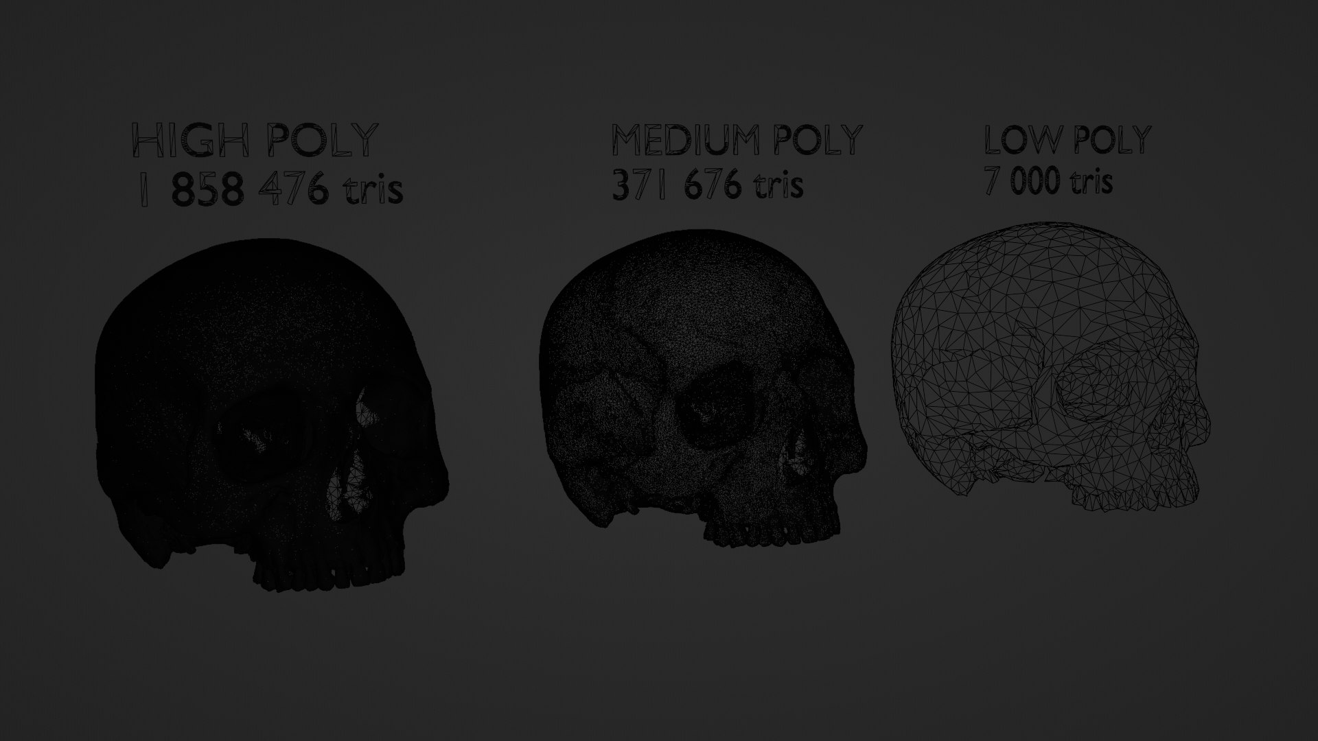
Principal Component Analysis (PCA) applied to GPA datasets allows researchers to map cranial shape variations cleanly, separating ancient lineages from recent modern human variations along distinct mathematical axes. High-resolution photogrammetry and structured-light laser scanners generate sub-millimeter 3D surface models required for these computations without damaging fragile bone.
High-Resolution Micro-Computed Tomography (micro-CT)
Micro-computed tomography (micro-CT) has transformed bioarchaeological studies by permitting non-invasive inspection of internal cranial structures.
Micro-CT applications include:
- Endocranial Casts (Endocasts): Scanning the interior surface of the neurocranium generates digital 3D models of the brain's outer anatomy. Researchers analyze middle meningeal artery branching patterns, frontal lobe asymmetry, and cerebellar volume changes across ancient and modern populations.
- Inner Ear Labyrinth: The bony labyrinth of the inner ear (semicircular canals) is embedded deep within the petrous part of the temporal bone. Semicircular canal geometry is morphologically stable, highly heritable, and species-specific, serving as a reliable taxonomic marker to differentiate archaic hominins, fossil Homo sapiens, and modern populations.
- Paranasal Sinus Morphology: Internal sinus volume, branching complexity, and septal positions within the frontal and sphenoid bones offer key insights into evolutionary thermal regulation and structural skull reinforcement.
Biomolecular Analysis: aDNA and Isotope Biogeochemistry
When physical morphology alone yields ambiguous results due to fragmentation or taphonomic deformation, researchers turn to biomolecular extraction.
- Ancient DNA (aDNA) Sampling: The petrous portion of the temporal bone (pars petrosa), specifically the dense osseous capsule surrounding the inner ear cochlea, yields up to 50 times more endogenous DNA than other skeletal elements. High-throughput next-generation sequencing (NGS) allows researchers to quantify archaic genomic admixture (e.g., Neanderthal or Denisovan introgressed alleles) and establish precise evolutionary divergence dates.
- Stable Isotope Biogeochemistry: Carbon ($^{13}\text{C}/^{12}\text{C}$) and Nitrogen ($^{15}\text{N}/^{14}\text{N}$) isotope ratios extracted from bone collagen and tooth dentin allow researchers to reconstruct dietary profiles. Ancient hunter-gatherers demonstrate distinct nitrogen values reflecting high terrestrial animal or marine protein consumption, differing markedly from modern agricultural and post-industrial diets. Strontium ($^{87}\text{Sr}/^{86}\text{Sr}$) isotopes in enamel record geographic mobility and geological origins during early childhood development.
Ethical, Legal, and Methodological Protocols for Researchers
Working with ancient and historic modern human remains requires strict adherence to legal compliance frameworks, institutional oversight, and non-destructive analysis protocols.
Skeletal Material Protocol Checklist
- Step 1: Establish Legal Jurisdiction (NAGPRA, National Heritage Laws)
- Step 2: Non-Destructive Digital Scanning (Photogrammetry, Micro-CT)
- Step 3: Macroscopic Osteological Recording (AAPA/Standards Guidelines)
- Step 4: Minimal-Invasive Sampling Protocol (Petrous / Enamel Core)
- Step 5: Secure Data Archiving (Open-Access Digital Repository)
Preservation and Non-Destructive Sampling Protocols
Due to the finite nature of ancient skeletal remains, physical intervention must follow strict protocols:
- Digital Preservation First: Before performing any destructive sampling for aDNA or isotopic analysis, the skull must undergo complete 3D surface scanning and high-resolution micro-CT archiving to capture original morphological data permanently.
- Targeted Minimal-Invasive Sampling: When destructive sampling is approved, micro-drilling techniques targeting the petrous temporal bone or dental root dentin must use low-RPM equipment to prevent thermal degradation of endogenous proteins and DNA sequences.
- Environmental Controls: Research facilities must store ancient crania in climate-controlled environments with relative humidity maintained between 35% and 55% and temperature strictly regulated between 18°C and 22°C to prevent micro-fracturing of brittle bone matrix.
Provenance, NAGPRA, and International Repatriation Frameworks
In the United States, researchers analyzing human remains must comply with the Native American Graves Protection and Repatriation Act (NAGPRA) guidelines, updated significantly in regulatory frameworks leading into 2026. NAGPRA mandates consultation with culturally affiliated Indian Tribes and Native Hawaiian Organizations prior to conducting research, destructive testing, or inventory updates on ancestral remains.
Internationally, research on ancient human remains is governed by national heritage protection acts (such as the UNESCO 1970 Convention guidelines) and local indigenous governance boards. Researchers must verify clear legal provenance and export/import documentation prior to transporting skeletal samples across international boundaries. For detailed methodological standards in osteology, researchers frequently consult publications indexed on the National Center for Biotechnology Information (NCBI).
Common Analytical Pitfalls and Diagnostic Errors
Distinguishing ancient from modern skulls is often complicated by environmental degradation and biological variation. Researchers must remain vigilant against several recurring analytical errors:
- Confusing Taphonomic Distortion with Morphology: Soil pressure, ground movement, and thermal cracking can warp fossilized or buried skulls. Asymmetrical crushing of the frontal or parietal bones can mimic platycephaly or abnormal cranial vault height. Researchers must apply virtual reconstruction software (e.g., retrodeformation algorithms) to micro-CT data before drawing morphometric conclusions.
- Pathological Hyperostosis vs. Ancient Cortical Thickness: Modern human crania affected by metabolic diseases such as Paget’s disease, acromegaly, or fibrous dysplasia can develop thick cortical tables and pronounced brow ridges that simulate ancient or archaic hominin features. Pathological bone typically exhibits irregular, porous diploic expansion distinguishable via radiographic cross-sections.
- Over-relying on a Single Morphological Marker: Pronounced supraorbital ridges or dolichocephalic head shapes occasionally occur within normal population variation in gracile modern humans. Anthropologists must avoid relying on individual isolated traits, instead using multi-trait batteries, 3D geometric landmark matrices, and genomic verification.
- Misinterpreting Wear Patterns on Modern Teeth: Modern individuals with severe bruxism (teeth grinding) or industrial abrasive exposure can display dental wear simulating ancient hunter-gatherer attrition. However, bruxism wear lacks the rounded, perimeter-smoothed dentin exposure patterns characteristic of abrasive ancient diets.
Frequently Asked Questions
What is the most reliable skeletal marker to distinguish an ancient skull from a modern one?
The presence of a true inverted T-shaped mental protuberance (chin) on the mandible and a deep canine fossa on the maxilla are two of the most reliable autapomorphies defining modern Homo sapiens. If the mandible is missing, 3D geometric morphometrics of neurocranial globularity and petrous bone labyrinth shape serve as highly reliable diagnostic indicators.
Do ancient human skulls always have larger brain capacities than modern human skulls?
Not necessarily. While early modern Homo sapiens and Neanderthals had mean cranial capacities (1,400 to 1,600 cc) that equaled or exceeded the average modern human cranial capacity (1,300 to 1,350 cc), total volume does not indicate structural organization. Modern skulls feature elevated, globular parietals and expanded frontal regions, whereas ancient skulls held equal or larger brain volumes within longer, lower, and wider neurocrania.
How does diet impact the appearance of a modern skull compared to an ancient one?
Diet dramatically impacts jaw development and dental health. Ancient diets required heavy masticatory force, stimulating lateral bone growth, thick mandibular bodies, flat dental wear, and wider dental arches that accommodated wisdom teeth. Modern soft diets require minimal chewing force, causing underdevelopment of the maxilla and mandible, leading to dental crowding, malocclusion, impaction, and thinner muscle attachment sites.
Can ancient DNA be extracted from any part of a modern or ancient human skull?
While aDNA can theoretically be extracted from various bones, success rates vary drastically. The petrous portion of the temporal bone (pars petrosa), specifically the dense osseous capsule surrounding the inner ear cochlea, provides the highest yield and best preservation of endogenous DNA compared to any other skeletal site. Tooth dentin serves as the secondary preferred target.
What digital tools are standard for comparing ancient and modern skulls today?
Researchers primarily use 3D surface scanning (structured-light laser scanning and high-resolution photogrammetry), micro-CT (micro-computed tomography), and 3D Geometric Morphometrics software (such as R packages geomorph and Morpho, or Stratovan Checkpoint) to capture geometric landmarks, analyze cortical density non-destructively, and virtually repair taphonomic warping.
Actionable Next Steps for Osteological Research
To execute a rigorous comparative osteological analysis between ancient and modern cranial specimens, follow this structured research pathway:
- Establish Legal and Ethical Clearances: Secure institutional review board (IRB) approvals, NAGPRA consultation clearances, and native community permissions before handling skeletal remains.
- Conduct High-Resolution 3D Scanning: Generate digital surface representations using structured-light scanning or photogrammetry. Store raw point-cloud and mesh files in open-access scientific repositories (e.g., MorphoSource) for reproducible open science.
- Perform Non-Destructive Internal Radiography: Utilize micro-CT to assess inner ear bony labyrinth geometry, cranial vault table-to-diploë ratios, and paranasal sinus boundaries.
- Apply Multi-Trait Morphometric Assessments: Collect homologous 3D coordinate landmarks across the neurocranium and splanchnocranium. Run Generalized Procrustes Analysis (GPA) alongside Principal Component Analysis (PCA) to map individual specimen shapes relative to verified archaic and modern reference datasets.
- Evaluate Biomolecular Feasibility: If destructive analysis is legally permitted and scientifically necessary, isolate the petrous temporal bone or intact dental roots for targeted aDNA sequencing and stable isotope analysis.