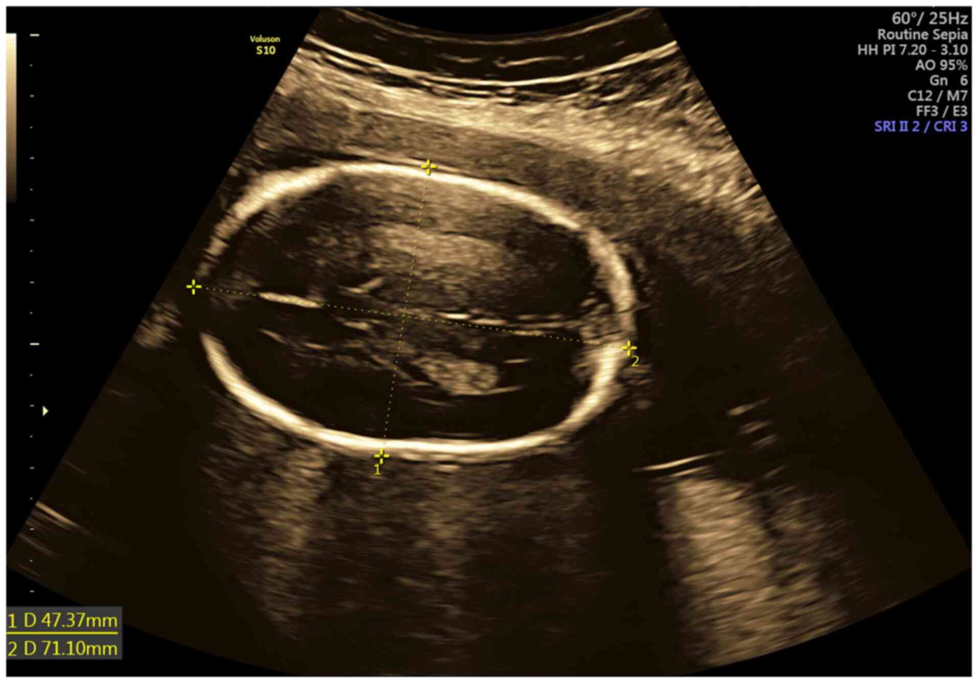
The 20-week ultrasound, often referred to as the anatomy scan or anomaly survey, is a pivotal diagnostic milestone in pregnancy. These detailed pictures allow sonographers and obstetricians to evaluate the structural development of the fetus, check the position of the placenta, and ensure that growth parameters align with the gestational age.
Understanding the Mid-Trimester Anatomy Scan
By the time an expectant parent reaches the 20th week of pregnancy in 2026, the fetus has transformed from a cluster of cells into a complex, recognizable human form. The 20-week ultrasound is not merely an opportunity to see the baby’s face or determine its sex; it is a comprehensive medical examination. This scan is typically performed between 18 and 22 weeks of gestation because the fetal organs are large enough to be visualized clearly, yet the bones have not yet fully ossified (hardened), allowing sound waves to pass through and reveal internal structures.
During this session, a registered diagnostic medical sonographer uses high-frequency sound waves to create real-time images. These images, or "pictures," are more than keepsakes. They serve as a visual record of the baby’s health, providing a baseline for the remainder of the pregnancy. The level of detail achieved in 2026 is significantly higher than in previous decades, thanks to advancements in transducer sensitivity and image processing software, which reduce "noise" and enhance the clarity of soft tissue.
What Every Image Represents: A Checklist of Organs
When you view 20-week ultrasound pictures, you are looking at a systematic review of fetal anatomy. The sonographer follows a strict protocol to ensure no major system is overlooked.
The Fetal Brain and Head
One of the first things a sonographer examines is the skull and brain. They look for the "butterfly" shape of the choroid plexus and ensure the two hemispheres of the brain are symmetrical. Key structures include:
- The Ventricles: These are fluid-filled spaces in the brain. Measuring them helps rule out ventriculomegaly, which can be an indicator of underlying developmental issues.
- The Cerebellum: Located at the back of the brain, this structure is responsible for muscle coordination.
- The Cisterna Magna: The space behind the cerebellum is measured to ensure it falls within a healthy range.
The Heart and Chest
The 20-week scan provides a detailed look at the fetal heart, which is approximately the size of a grape. The sonographer confirms the "four-chamber view," ensuring that the left and right atria and ventricles are proportional and that the heart is tilting toward the left side of the chest. In 2026, many facilities also use color Doppler imaging to visualize the flow of blood through the valves and the great vessels (the aorta and pulmonary artery). This helps in detecting congenital heart defects early.
The Spine and Limbs
The spine is viewed in both longitudinal and transverse planes. The goal is to see two parallel lines of vertebrae that meet at the tailbone, ensuring the skin covers the entire spine to rule out spina bifida. Additionally, the sonographer will capture pictures of the arms and legs, counting the bones (humerus, radius, ulna, femur, tibia, and fibula) and checking for the presence of ten fingers and ten toes.
Biometric Measurements: Tracking Growth and Development
Beyond looking for structural anomalies, the 20-week ultrasound pictures are used to take specific measurements known as fetal biometry. These numbers are compared to standardized growth charts to ensure the baby is developing at the expected rate.
| Measurement Type | Abbreviation | Description | Importance |
|---|---|---|---|
| Biparietal Diameter | BPD | The diameter of the fetal head from ear to ear. | Evaluates head growth and brain development. |
| Head Circumference | HC | The total distance around the baby's head. | Used to estimate gestational age and detect growth restriction. |
| Abdominal Circumference | AC | The distance around the baby's abdomen at the level of the liver. | The most sensitive indicator of fetal weight and nutrition. |
| Femur Length | FL | The length of the thigh bone. | Reflects longitudinal growth and can flag skeletal dysplasias. |
| Estimated Fetal Weight | EFW | A calculation based on BPD, HC, AC, and FL. | Helps clinicians monitor if the baby is too small (IUGR) or too large (Macrosomia). |
These measurements are critical because a significant deviation from the norm may prompt further testing, such as a follow-up growth scan or a consultation with a maternal-fetal medicine (MFM) specialist.
The Role of the Placenta and Amniotic Fluid
The 20-week ultrasound pictures reveal more than just the baby; they provide essential data on the fetal "life-support system."
Placental Location
The sonographer identifies where the placenta is attached to the uterine wall. If the placenta is located low in the uterus, covering or near the cervix, it is labeled as placenta previa. While many low-lying placentas "migrate" upward as the uterus expands, knowing the position at 20 weeks is vital for planning a safe delivery.
Amniotic Fluid Index (AFI)
The fluid surrounding the baby allows for movement, lung development, and temperature regulation. The sonographer looks for "pockets" of fluid. Too little fluid (oligohydramnios) or too much fluid (polyhydramnios) can indicate issues with the baby's kidneys or swallowing reflexes, or potential complications with the mother's health, such as gestational diabetes or dehydration.
The Umbilical Cord
The umbilical cord is checked to ensure it contains three vessels: two arteries and one vein. A "two-vessel cord" (single umbilical artery) is a variation that occurs in about 1% of pregnancies and usually requires additional monitoring of the baby's growth.
Identifying Congenital Abnormalities and Soft Markers
One of the primary goals of the mid-trimester scan is the early detection of structural abnormalities. However, sonographers also look for "soft markers." These are not birth defects themselves, but findings that may slightly increase the statistical risk for chromosomal conditions like Down syndrome (Trisomy 21) or Edwards syndrome (Trisomy 18).
Common soft markers include:
- Echogenic Intracardiac Focus (EIF): A small bright spot in the heart, usually representing a tiny calcium deposit. In the absence of other markers, this is often a normal variation.
- Choroid Plexus Cysts (CPC): Small fluid-filled spaces in the brain that typically disappear by the third trimester.
- Echogenic Bowel: When the baby's bowel appears as bright as bone on the ultrasound.
If soft markers are found, your healthcare provider will review them in the context of your previous screening results, such as NIPT (Non-Invasive Prenatal Testing) or first-trimester blood work. In many cases, these markers are "isolated findings" and do not result in any health issues for the baby.
The Difference Between 2D, 3D, and 4D Imaging in 2026
While the medical diagnostic portion of the scan is almost always performed in 2D (two-dimensional), many parents are eager for 3D or 4D images.
- 2D Ultrasound: This is the "gold standard" for medical diagnosis. It produces a cross-sectional "slice" of the baby, allowing doctors to see internal organs and skeletal structures clearly.
- 3D Ultrasound: This technology captures multiple 2D images at different angles and stitches them together to create a three-dimensional volume. It is excellent for viewing surface features, such as the shape of the nose and lips, which can help detect a cleft lip.
- 4D Ultrasound: This is essentially 3D imaging in motion. It allows you to see the baby yawning, sucking their thumb, or kicking in real-time.
In 2026, most clinical practices prioritize the 2D anatomy scan for its diagnostic accuracy but may switch to 3D for a few moments at the end of the session to give parents a clearer "picture" of their baby's face.
Preparing for Your Appointment: What to Expect
To get the best possible 20-week ultrasound pictures, some preparation is helpful. Most clinics recommend arriving with a moderately full bladder. A full bladder pushes the uterus upward and moves the bowel out of the way, providing a clearer "window" for the sound waves to travel through.
The scan typically lasts between 30 and 60 minutes. The sonographer will apply a warm gel to your abdomen and move a transducer over the skin. You may feel some pressure as they try to get specific angles, especially if the baby is in a difficult position or is very active. It is important to remember that the sonographer's primary job is to complete the medical checklist; they may be quiet during parts of the scan while they concentrate on complex measurements.
Navigating Results and Follow-Up Care
In most cases, the 20-week ultrasound provides reassuring news that the baby is developing normally. However, it is not uncommon for a "follow-up" scan to be requested. This does not always indicate a problem. Common reasons for a repeat scan include:
- Fetal Position: The baby may have been curled in a way that hid the face, spine, or heart.
- Maternal Habitose: Depending on the mother's body composition, sound waves may have difficulty penetrating tissue to get a high-resolution image of certain organs.
- Early Gestation: If the scan was done at exactly 18 weeks, some structures might be more clearly visualized two weeks later.
According to the American Institute of Ultrasound in Medicine (AIUM), ultrasound is considered safe when used for medical purposes, but it should always be performed by trained professionals. If a structural concern is identified, you will likely be referred to a perinatologist for a specialized "Level II" ultrasound.
FAQ
Can the 20-week ultrasound determine the baby’s sex with 100% accuracy?
While the 20-week scan is highly accurate for gender determination (usually over 99%), it is not 100%. The accuracy depends on the baby’s position. If the baby’s legs are crossed or the umbilical cord is between the legs, the sonographer might not be able to get a clear view of the genitalia.
Is it normal if the sonographer doesn't tell me the results immediately?
Yes. In many clinical settings, the sonographer is trained to capture the images but is not legally permitted to provide a diagnosis. The images must be reviewed by a radiologist or your OB-GYN. You will typically receive a full report within a few days.
What if I have a high BMI? Will the pictures be clear?
Higher maternal BMI can sometimes make ultrasound imaging more challenging because adipose tissue can scatter the sound waves. However, modern 2026 ultrasound technology includes "tissue harmonic imaging" and other software enhancements specifically designed to improve clarity in these situations.
Does a normal 20-week scan guarantee a healthy baby?
While a normal anatomy scan is a very positive sign, it cannot rule out every possible condition. Ultrasound is excellent for detecting structural issues (like heart defects or limb abnormalities) but cannot detect most genetic disorders, metabolic conditions, or functional issues like autism or learning disabilities.
Can I bring family members to the scan?
Policies vary by clinic. Many facilities allow one or two guests to join for the "viewing" portion of the scan, but some may ask guests to wait in the lobby during the technical measurement phase to allow the sonographer to focus.
Practical Recommendation
If you are approaching your 20-week milestone in 2026, the best course of action is to stay hydrated in the days leading up to your appointment, as good hydration can improve the clarity of the amniotic fluid and the resulting pictures. Prepare a list of questions for your doctor regarding placental health and growth percentiles. Most importantly, remember that while the "pictures" are a beautiful way to bond with your baby, the medical data they provide is the most valuable asset for a healthy pregnancy outcome. If any findings are unclear, do not hesitate to ask for a detailed explanation of the "Level I" report from your primary healthcare provider.