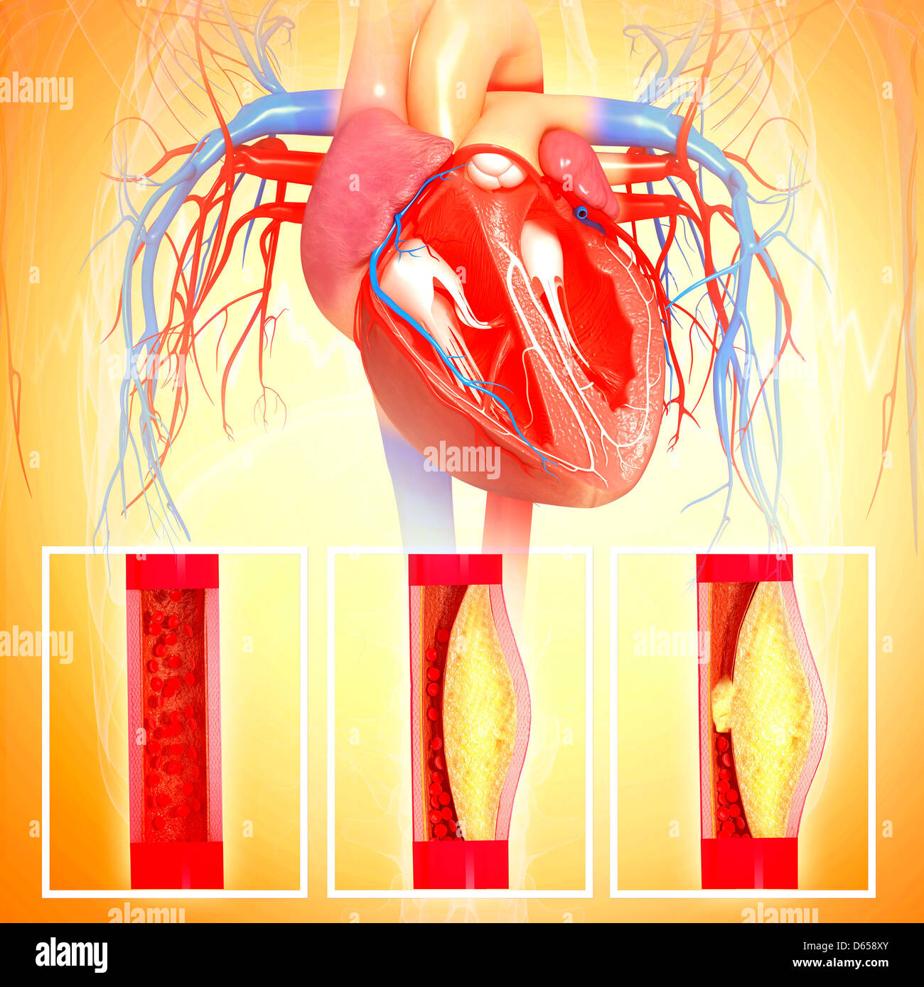
Atherosclerosis of the aorta is a chronic vascular condition where plaque—a buildup of fats, cholesterol, and calcium—hardens and narrows the body's largest artery. This progression can lead to life-threatening complications, including aneurysms, strokes, or peripheral embolization. Management involves aggressive lifestyle modification, pharmacological intervention, and, in advanced cases, surgical repair.
The Pathophysiology of Aortic Atherosclerosis
The aorta is the primary conduit for oxygenated blood leaving the heart, making it subject to immense hemodynamic pressure. Atherosclerosis in this vessel is not merely a "clogging of the pipes" but a complex, chronic inflammatory response within the arterial wall.
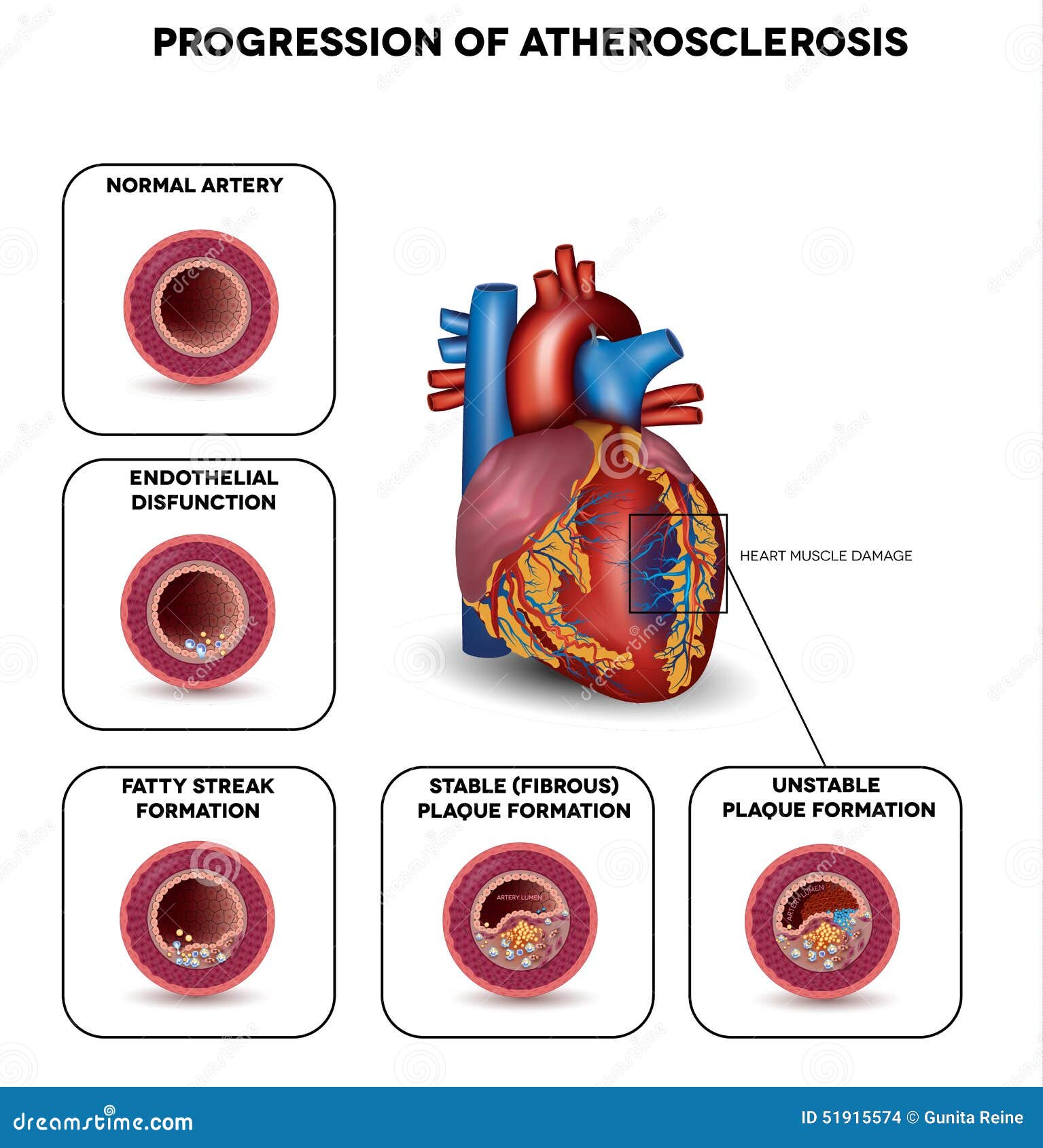
The process typically begins with endothelial dysfunction. The endothelium is the thin inner lining of the blood vessels. When it is damaged by factors such as high blood pressure, toxins from smoking, or high glucose levels, it becomes permeable. This allows Low-Density Lipoprotein (LDL) cholesterol to enter the subendothelial space, where it becomes oxidized.
In response, the immune system dispatches macrophages to ingest these lipids. These cells eventually become overloaded and transform into "foam cells," which aggregate to form fatty streaks. Over decades, these streaks evolve into complex plaques consisting of a lipid core covered by a fibrous cap. In the aorta, these plaques can become quite extensive, often involving the entire length of the vessel from the thoracic cavity down to the abdominal bifurcation.
Classification: Thoracic vs. Abdominal Involvement
Medical professionals categorize aortic atherosclerosis based on its location, as the clinical implications and risks vary significantly between segments.
Thoracic Aortic Atherosclerosis (TAA)
Planes of plaque in the thoracic aorta are most commonly associated with embolic events. Because the thoracic aorta provides the blood supply to the brain through the carotid and vertebral arteries, a piece of plaque that breaks off (an embolus) can travel directly to the brain, causing a transient ischemic attack (TIA) or a full ischemic stroke. Cardiologists often grade thoracic plaque thickness; plaques larger than 4mm or those with mobile components are considered "high risk."
Abdominal Aortic Atherosclerosis (AAA)
The abdominal segment of the aorta is the most frequent site for atherosclerotic development. Unlike the thoracic segment, atherosclerosis here is the primary driver of Abdominal Aortic Aneurysms. The chronic inflammation weakens the structural integrity of the aortic wall (the media and adventitia layers), causing the vessel to bulge outward. If left unchecked, the pressure can cause the vessel to rupture, which is a surgical emergency with a high mortality rate.
Key Risk Factors and Causes
As of 2026, the primary drivers of aortic atherosclerosis remain consistent with long-term epidemiological data, though our understanding of genetic markers has improved.
- Hypertension (High Blood Pressure): Chronic pressure creates mechanical stress on the aortic wall, accelerating endothelial damage.
- Dyslipidemia: Elevated levels of LDL and triglycerides, combined with low HDL, provide the raw materials for plaque formation.
- Tobacco Use: Smoking is perhaps the single most significant modifiable risk factor. The chemicals in tobacco smoke promote oxidation of lipids and induce systemic inflammation.
- Diabetes Mellitus: High blood glucose levels cause glycation of proteins and lipids, which interferes with the vessel's ability to repair itself.
- Age and Genetics: Risk increases significantly after age 50. Family history of premature cardiovascular disease often indicates a genetic predisposition to aggressive plaque buildup.
Clinical Presentation: The Silent Progression
Atherosclerosis of the aorta is notoriously asymptomatic in its early and middle stages. Most patients are unaware of the condition until a secondary event occurs. When symptoms do manifest, they are usually related to the complications of the disease rather than the plaque itself.
- Embolization Signs: Small pieces of plaque or associated blood clots may break off and travel to the extremities. This can cause "Blue Toe Syndrome," where the toes suddenly become painful and discolored due to a lack of blood flow.
- Aneurysmal Pain: If the aorta begins to dilate significantly, a patient might feel a pulsing sensation in the abdomen or persistent back/flank pain.
- Claudication: While usually associated with the iliac or femoral arteries, severe aortic atherosclerosis at the bifurcation (where the aorta splits to the legs) can cause pain in the buttocks or thighs during exercise.
Diagnostic Protocols and Imaging
Because the condition is often "silent," diagnosis frequently occurs during routine screenings or imaging for unrelated issues. However, when aortic atherosclerosis is suspected, several specialized tools are used to assess the severity.
| Imaging Modality | Primary Use | Pros | Cons |
|---|---|---|---|
| Transthoracic Ultrasound | Screening for AAA | Non-invasive, no radiation, cost-effective. | Limited view of the thoracic aorta. |
| CT Angiography (CTA) | Definitive mapping | Excellent detail of plaque volume and calcification. | Requires IV contrast and involves radiation. |
| MRI / MRA | Detailed tissue analysis | Superior for viewing plaque composition without radiation. | Expensive, time-consuming, not for patients with certain implants. |
| Transesophageal Echo (TEE) | Thoracic assessment | Best for viewing the aortic arch and identifying mobile plaques. | Invasive; requires sedation. |
Management and Treatment Strategies in 2026
The goal of treating aortic atherosclerosis is two-fold: stabilizing existing plaque to prevent embolization and slowing the progression of new plaque formation.
Pharmacological Interventions
Medical therapy is the cornerstone of management. In 2026, the standard of care includes:
- High-Intensity Statins: Drugs like Atorvastatin or Rosuvastatin are used not just to lower LDL, but for their "pleiotropic effects"—their ability to reduce inflammation and stabilize the fibrous cap of the plaque.
- PCSK9 Inhibitors: For patients who cannot reach target cholesterol levels with statins alone, these injectable medications offer profound LDL reduction.
- Antiplatelet Therapy: Aspirin or clopidogrel are often prescribed to prevent blood clots from forming on the surface of ulcerated plaques.
- Antihypertensives: ACE inhibitors or ARBs are preferred because they help protect the vascular endothelium.
Lifestyle Optimization
No medical intervention is fully effective without addressing behavioral factors. Patients are encouraged to adopt the Mediterranean Diet, which is rich in anti-inflammatory fats and antioxidants. Smoking cessation is mandatory; even a "low" amount of tobacco use can negate the benefits of medication. Exercise regimens, specifically aerobic activity for at least 150 minutes per week, help improve vascular compliance.
Surgical and Interventional Procedures
Surgery is generally reserved for complications such as significant aneurysms or severe occlusive disease.
- EVAR (Endovascular Aneurysm Repair): A minimally invasive procedure where a stent-graft is inserted through the femoral arteries to reinforce the weakened aortic wall.
- Open Surgical Repair: In cases where the anatomy is not suitable for a stent, the diseased portion of the aorta is replaced with a synthetic graft.
- Endarterectomy: Occasionally, if the plaque is causing severe blockage, surgeons may physically "clean out" the inner lining of the aorta, though this is more common in the carotid arteries than the aorta itself.
Complications: What to Watch For
The most feared complication of aortic atherosclerosis is Aortic Dissection. This occurs when a tear in the inner layer of the aorta allows blood to flow between the layers of the aortic wall, forcing them apart. This is a catastrophic event characterized by a "tearing" sensation in the chest or back and requires immediate emergency surgery.
Another concern is Renal Artery Stenosis. If plaque accumulates near the point where the renal arteries branch off the aorta, it can reduce blood flow to the kidneys, leading to resistant hypertension and eventual kidney failure.
Frequently Asked Questions
Can aortic plaque be reversed?
While it is difficult to completely "dissolve" established, calcified plaque, intensive medical therapy (especially high-dose statins) has been shown to cause plaque regression. More importantly, these treatments can shrink the lipid core and thicken the fibrous cap, making the plaque much less likely to rupture or cause a stroke.
Is aortic atherosclerosis the same as heart disease?
They are related but occur in different vessels. Coronary artery disease affects the small vessels supplying the heart muscle, while aortic atherosclerosis affects the main "highway" of the circulatory system. However, if you have atherosclerosis in your aorta, there is a very high probability that you also have it in your coronary arteries.
What is the life expectancy for someone with this condition?
Life expectancy depends heavily on risk factor control. With modern medications and early detection of aneurysms, many individuals live a normal lifespan. However, untreated aortic atherosclerosis significantly increases the risk of premature death from cardiovascular events.
Does a "calcified aorta" mean I need surgery?
Not necessarily. Calcification is a sign of long-standing atherosclerosis. While it indicates the presence of disease, surgery is typically only required if the aorta has dilated into an aneurysm of a certain size (usually >5.0cm to 5.5cm) or if the plaque is causing a significant blockage of blood flow.
Next Steps for Patients
If you have been diagnosed with atherosclerosis of the aorta or have significant risk factors, the following actions are recommended:
- Baseline Imaging: Ensure you have had an ultrasound or CT scan to measure the diameter of your aorta to rule out an aneurysm.
- Laboratory Analysis: Get a comprehensive lipid panel and an A1c test to check for underlying diabetes.
- Blood Pressure Monitoring: Maintain a home log of blood pressure readings. The goal for most patients with vascular disease is a reading consistently below 130/80 mmHg.
- Specialist Consultation: Consult with a vascular surgeon or a cardiologist who specializes in "preventive cardiology" to tailor a medication and monitoring plan.