To receive a formal diagnosis of Neurofibromatosis Type 1 (NF1), an individual must meet at least two of the seven established clinical and genetic criteria. These include six or more café-au-lait spots, two or more neurofibromas (or one plexiform neurofibroma), skin-fold freckling, optic pathway gliomas, specific eye markers like Lisch nodules, distinctive bone lesions, or a confirmed pathogenic NF1 genetic mutation.
Neurofibromatosis Type 1, historically known as von Recklinghausen disease, is a complex multisystem genetic disorder that primarily affects the skin, nervous system, and skeleton. Because the condition is progressive and symptoms vary significantly between individuals—even within the same family—clinicians rely on a standardized set of diagnostic criteria. These criteria were significantly refined by the International Consensus Group in 2021 to incorporate modern genetic testing and updated clinical observations, which remain the gold standard of practice in 2026.
The Evolution of NF1 Diagnostic Standards
The journey toward a precise NF1 diagnosis has changed over the decades. For many years, the medical community relied on the 1987 National Institutes of Health (NIH) Consensus Development Conference criteria. However, as our understanding of molecular genetics grew, it became clear that clinical observation alone sometimes failed to distinguish NF1 from similar conditions, such as Legius syndrome.
In 2021, an international panel of experts updated these criteria to improve diagnostic accuracy, particularly in children who may not yet exhibit all the classic physical signs. As of 2026, these revised criteria are the universal benchmark used by geneticists, neurologists, and pediatricians worldwide. The most significant change in the modern era is the formal inclusion of genetic testing as a standalone diagnostic criterion, alongside clinical manifestations.
The Seven Core Criteria for NF1 Diagnosis
A diagnosis of NF1 is confirmed if two or more of the following seven features are present. While many people search for the "4 criteria" of NF1, referring to the most visible symptoms, the clinical reality involves these seven specific markers:
1. Café-au-lait Macules (CALMs)
These are flat, pigmented birthmarks that are often the first sign of NF1. For this to count as a diagnostic criterion, the patient must have:
- Six or more macules.
- A diameter of more than 5 mm in prepubertal individuals.
- A diameter of more than 15 mm in postpubertal individuals.
These spots are usually "Coast of California" in appearance, meaning they have smooth, regular borders. While one or two café-au-lait spots are common in the general population, the presence of six or more is highly suggestive of NF1. In 2026, clinicians use polarized light dermatoscopy to better visualize these lesions in fair-skinned infants.
2. Skin-fold Freckling (Crowe Sign)
Unlike common freckles caused by sun exposure, NF1-related freckling occurs in areas where skin rubs against skin. This includes:
- Axillary freckling: Freckles in the armpits.
- Inguinal freckling: Freckles in the groin area.
- Freckling can also occur under the breasts or in the folds of the neck.
This criterion is particularly useful because it typically appears between ages 3 and 5, providing a secondary clinical marker shortly after the appearance of café-au-lait spots.
3. Neurofibromas
Neurofibromas are benign tumors of the peripheral nerve sheath. To meet this criterion, a patient needs:
- Two or more neurofibromas of any type (cutaneous or subcutaneous).
- OR one plexiform neurofibroma.
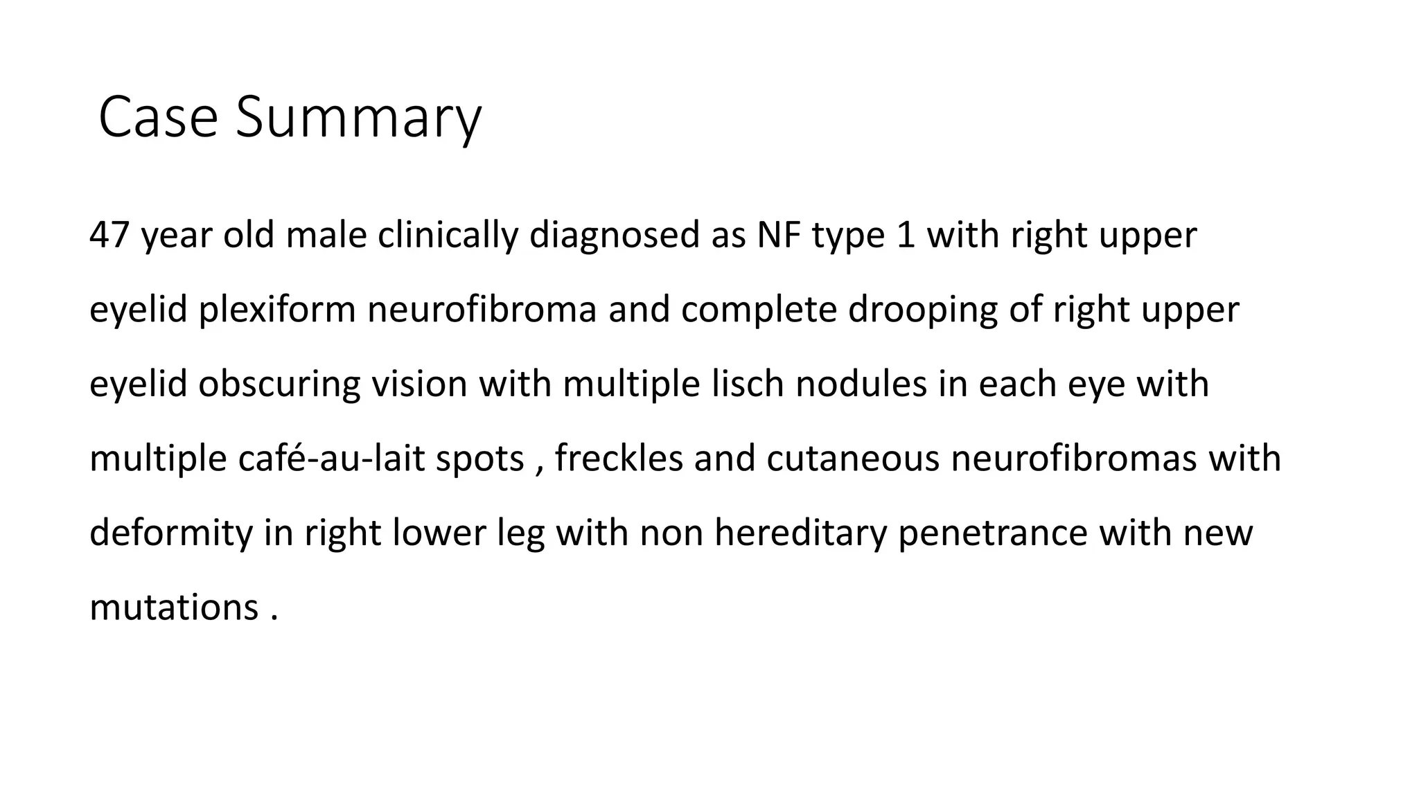
Cutaneous neurofibromas are small, soft bumps on or under the skin that usually develop during adolescence or pregnancy. Plexiform neurofibromas are more complex; they grow along the length of a nerve and can involve multiple nerve branches. These are often congenital (present at birth) and can cause significant cosmetic or functional issues.
4. Optic Pathway Glioma (OPG)
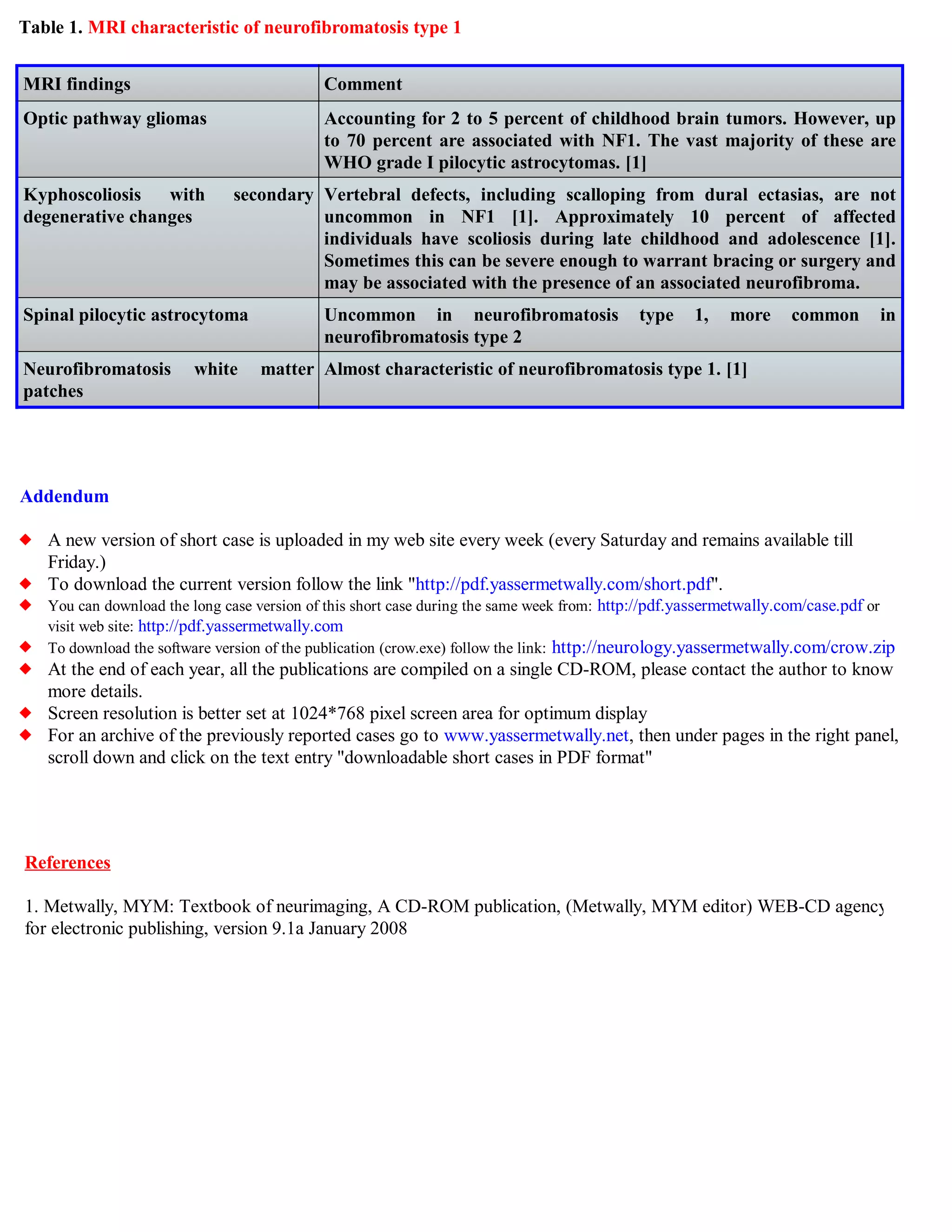
An optic pathway glioma is a low-grade tumor (usually a pilocytic astrocytoma) that grows along the optic nerve, optic chiasm, or optic tracts. While many OPGs are asymptomatic, they can cause vision loss or precocious puberty. In 2026, routine ophthalmologic screening remains mandatory for children with NF1 until age 10 to monitor for these growths.
5. Lisch Nodules or Choroidal Abnormalities
The eyes provide critical diagnostic clues for NF1:
- Lisch Nodules: These are small, dome-shaped yellowish-brown elevations on the iris (the colored part of the eye). They do not affect vision but are present in over 90% of adults with NF1.
- Choroidal Abnormalities: Added in the 2021 revision, these are bright patches on the choroid (the vascular layer of the eye) seen during Optical Coherence Tomography (OCT) imaging.
The inclusion of choroidal abnormalities has been a breakthrough for early diagnosis, as they can often be detected in younger children before Lisch nodules become visible under a slit-lamp exam.
6. Distinctive Osseous (Bone) Lesions
NF1 can affect bone development. The specific lesions recognized for diagnosis include:
- Sphenoid Dysplasia: A malformation of the bone behind the eye socket.
- Anterolateral bowing of the tibia: A characteristic curve in the shin bone, which may lead to a "pseudoarthrosis" (false joint) if the bone fractures and fails to heal properly.
These bone abnormalities are usually detected in infancy or early childhood and are often present at birth.
7. A Heterozygous Pathogenic NF1 Variant
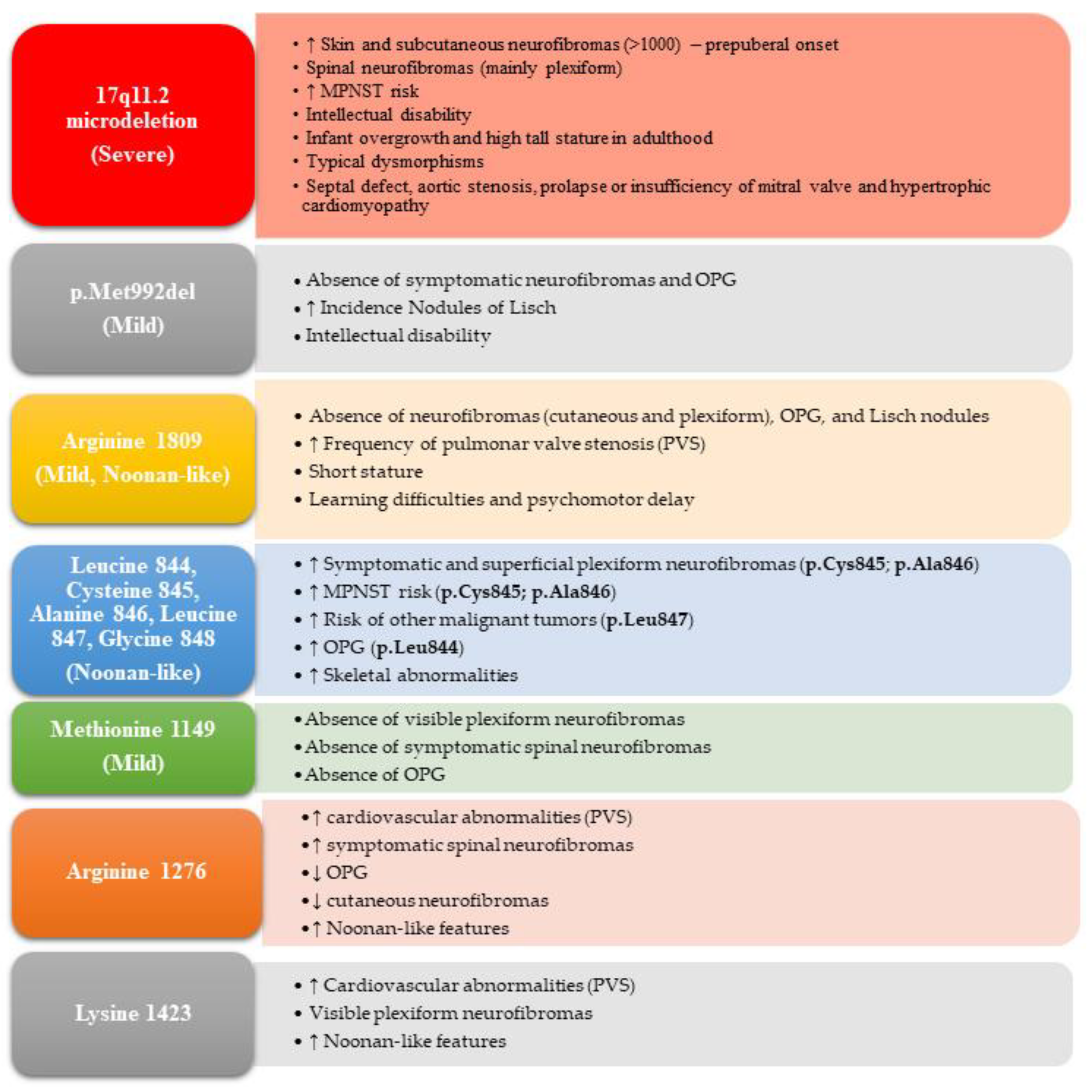
The most definitive modern criterion is the identification of a mutation in the NF1 gene through molecular genetic testing. If a patient has a confirmed pathogenic variant in the NF1 gene with an allele fraction of approximately 50% in healthy tissue, this counts as one of the two required criteria. This has drastically reduced the "wait and see" period for parents of infants who only show café-au-lait spots.
Comparative Summary of NF1 Diagnostic Markers
The following table summarizes the typical age of onset and the methods clinicians use to verify each criterion as of 2026.
| Criterion | Typical Age of Onset | Verification Method |
|---|---|---|
| Café-au-lait Spots | Birth to 2 years | Physical exam / Dermatoscopy |
| Skin-fold Freckling | 3 to 5 years | Physical exam |
| Neurofibromas | Adolescence / Adulthood | Physical exam / MRI for Plexiform |
| Optic Glioma | Birth to 6 years | MRI of the brain and orbits |
| Lisch Nodules | 5 to 10+ years | Slit-lamp ophthalmologic exam |
| Bone Lesions | Infancy (often congenital) | X-ray or CT scan |
| Genetic Mutation | Any age (Congenital) | Blood or saliva DNA sequencing |
Why the "2 out of 7" Rule Matters
The requirement of at least two criteria is a safeguard against misdiagnosis. Several other conditions can mimic aspects of NF1. For instance, Legius Syndrome (caused by a mutation in the SPRED1 gene) often presents with café-au-lait spots and freckling but does not involve neurofibromas or Lisch nodules. Without the "2 out of 7" rule—and specifically the inclusion of genetic testing—a patient with Legius Syndrome might be incorrectly diagnosed with NF1, leading to unnecessary anxiety and medical surveillance for tumors that will never develop.
Furthermore, if a child has a parent who is already diagnosed with NF1 according to these criteria, the child only needs one additional criterion from the list to be formally diagnosed. This reflects the 50% chance of inheritance (autosomal dominant pattern) and allows for earlier intervention and monitoring.
Diagnostic Challenges in 2026: Mosaicism
A complicating factor in 2026 clinical practice is Segmental or Mosaic NF1. This occurs when the NF1 mutation happens after conception, affecting only a specific part of the body (e.g., one limb or one side of the trunk). Individuals with mosaic NF1 may meet the criteria locally—for example, having many café-au-lait spots and neurofibromas on the right arm—but may test negative for the mutation in a standard blood test. In these cases, doctors may need to biopsy a neurofibroma or a skin spot to find the genetic variant and confirm the diagnosis.
Next Steps Following an NF1 Diagnosis
Once a diagnosis is confirmed using the criteria mentioned above, the focus shifts from identification to management. Because NF1 is a life-long condition, the American Academy of Pediatrics and other global health bodies recommend a multidisciplinary approach.
- Annual Exams: Regular check-ups with a neurologist or geneticist specializing in neurofibromatosis.
- Ophthalmology: Yearly eye exams until age 10, then less frequently, to monitor for OPGs and vision changes.
- Blood Pressure Monitoring: NF1 patients have an increased risk of hypertension, sometimes caused by renal artery stenosis or pheochromocytoma.
- Developmental Screening: Many children with NF1 face learning disabilities, ADHD, or executive functioning challenges that require early educational intervention.
- Dermatology: Monitoring neurofibromas for rapid growth, pain, or changes in texture, which could indicate a rare transformation into a Malignant Peripheral Nerve Sheath Tumor (MPNST).
Frequently Asked Questions
Can you have NF1 with only café-au-lait spots?
While café-au-lait spots are often the first sign, they are not enough for a diagnosis on their own. Under the 2026 guidelines, a child with only these spots is considered "at risk" but not yet diagnosed unless a genetic test confirms an NF1 mutation. Many children eventually develop a second criterion, such as freckling or Lisch nodules, as they age.
Is genetic testing mandatory for an NF1 diagnosis?
Genetic testing is not strictly mandatory if a patient already meets two or more clinical criteria (such as spots and freckling). However, it is highly recommended in 2026 to confirm the diagnosis early, rule out Legius syndrome, and assist with family planning. A negative blood test does not 100% rule out NF1 if mosaicism is suspected.
Are neurofibromas cancerous?
The vast majority of neurofibromas are benign (non-cancerous). However, plexiform neurofibromas have a small risk (roughly 8-15% over a lifetime) of transforming into a Malignant Peripheral Nerve Sheath Tumor (MPNST). Regular monitoring by a specialist is essential to catch any suspicious changes early.
At what age are the criteria usually met?
Most children with NF1 meet the diagnostic criteria by age 8. Café-au-lait spots are usually present at birth or appear in the first year. Freckling and OPGs typically appear by age 6. Lisch nodules and cutaneous neurofibromas often emerge during the transition into puberty.
Does a diagnosis of NF1 mean the symptoms will be severe?
No. NF1 is characterized by "variable expressivity." This means that even people with the exact same genetic mutation can have very different experiences. One person may only have skin spots and Lisch nodules, while another may have significant bone issues or internal tumors. Currently, in 2026, we cannot accurately predict the severity of the condition based solely on the initial diagnosis.
Practical Recommendations for Patients and Caregivers
If you or your child meet one of the criteria, such as the presence of multiple café-au-lait spots, the first step is to consult a pediatric geneticist or a specialized NF Clinic. Early identification through these seven criteria allows for proactive management that can prevent complications and improve quality of life.
In 2026, many specialized centers now offer "whole-body MRI" for patients with plexiform neurofibromas to monitor tumor volume, though this is not yet a standard requirement for an initial diagnosis. Stay informed through reputable organizations like the Children's Tumor Foundation (CTF) or the Neurofibromatosis Network to keep up with the latest therapeutic advancements, such as MEK inhibitors, which have revolutionized the treatment of plexiform tumors in recent years.
For more information on the specific clinical manifestations, you may refer to the American College of Medical Genetics and Genomics (ACMG) or the Journal of the American Medical Association (JAMA) for the full text of the revised diagnostic consensus.