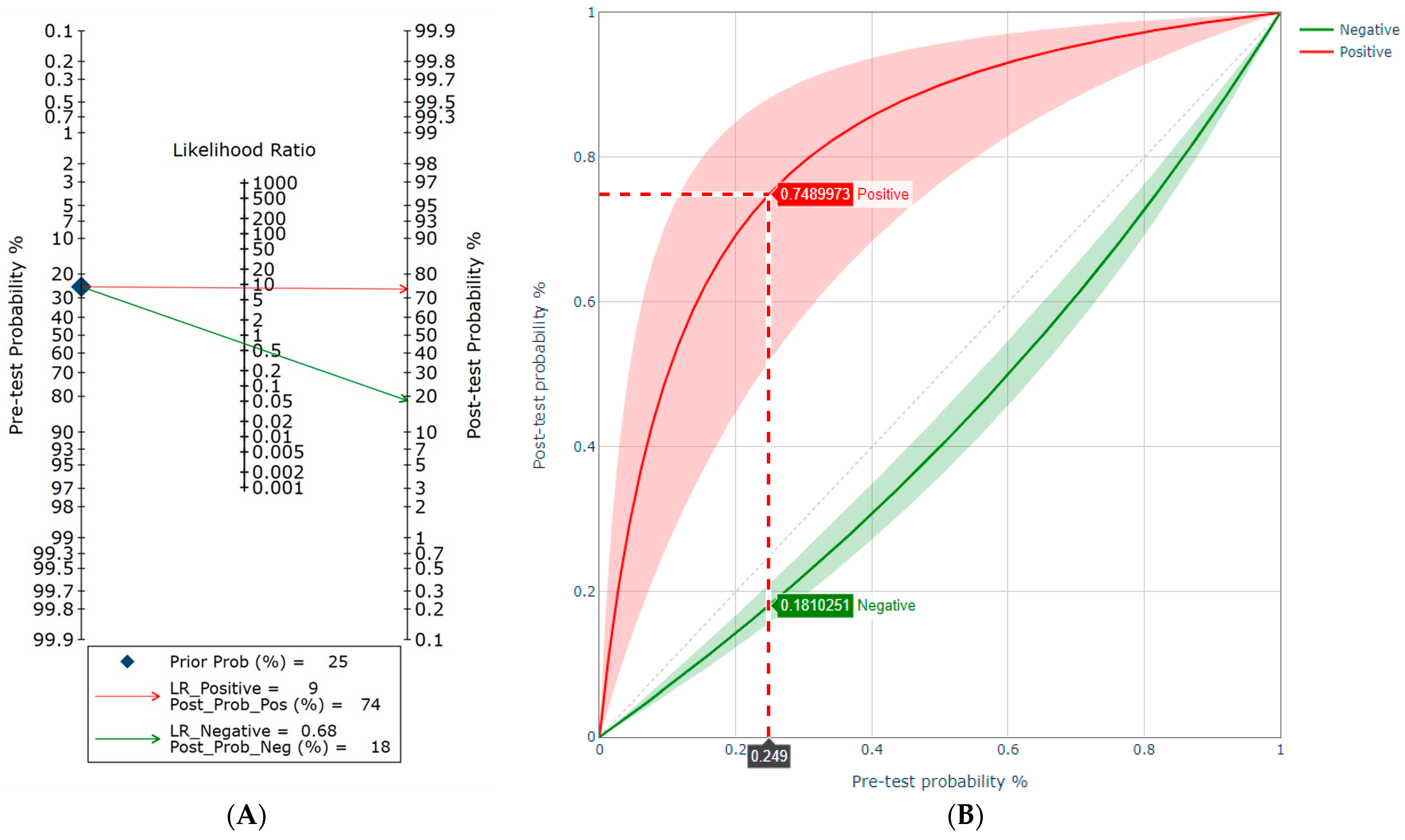
Anti-proteinase 3 (PR3) antibodies, commonly referred to as c-ANCA (cytoplasmic antineutrophil cytoplasmic antibodies), are specific autoantibodies that play a central role in the pathogenesis and clinical diagnosis of certain systemic vasculitides. These antibodies target proteinase 3, a serine protease enzyme located within the azurophilic granules of neutrophils. When these antibodies bind to PR3, they trigger an inflammatory cascade that leads to the destruction of small blood vessels, most notably in conditions categorized as ANCA-associated vasculitis (AAV). Understanding how these markers are measured, what they signify in clinical reports, and how they correlate with disease activity is essential for managing chronic autoimmune conditions.
The Biological Mechanism of Anti-PR3 Antibodies
At the cellular level, the immune system typically utilizes proteinase 3 (PR3) as a defensive tool. Within healthy neutrophils, PR3 remains stored within intracellular granules. However, in individuals with specific immune dysregulation, the body produces autoantibodies—anti-PR3—that recognize this enzyme as a foreign threat.
When these autoantibodies encounter neutrophils in the bloodstream, they bind to PR3, which may be expressed on the surface of the cell under specific inflammatory conditions. This binding event activates the neutrophil, causing it to undergo "respiratory burst" and degranulation. During this process, the neutrophil releases a toxic cocktail of enzymes and reactive oxygen species into the surrounding vascular endothelium. Because this occurs within the walls of small vessels, it results in necrosis, inflammation, and eventual organ damage. This process is the hallmark of Granulomatosis with Polyangiitis (GPA), formerly known as Wegener’s granulomatosis.
Diagnostic Procedures and Testing Methods
In a clinical setting, detecting anti-PR3 antibodies involves standardized serological assays. Because the presence of these antibodies is a significant indicator of autoimmune disease, accuracy is paramount. Laboratories typically employ a multi-step testing strategy to ensure both sensitivity and specificity.
Indirect Immunofluorescence (IIF)
The traditional gold standard for initial screening is indirect immunofluorescence. In this test, patient serum is applied to a substrate of ethanol-fixed human neutrophils. If anti-PR3 antibodies are present, they produce a distinct "cytoplasmic" staining pattern (c-ANCA) under a microscope. While IIF is excellent for detecting the presence of ANCA, it does not confirm the target antigen; therefore, it requires further verification.
Enzyme-Linked Immunosorbent Assay (ELISA)
To confirm the presence of PR3-specific antibodies, clinicians use an antigen-specific ELISA. This highly sensitive method uses purified or recombinant proteinase 3 to capture and quantify the anti-PR3 levels in the blood. In 2026, modern clinical guidelines recommend that for patients with suspected small-vessel vasculitis, a positive IIF result should always be confirmed by an antigen-specific assay like ELISA to minimize the risk of false positives.
Comparative Diagnostic Standards
| Test Method | Primary Use | Specificity | Limitations |
|---|---|---|---|
| Indirect Immunofluorescence | Initial Screening | Moderate | Requires experienced interpretation |
| Antigen-Specific ELISA | Confirmation/Quantification | High | Does not identify all ANCA types |
| Multiplex Immunoassays | Large-scale screening | High | High cost per test |
Clinical Significance in Vasculitis Management
The interpretation of anti-PR3 levels is not as simple as a binary positive or negative result. While the presence of anti-PR3 is highly suggestive of Granulomatosis with Polyangiitis, it is not diagnostic in isolation. Clinicians must synthesize the laboratory data with physical symptoms, imaging studies, and, in many cases, tissue biopsies.
The Role of Titer Monitoring
A frequent question among patients and practitioners is whether fluctuating levels of anti-PR3 indicate a disease flare. Current evidence suggests that while sustained increases in titers may precede clinical relapses in some patients, there is significant inter-patient variability. In clinical practice, serial measurements are used as one component of a broader monitoring strategy rather than the sole indicator for adjusting immunosuppressive therapy.
False Positives and Other Associations
It is crucial to recognize that anti-PR3 is not exclusively found in GPA. While it is the defining marker for that condition, positive results can occasionally occur in other systemic diseases or infections, such as endocarditis or drug-induced vasculitis. Understanding this nuance helps prevent the misattribution of symptoms to an autoimmune cause when an underlying infection might be the primary driver.
Clinical Presentations and Symptom Correlation
Understanding anti-PR3 is most useful when mapped against the clinical symptoms of vasculitis. Patients often present with a constellation of symptoms that reflect systemic involvement of small vessels. These may include:
- Upper Respiratory Tract: Chronic sinusitis, nasal congestion, or crusting, often mistaken for allergies or recurrent infections.
- Lower Respiratory Tract: Persistent cough, hemoptysis (coughing up blood), or abnormal findings on chest X-rays.
- Renal Involvement: Changes in urine output, hematuria (blood in the urine), or rising creatinine levels, indicating glomerulonephritis.
- Systemic Constitutional Symptoms: Unexplained fatigue, weight loss, night sweats, or low-grade fevers.
When these symptoms appear alongside a positive anti-PR3 result, it provides a strong diagnostic lead. However, the absence of anti-PR3 does not strictly rule out vasculitis. A subset of patients, particularly those with localized disease or certain variants, may remain ANCA-negative.
Treatment Considerations and Immunomodulation
Once a diagnosis is confirmed, the management of anti-PR3-associated vasculitis generally follows a two-phased approach: induction of remission and maintenance of remission.
Induction of Remission
The goal in the initial phase is to suppress the overactive immune system rapidly to prevent irreversible organ damage. This typically involves the use of high-dose corticosteroids in combination with potent immunosuppressants such as cyclophosphamide or rituximab. In 2026, the use of B-cell depletion therapy like rituximab has become a standard of care for many, often showing superior efficacy in preventing relapses compared to older regimens.
Maintenance Therapy
Once remission is achieved, the objective shifts to preventing the recurrence of disease. This phase involves lower, more manageable doses of immunosuppressive medications. Because anti-PR3 production is driven by memory B-cells, maintenance therapy is essential for keeping the underlying immune dysfunction in check.
Potential Challenges and Troubleshooting
Patients undergoing monitoring for anti-PR3 often encounter concerns regarding laboratory variability. Different laboratories may use different "cut-off" values for defining a positive result, and assays from different manufacturers may yield slightly different numerical titers.
- Laboratory Consistency: It is highly recommended to have serial tests performed at the same laboratory facility to ensure the results are comparable over time.
- Clinical Correlation: Always prioritize the clinical picture. A rise in anti-PR3 levels in an asymptomatic patient may warrant closer surveillance but does not always necessitate an immediate change in medication.
- Medication Interference: Certain medications or concurrent illnesses can skew laboratory findings. Always disclose all current prescriptions and supplements to the rheumatologist or immunologist overseeing the care plan.
Frequently Asked Questions
Does a high anti-PR3 level always mean the disease is active? No, not necessarily. While a significant rise in titers can be a warning sign of a potential flare, many factors influence these levels. Clinical symptoms, physical examination, and other laboratory markers—such as inflammatory markers (ESR/CRP) and kidney function tests—are just as critical as the antibody titer itself.
Can anti-PR3 disappear after treatment? Yes, successful treatment can lead to a significant decline or the disappearance of detectable anti-PR3 antibodies. However, even if the antibody level drops to undetectable, the risk of recurrence remains, requiring ongoing clinical vigilance and regular follow-up.
Is anti-PR3 the same as p-ANCA? No. Anti-PR3 is typically associated with a c-ANCA (cytoplasmic) pattern on immunofluorescence. p-ANCA (perinuclear) is more commonly associated with antibodies against myeloperoxidase (MPO). These represent different clinical syndromes and require different diagnostic approaches.
What should I do if my test results are "borderline"? Borderline results can be frustrating. Often, clinicians will repeat the test in a few weeks to look for a trend. They may also order different tests to check for alternative antibodies or recommend consultation with a sub-specialist in vasculitis to evaluate clinical symptoms further.
Are there dietary changes that lower anti-PR3? There is no evidence that diet alone can influence the production of specific autoantibodies like anti-PR3. While a healthy, anti-inflammatory diet is generally beneficial for overall wellness and the management of chronic conditions, it is not a substitute for standard medical therapy in autoimmune vasculitis.
Next Steps for Patients
If you have received an anti-PR3 positive result, the most important step is to establish or maintain a close relationship with a rheumatologist. The management of ANCA-associated vasculitis is complex and requires specialized expertise. Ensure that your medical team has a clear timeline of your symptoms and that you are having your blood work monitored at consistent intervals. Keep a symptom journal, noting any changes in energy, breathing, or physical comfort, as this subjective data is invaluable to your physician.
Medical information regarding autoimmune markers and vasculitis is subject to ongoing research. Always consult with your primary healthcare provider or a specialist before making decisions regarding medication changes or treatment plans. This article is intended for educational purposes and does not constitute medical advice or a substitute for professional clinical diagnosis.