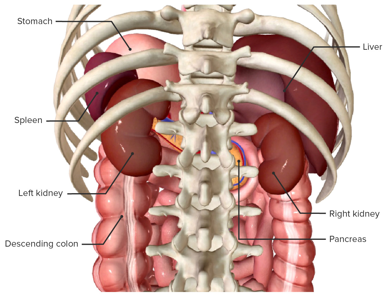
While the back is primarily defined by the vertebral column, spinal cord, and dense muscular layers, several major internal organs lie directly adjacent to its posterior wall. These structures, primarily situated within the retroperitoneal space and the posterior thoracic cavity, include the kidneys, adrenal glands, pancreas, major blood vessels, and posterior lung segments.
Disclaimer: This article is intended strictly for educational and informational purposes and does not constitute medical advice. If you are experiencing severe, sudden, or unexplained back pain, consult a qualified healthcare professional immediately.
Clarifying "Back Organs": Anatomical Cavities and Spaces
When discussing the organs of the back, anatomical precision requires distinguishing between the thoracic cavity, the peritoneal cavity, and the retroperitoneal space. Strictly speaking, no major visceral organ resides inside the skeletal tissue or musculature of the back. Instead, "back organs" refers to internal abdominal and thoracic organs located immediately adjacent to the posterior body wall.
- POSTERIOR BODY WALL
- Thoracic Cavity (Posterior)
- Lower Lungs
- Esophagus
- Thoracic Aorta
- Retroperitoneal Space
- Kidneys
- Adrenal Glands
- Ureters
- Pancreas
- Duodenum
- Asc/Desc Colon
- Abdominal Aorta
- Thoracic Cavity (Posterior)
The Retroperitoneal Space
The retroperitoneal space (or retroperitoneum) is the anatomical area located behind the posterior layer of the peritoneum—the continuous membrane that lines the abdominal cavity. Organs situated within this space are cushioned between the parietal peritoneum anteriorly and the posterior abdominal muscles (such as the psoas major, quadratus lumborum, and transversus abdominis) posteriorly.
Anatomists classify retroperitoneal organs into two distinct sub-categories:
- Primary Retroperitoneal Organs: Structures that developed outside the peritoneal cavity from initial embryological formation and remained retroperitoneal throughout development. Key examples include the kidneys, adrenal glands, ureters, abdominal aorta, and inferior vena cava.
- Secondary Retroperitoneal Organs: Structures that originally developed within the peritoneal membrane suspended by mesentery, but subsequently migrated posteriorly and fused to the posterior abdominal wall during embryonic development. Key examples include the pancreas (head, body, and neck), parts 2 through 4 of the duodenum, and the ascending and descending colon.
The Posterior Thorax
Superior to the diaphragm, within the chest cavity, the posterior thoracic space houses the lungs (specifically the basal and posterior segments of the inferior lobes), the thoracic esophagus, the vagus nerves, and the descending thoracic aorta. These structures lie close to the thoracic vertebrae and posterior rib cage, meaning disorders affecting them often project symptoms into the upper and mid-back.
Posterior Thoracic Organs (Upper and Mid-Back)
The upper and middle regions of the back correspond posteriorly to the thoracic spine (vertebrae T1 through T12) and the posterior rib cage. The internal organs adjacent to this region are essential for respiratory, cardiovascular, and digestive functions.
| Organ | Anatomical Location | Spinal Level |
|---|---|---|
| Inferior Lobes (Lungs) | Posterior pleural cavity | T4 - T12 |
| Thoracic Aorta | Left anterior vertebral body | T4 - T12 |
| Esophagus | Posterior mediastinum | T1 - T10 |
1. Inferior Lobes of the Lungs
The lungs occupy the bilateral pleural cavities of the thorax. Anatomically, the posterior aspect of the chest wall is predominantly covered by the inferior lobes of both the right and left lungs.
- Anatomical Boundaries: The posterior upper margin of the inferior lobes begins near the level of the T3 or T4 spinous process and extends downward to the level of T10 during normal quiet breathing, reaching T12 during deep inspiration.
- Tissue Structure: The posterior segments are wrapped by the visceral pleura, which glides against the parietal peritoneum lining the inner surface of the ribs and intercostal muscles.
- Clinical Correlation: Inflammatory conditions of the lung parenchyma or pleural lining—such as lower-lobe pneumonia, pleurisy, or pulmonary embolism—frequently manifest as acute, sharp mid-to-upper back pain that expands or worsens upon deep inhalation (pleuritic chest/back pain).
2. Thoracic Esophagus
The esophagus is a muscular tube approximately 25 centimeters in length that transports food and liquids from the pharynx to the stomach. It traverses the posterior mediastinum, running directly anterior to the thoracic vertebral column.
- Spatial Position: It enters the thorax at the T1 level, descends behind the trachea and heart, and pierces the diaphragm at the esophageal hiatus around the T10 level.
- Nerve Supply: Surrounded by the esophageal plexus, formed by branches of the vagus nerve (Cranial Nerve X) and sympathetic fibers.
- Clinical Correlation: Conditions such as severe gastroesophageal reflux disease (GERD), esophageal spasms, or esophageal perforation often cause deep retrosternal pain that radiates straight through to the interscapular region of the back.
3. Descending Thoracic Aorta
The thoracic aorta is the primary arterial conduit carrying oxygenated blood from the left ventricle of the heart to the systemic circulation.
- Path: Originating at the aortic arch (level T4), the descending thoracic aorta runs along the left side of the vertebral bodies, gradually shifting toward the midline as it descends toward the aortic hiatus of the diaphragm at level T12.
- Structural Importance: It gives off paired intercostal arteries that supply the rib cage, intercostal muscles, and spinal cord segmentally.
- Clinical Significance: A thoracic aortic aneurysm or acute thoracic aortic dissection represents a life-threatening medical emergency. Dissection typically presents as a sudden, tearing, or ripping pain between the shoulder blades.
Retroperitoneal Organs (Mid to Lower Back)
The lower back region, corresponding to lumbar vertebrae L1 through L5, is anatomically backed by the retroperitoneal space. The organs in this space perform metabolic, excretory, endocrine, and digestive operations.
- RETROPERITONEAL CAVITY
- Excretory & Endocrine
- Kidneys
- Adrenal Glands
- Ureters
- Gastrointestinal Systems
- Pancreas
- Duodenum (2-4)
- Asc/Desc Colon
- Excretory & Endocrine
1. The Kidneys (Renes)
The kidneys are paired, bean-shaped excretory organs located on either side of the vertebral column in the deep retroperitoneal space.
- Anatomical Coordinates: The right kidney sits slightly lower (level L1 to L3) than the left kidney (level T12 to L2) due to the large mass of the liver occupying the upper right quadrant of the abdomen. The upper poles of both kidneys are partially protected by the 11th and 12th ribs posteriorly.
- Fascial Encasement: Each kidney is encapsulated by three distinct layers:
- Fibrous Renal Capsule: The innermost layer directly touching the parenchyma.
- Perirenal Fat Pad (Adipose Capsule): A dense layer of protective fat surrounding the fibrous capsule.
- Renal Fascia (Gerota's Fascia): A dense connective tissue layer anchoring the kidney and adrenal gland to the abdominal wall and diaphragm.
- Functional Anatomy: The kidneys filter metabolic waste from the blood, regulate systemic arterial pressure via the renin-angiotensin-aldosterone system (RAAS), maintain fluid and electrolyte homeostasis, and produce erythropoietin.
- Clinical Relevance: Kidney pathology often presents as pain in the flank—the region between the lower rib margin and the iliac crest. Conditions such as pyelonephritis (kidney infection) or nephrolithiasis (kidney stones) generate pain in the costovertebral angle (CVA), often radiating downward toward the groin.
2. Adrenal Glands (Glandulae Suprarenales)
The adrenal glands are small, yellow, pyramidal endocrine glands situated immediately superior and anterior to the upper pole of each kidney.
- Anatomical Boundary: Positioned at the level of the 11th and 12th thoracic vertebrae, enclosed within Gerota’s fascia, separated from the kidney by a thin septum of perirenal fat.
- Structural Division:
- Adrenal Cortex: The outer structural zone, which synthesizes steroid hormones, including aldosterone (mineralocorticoid), cortisol (glucocorticoid), and androgens.
- Adrenal Medulla: The inner neuroendocrine core, which synthesizes and secretes catecholamines (epinephrine and norepinephrine) under direct control of the sympathetic nervous system.
- Clinical Relevance: While small, large adrenal tumors (such as adrenal cortical carcinomas or pheochromocytomas) or adrenal hemorrhages can produce local retroperitoneal pressure, causing vague back or flank discomfort alongside systemic hormone dysregulation.
3. Ureters
The ureters are muscular tubes measuring approximately 25 to 30 centimeters in length that transport urine from the renal pelvis of each kidney down to the urinary bladder.
- Pathways: They descend vertically along the anterior surface of the psoas major muscle within the retroperitoneum, crossing the pelvic brim near the bifurcation of the common iliac arteries.
- Constriction Sites: The ureters have three natural anatomical narrowings where kidney stones frequently become lodged:
- The ureteropelvic junction (UPJ).
- The crossing of the iliac vessels at the pelvic brim.
- The ureterovesical junction (UVJ) entering the bladder wall.
- Clinical Relevance: Obstruction by a ureteral calculus triggers smooth muscle spasms (ureteral colic), resulting in severe, sharp back and flank pain that fluctuates in intensity and radiates along the dermatomal pathway of the T11–L2 nerves to the lower abdomen and labia/testicles.
- Renal Pelvis
- Ureteropelvic Junction - UPJ
- Upper Ureter
- Iliac Vessel Crossing
- Lower Ureter
- Ureterovesical Junction - UVJ
- Urinary Bladder
4. Pancreas
The pancreas is an elongated, soft retroperitoneal gland lying transversely across the posterior abdominal wall at approximately the L1 to L2 vertebral level.
- Anatomical Structure: Divided into a head, neck, body, and tail. The head of the pancreas rests within the C-shaped curvature of the duodenum on the right side. The body crosses the midline anterior to the aorta and L1 vertebra, while the tail extends horizontally to touch the hilum of the spleen on the left.
- Dual Function:
- Exocrine: Synthesizes digestive enzymes (lipase, amylase, trypsinogen) secreted into the duodenum via the pancreatic duct.
- Endocrine: Islets of Langerhans synthesize insulin, glucagon, and somatostatin directly into the bloodstream.
- Clinical Relevance: Because the pancreas lies directly against the retroperitoneal nerves and vertebral column, acute or chronic pancreatitis causes severe epigastric abdominal pain that classically radiates directly through to the upper lumbar back.
5. Duodenum, Ascending Colon, and Descending Colon
Parts of the gastrointestinal tract are anchored to the posterior abdominal wall, placing them in close structural proximity to the muscles of the lower back.
- Duodenum: The 2nd (descending), 3rd (horizontal), and 4th (ascending) parts of the duodenum are retroperitoneal. They hug the inferior vena cava and aorta at levels L1 to L3. Duodenal ulcers penetrating the posterior wall can cause referred pain to the back.
- Ascending and Descending Colon: The right (ascending) and left (descending) sides of the large intestine are retroperitoneal structures bound to the posterior abdominal wall by the fusion fascia of Toldt. Massive colonic distension, diverticulitis, or retrocecal appendicitis can cause deep lumbar or flank pain.
Vascular and Neuromuscular Structures of the Posterior Wall
In addition to visceral organs, major circulatory vessels and neural networks course along the anterior surface of the spine within the retroperitoneal space.
| Structure | Anatomical Alignment | Key Branches / Connections |
|---|---|---|
| Abdominal Aorta | Left of midline (L1-L4) | Celiac, SMA, IMA, Renal Arts |
| Inferior Vena Cava | Right of midline (L5-T8) | Renal, Hepatic, Iliac Veins |
| Lumbar Plexus | Psoas major substance | Femoral, Obturator Nerves |
| Sympathetic Trunk | Bilateral anterolateral vertebral bodies | Splanchnic Nerves (T5-L2) |
Abdominal Aorta
The abdominal aorta begins at the aortic hiatus of the diaphragm (T12) and descends anterior to the lumbar vertebrae until it bifurcates into the common iliac arteries at the L4 level.
- Key Branches: It gives rise to visceral branches (celiac trunk, superior mesenteric artery, inferior mesenteric artery, renal arteries, suprarenal arteries) and parietal branches (lumbar arteries supplying the posterior wall).
- Clinical Relevance: An Abdominal Aortic Aneurysm (AAA) is a localized ballooning of the abdominal aortic wall. As it expands, it exerts pressure on the lumbar spine and surrounding nerves, causing a constant, deep, throbbing back pain. Rupture of an AAA leads to retroperitoneal hemorrhage and is a severe medical emergency.
Inferior Vena Cava (IVC)
The IVC is a large vein that runs parallel to the abdominal aorta on its right side, extending from its formation at the L5 vertebral level (by the junction of the common iliac veins) up through the vena caval foramen of the diaphragm at T8. It returns deoxygenated blood from the lower extremities, kidneys, and abdominal organs to the right atrium of the heart.
The Lumbar Plexus and Sympathetic Chain
- Lumbar Plexus: Formed by the anterior rami of L1 through L4 spinal nerves, embedded within the substance of the psoas major muscle. It innervates the lower abdominal wall and anterior thigh.
- Sympathetic Trunk: Paired nerve fibers running down the bilateral anterolateral aspects of the vertebral bodies, carrying autonomic signals that modulate vascular tone and visceral organ function throughout the thorax and abdomen.
Anatomical Comparison of Primary Back Organs
The following table summarizes the structural characteristics, anatomical locations, primary physiological roles, and common clinical pain presentations of the organs associated with the back.
| Organ / Structure | Primary Anatomical Space | Vertebral Level Range | Primary Function | Clinical Referred Pain Area |
|---|---|---|---|---|
| Inferior Lungs | Posterior Thoracic / Pleural Cavity | T4 – T12 | Gas exchange (respiration) | Mid-to-upper back, pleuritic thoracic pain |
| Thoracic Aorta | Posterior Mediastinum | T4 – T12 | High-pressure oxygenated blood transport | Sharp, tearing interscapular back pain |
| Esophagus | Posterior Mediastinum | T1 – T10 | Transport of food/fluids to stomach | Mid-thoracic retrosternal and interscapular back pain |
| Kidneys | Primary Retroperitoneal | T12 – L3 | Filtration, blood pressure regulation, fluid balance | Flank, costovertebral angle (CVA), radiating to groin |
| Adrenal Glands | Primary Retroperitoneal | T11 – L1 | Steroid hormone and catecholamine secretion | Deep upper flank discomfort (rarely localized) |
| Ureters | Primary Retroperitoneal | L2 – S2 | Urine transport to bladder | Flank to groin, lower back, inner thigh |
| Pancreas | Secondary Retroperitoneal | L1 – L2 | Digestive enzyme secretion & glucose homeostasis | Epigastrium radiating straight through to upper lumbar back |
| Abdominal Aorta | Primary Retroperitoneal | T12 – L4 | Systemic blood delivery to abdomen and legs | Deep, pulsating lower back or lumbar pain |
Clinical Relevance: Neurological Pathways of Referred Back Pain
A critical challenge in clinical medicine is distinguishing between somatic back pain (originating from spine joints, intervertebral discs, ligaments, and paraspinal muscles) and visceral referred pain (originating from internal back organs).
- Visceral Organ Pain Stimulus
- Visceral Afferent Nerve Fibers
- Dorsal Horn of Spinal Cord (T1-L2) (Convergence with Somatic Nerves)
- Brain Perceives Pain as Somatic (Skin / Muscle / Back Wall)
Mechanism of Visceral Referral
Visceral organs lack the precise tactile sensory receptors found in the skin and skeletal muscle. Instead, sensory signals from internal organs travel along autonomic (sympathetic) afferent nerve fibers back to the spinal cord.
These visceral afferent fibers enter the dorsal horn of the spinal cord at the same segmental levels (dermatomes) as somatic sensory fibers coming from the skin and muscles of the back:
- T5 – T9 Spinal Segments: Receive sensory inputs from the stomach, liver, gallbladder, and pancreas. Pathology in these organs can be perceived as mid-back or lower scapular pain.
- T10 – T11 Spinal Segments: Receive inputs from the kidneys, ureters, and small intestine. Pathology here refers pain to the lower thoracic and upper lumbar flank regions.
- T12 – L2 Spinal Segments: Receive inputs from the lower colon, rectum, bladder, and vascular structures like the abdominal aorta. Pathology manifests in the lower lumbar and sacral back regions.
Because the central nervous system cannot reliably distinguish between visceral and somatic afferent inputs arriving at the same spinal segment, the brain interprets signals from an inflamed retroperitoneal organ as originating from the somatic back wall.
Diagnostic Imaging and Evaluation Procedures
When healthcare providers suspect that back symptoms originate from internal organ pathology rather than a musculoskeletal sprain or disc herniation, specific diagnostic modalities are used to evaluate the retroperitoneal and thoracic spaces.
- Clinical Presentation: Flank / Back Pain
- Lab Evaluations
- Complete Blood Count
- Urinalysis & Microscopy
- Serum Creatinine / BUN
- Amylase / Lipase Enzymes
- Diagnostic Imaging
- Diagnostic Ultrasound
- Computed Tomography (CT)
- Magnetic Resonance Imaging (MRI)
- Chest & Abdominal X-Rays
- Lab Evaluations
1. Ultrasonography (US)
- Application: Ultrasound is the primary non-invasive modality used to evaluate retroperitoneal solid organs.
- Utility: Excellent for detecting renal hydronephrosis (kidney swelling due to obstruction), gallstones, renal cysts, abdominal aortic aneurysm diameter, and free fluid accumulation. It avoids ionizing radiation.
2. Computed Tomography (CT)
- Application: The gold-standard diagnostic tool for evaluating acute retroperitoneal pathology.
- Utility: High-resolution non-contrast CT easily identifies calcium-containing kidney and ureteral stones. Contrast-enhanced CT scans assess pancreatic necrosis in pancreatitis, detect aortic dissections or aneurysms, and evaluate traumatic injury to retroperitoneal structures.
3. Magnetic Resonance Imaging (MRI)
- Application: Used for detailed soft-tissue characterization when CT is contraindicated (e.g., severe iodinated contrast allergy or renal insufficiency).
- Utility: Magnetic Resonance Cholangiopancreatography (MRCP) provides non-invasive visualization of the pancreatic and biliary ductal systems. MRI is also helpful for evaluating retroperitoneal soft-tissue masses or sarcoma extensions.
4. Laboratory Diagnostics
Clinical evaluation of back organs relies heavily on targeted laboratory testing:
- Urinalysis: Assesses hematuria (blood in urine), leukocyte esterase, and nitrites to diagnose urolithiasis or urinary tract infections.
- Serum Chemistry: Serum creatinine and Blood Urea Nitrogen (BUN) measure renal filtration capacity.
- Pancreatic Enzymes: Serum lipase and amylase elevations confirm acute pancreatitis.
- Inflammatory Markers: Elevated C-reactive protein (CRP) and white blood cell counts point toward infectious processes like pyelonephritis or retroperitoneal abscesses.
Frequently Asked Questions
Which specific organs are located directly in the lower back?
Strictly speaking, organs are situated in the retroperitoneal space anterior to the lower back muscles. The principal organs in this region are the left and right kidneys, the adrenal glands, the ureters, the pancreas (body and tail), parts of the duodenum, the ascending and descending colon, the abdominal aorta, and the inferior vena cava.
How can I tell if my back pain is muscular or from an organ?
Muscular back pain typically changes with physical movement—bending, twisting, sitting, or heavy lifting—and often features localized tenderness when the muscle is pressed. Pain originating from an organ (visceral pain) is often described as a constant, deep ache, throbbing, or severe spasmodic cramping that does not resolve with position changes. Organ-related back pain is frequently accompanied by systemic symptoms such as fever, nausea, vomiting, blood in the urine, changes in urination, or lightheadedness.
Why do kidney infections cause severe back pain?
The kidneys are encased in a tough fibrous capsule (capsula fibrosa) and surrounded by Gerota's fascia. When infected (pyelonephritis), the kidney tissue becomes inflamed and swells. This swelling stretches the sensory nerve fibers in the renal capsule, sending strong pain signals to the T10–L1 spinal cord segments, which the brain perceives as intense pain in the costovertebral angle of the lower back.
Can heart or major blood vessel issues cause mid-back pain?
Yes. The descending thoracic aorta and abdominal aorta run along the anterior column of the spine. A thoracic aortic dissection typically causes sudden, severe, tearing pain in the mid-back between the shoulder blades. An abdominal aortic aneurysm (AAA) causes continuous, deep, throbbing lower back pain. Both conditions are life-threatening medical emergencies.
Does the liver sit in the back wall of the abdomen?
The liver is an intraperitoneal organ situated in the right upper quadrant of the abdomen beneath the diaphragm. While most of it is anterior, its posterior border rests against the right kidney, right adrenal gland, and inferior vena cava. Diseases of the liver or gallbladder (such as cholecystitis) frequently refer pain to the right lower scapula or upper right back via the phrenic nerve and thoracic spinal nerves.
Can stomach or intestinal disorders radiate pain to the back?
Yes. Secondary retroperitoneal portions of the digestive system—such as the posterior wall of the duodenum and the colon—lie adjacent to the posterior abdominal wall. A penetrating posterior duodenal ulcer can erode toward the retroperitoneum, causing intense back pain. Similarly, severe intestinal distension, diverticulitis, or retrocecal appendicitis can radiate pain into the lower lumbar or flank regions.
Recommended Next Steps
Understanding the anatomical layout of back organs helps distinguish between benign mechanical back strain and serious visceral conditions. If you or someone under your care is experiencing back symptoms, consider the following practical guidance:
- Conduct a Symptom Assessment: Take note of whether the pain fluctuates with body movement or posture. Physical movements that aggravate or relieve pain strongly suggest a musculoskeletal origin.
- Identify Warning Signs: Seek emergency medical evaluation immediately if back pain is accompanied by high fever, severe nausea or vomiting, sudden abdominal pulsatile sensations, visible blood in the urine, inability to urinate, unexplained lightheadedness, or sudden loss of bowel/bladder control.
- Seek Appropriate Diagnostic Testing: If back pain persists despite rest and physical therapy, consult a primary care physician. They can order non-invasive imaging, such as a renal ultrasound or abdominal CT scan, along with routine blood and urine tests to evaluate the retroperitoneal organs.