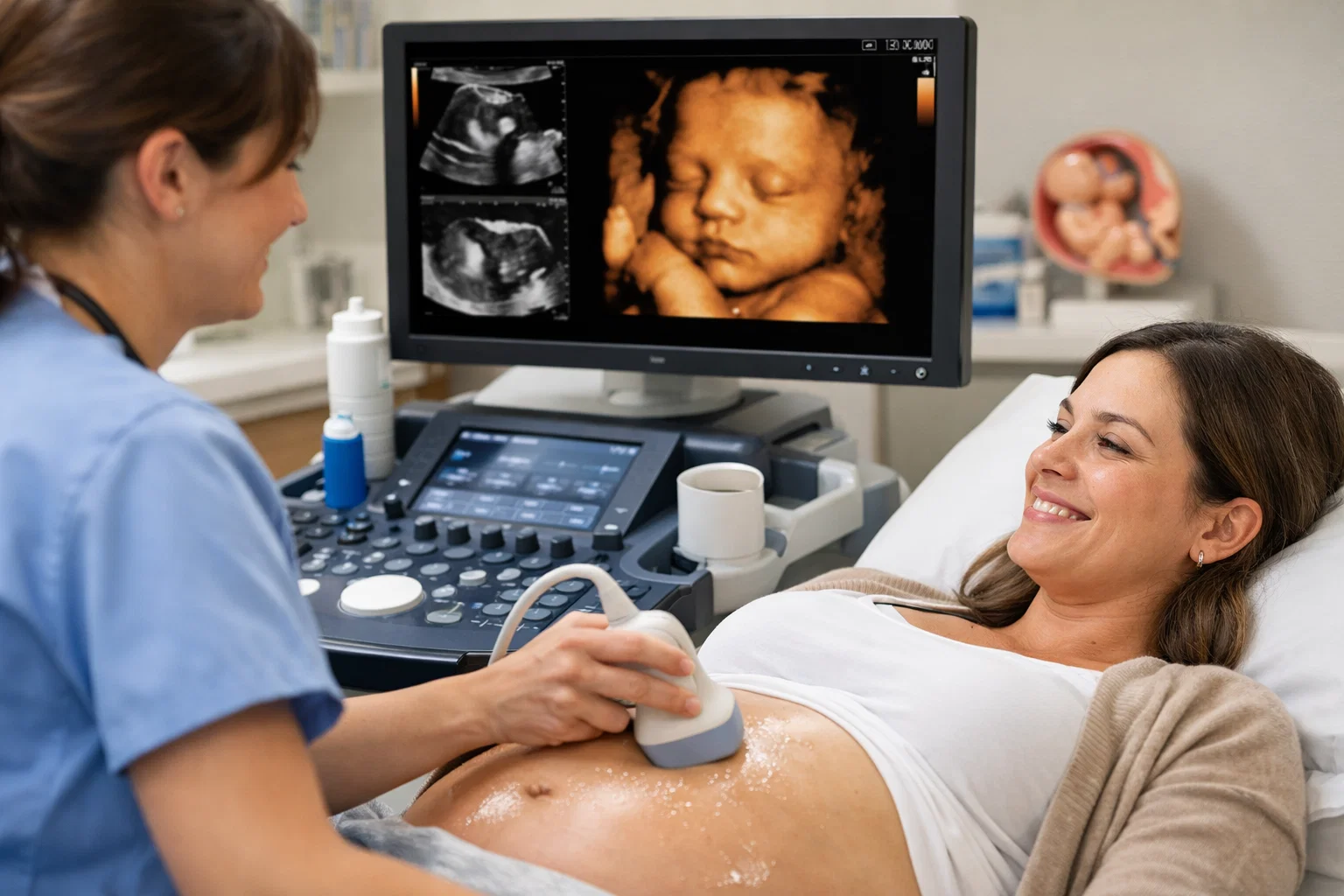
Ultrasound imaging is generally considered safe when performed by trained medical professionals using standard diagnostic settings, as it uses non-ionizing sound waves rather than radiation. Decades of clinical use indicate no confirmed long-term adverse health effects from standard diagnostic obstetric and medical sonograms. However, safe application depends on adhering to established medical necessity and operational guidelines, minimizing unnecessary exposure.
Navigating medical imaging procedures often raises questions regarding safety, efficacy, and potential biological impacts. Among the most common diagnostic tools in modern medicine is ultrasound technology, widely utilized across various specialties, most notably in obstetrics to monitor fetal development. Because patients and expectant parents frequently encounter conflicting information regarding the physical impact of high-frequency sound waves, a comprehensive examination of how the technology operates, what clinical guidelines dictate, and where risks may emerge provides necessary clarity.
Understanding Ultrasound Technology and Mechanics
To evaluate the safety of ultrasound imaging, it is essential to understand the underlying mechanics of how the technology functions. Unlike X-rays or computed tomography (CT) scans, which rely on ionizing radiation—a form of energy capable of altering chemical bonds and damaging cellular DNA—diagnostic ultrasound utilizes high-frequency sound waves. These sound waves, typically ranging from 2 to 18 megahertz, far exceed the upper limit of human hearing.
The imaging process begins with a handheld device known as a transducer. This device contains piezoelectric crystals that convert electrical energy into acoustic pulses. When the transducer is placed against the skin (often using a conductive gel to eliminate air pockets that impede sound transmission), these acoustic pulses travel into the body. As the sound waves encounter boundaries between different biological tissues—such as fluid, muscle, bone, or organ walls—they bounce back as echoes.
The transducer captures these returning echoes and converts them back into electrical signals. A specialized computer processes these signals instantaneously, translating the time delay and strength of the echoes into a real-time visual image displayed on a monitor. Because this process relies entirely on mechanical vibrations and pressure waves rather than electromagnetic radiation, it avoids the fundamental cellular risks associated with radiological imaging modalities.
Biological Effects and Thermal and Mechanical Indices
Even though ultrasound does not use ionizing radiation, it introduces energy into human tissue through mechanical waves. This energy interacts with biological material in two primary ways that researchers and regulatory bodies monitor closely: thermal effects and mechanical effects (cavitation).
Thermal Effects
As sound waves travel through tissue, a portion of the acoustic energy is absorbed and converted into heat. The degree of temperature elevation depends on several variables, including the frequency of the sound wave, the intensity of the beam, the duration of exposure, and the specific type of tissue being scanned (with bone absorbing more acoustic energy than soft tissue or fluid).
Modern ultrasound systems are equipped with standardized on-screen readouts known as the Thermal Index (TI). This index estimates the potential for tissue temperature elevation. The Thermal Index is further categorized based on the tissue type being examined, such as TIS (Thermal Index Soft tissue) and TIB (Thermal Index Bone). Regulatory standards, such as those overseen by the U.S. Food and Drug Administration (FDA) and international electro-technical commissions, mandate strict limits on acoustic output to prevent tissue heating from exceeding safe thresholds during diagnostic examinations.
Mechanical Effects and Cavitation
Mechanical effects involve the physical forces exerted by the acoustic wave on tissue. The primary concern in this category is acoustic cavitation, which occurs when microscopic gas bubbles are formed or agitated within a fluid medium due to pressure fluctuations. If these bubbles rapidly expand and collapse, they can generate localized high temperatures and intense shear stresses.
While inertial cavitation can cause cellular damage in experimental settings, it requires acoustic intensities significantly higher than those delivered by standard diagnostic ultrasound equipment. The Mechanical Index (MI) is displayed on modern ultrasound machines to help operators monitor the relative likelihood of non-thermal, mechanical bioeffects, ensuring they remain well within safe clinical boundaries.
| Metric | Purpose | Monitoring Threshold | Clinical Relevance |
|---|---|---|---|
| Thermal Index (TI) | Estimates potential tissue temperature elevation from acoustic absorption. | Kept below regulatory limits (typically designed to avoid local temperature increases >1°C above physiological norms). | Particularly relevant during obstetric scans involving developing fetal bone and the central nervous system. |
| Mechanical Index (MI) | Estimates the likelihood of mechanical bioeffects, specifically acoustic cavitation. | Maintained below specific FDA-cleared thresholds (generally < 1.9 for non-ophthalmic imaging). | Evaluates the risk of micro-bubble formation and fluid agitation in tissues. |
Safety in Obstetric Ultrasound
The most frequent application of diagnostic ultrasound occurs during pregnancy, where it serves as an indispensable tool for confirming viability, dating gestational age, screening for anatomical anomalies, evaluating amniotic fluid volume, and assessing placental placement. Because the developing fetus is uniquely sensitive to environmental factors, obstetric ultrasound has been subjected to rigorous scientific scrutiny for over half a century.
Extensive epidemiological studies and longitudinal clinical follow-ups involving children exposed to diagnostic ultrasound in utero have consistently found no conclusive evidence linking standard diagnostic sonography to adverse long-term outcomes. These studies have evaluated endpoints including birth weight, childhood physical development, cognitive function, neurological performance, and the incidence of childhood malignancies.
However, clinical organizations such as the American College of Obstetricians and Gynecologists (ACOG) and the American Institute of Ultrasound in Medicine (AIUM) emphasize that obstetric ultrasound should be performed only when there is a valid medical indication. This principle is guided by the ALARA (As Low As Reasonably Achievable) protocol.
The ALARA principle dictates that the acoustic output power and exposure time should be kept as low as possible while still obtaining the necessary diagnostic information. While the procedure is safe, indiscriminate or prolonged exposure without clear clinical justification is discouraged.
Commercial Keepsake Scans vs. Diagnostic Medical Sonography
A critical distinction in evaluating ultrasound safety is the difference between medically indicated diagnostic scans and non-diagnostic commercial keepsake videos or photographs. Commercial enterprise services, sometimes marketed as "baby bonding" studios, offer souvenirs such as 3D/4D photographs or keepsake video recordings of the fetus.
Major medical and regulatory organizations, including the FDA, strongly discourage elective ultrasound scans for non-medical purposes. While these commercial operators often use the same fundamental technology as medical equipment, several operational differences raise concern:
- Training and Certification: Commercial operators may lack the rigorous clinical training required of certified diagnostic medical sonographers, potentially leading to prolonged scanning times over sensitive fetal structures.
- Lack of Medical Oversight: Keepsake facilities do not provide diagnostic interpretations or medical screenings. If an anomaly is inadvertently visualized, personnel are generally not qualified to assess or communicate clinical findings.
- Prolonged Exposure: Commercial sessions often prioritize aesthetic results over clinical efficiency, resulting in extended exposure durations that contradict the ALARA principle without providing a health benefit.
Comparative Overview of Diagnostic Imaging Modalities
To better understand the safety profile of ultrasound, it is helpful to compare it against other common diagnostic imaging technologies. Each modality balances diagnostic capability, cost, availability, and physical safety risks differently.
| Imaging Modality | Underlying Energy Source | Primary Safety Consideration | Typical Clinical Application |
|---|---|---|---|
| Ultrasound | High-frequency acoustic sound waves (mechanical energy). | Minimal risk; thermal and mechanical indices monitored to prevent tissue heating or cavitation. | Obstetric monitoring, echocardiography, abdominal and pelvic organ assessment. |
| X-Ray | Ionizing radiation (electromagnetic waves). | Exposure to ionizing radiation carries a cumulative, low-level risk of cellular DNA damage. | Skeletal fractures, chest examinations for pulmonary conditions, dental evaluations. |
| Computed Tomography (CT) | Ionizing radiation (rotating X-ray beams processed by computer). | Higher radiation dose than standard X-rays; risk must be weighed against diagnostic necessity. | Detailed cross-sectional imaging of the brain, chest, abdomen, and trauma assessment. |
| Magnetic Resonance Imaging (MRI) | Strong magnetic fields and radiofrequency pulses. | No ionizing radiation; primary hazards involve metallic object projectile risks and acoustic noise. | Detailed soft-tissue imaging of the brain, spinal cord, joints, and internal organs. |
Potential Risks and Practical Limitations
While standard diagnostic ultrasound is exceptionally safe, practitioners and patients must remain aware of its operational limitations and specific localized risks.
First, ultrasound is operator-dependent. The diagnostic accuracy of the scan relies heavily on the skill, training, and technique of the sonographer and the interpreting physician. Incomplete training or improper equipment settings can lead to missed findings or, conversely, unnecessary follow-up procedures.
Second, although thermal and mechanical indices ensure that typical soft-tissue and bone heating remains negligible, certain anatomical regions require enhanced caution. For example, the developing embryonic eye and central nervous system are particularly sensitive to thermal changes. Consequently, equipment presets for early first-trimester scans and ocular examinations are specifically calibrated to minimize acoustic output and restrict the thermal index.
Finally, patients should understand that ultrasound has physical limitations regarding tissue penetration. Highly dense bone structures and excessive subcutaneous adipose tissue can attenuate or scatter sound waves, degrading image quality and occasionally necessitating alternative imaging modalities to secure a definitive diagnosis.
Next Actions and Patient Guidance
Patients scheduled for an ultrasound examination can take several practical steps to ensure an effective, safe experience while addressing any lingering concerns:
- Verify Facility Accreditation: Ensure that the imaging center or hospital department is accredited by recognized bodies such as the American College of Radiology (ACR) or the AIUM, which guarantees adherence to strict equipment maintenance and quality assurance standards.
- Confirm Clinical Justification: Discuss the specific medical purpose of the scan with the referring physician to align expectations and ensure the procedure is clinically warranted.
- Avoid Elective Keepsake Centers: Decline commercial or entertainment-based ultrasound packages that lack direct medical supervision, adhering strictly to medically prescribed imaging protocols.
- Communicate Sensitivities: Inform the sonographer of any prior surgeries, known tissue sensitivities, or specific discomforts so adjustments can be made promptly during the examination.
By combining rigorous regulatory oversight, adherence to established safety principles like ALARA, and the expertise of credentialed medical professionals, ultrasound remains a cornerstone of safe, non-invasive diagnostic medicine.