Understanding ALS mimics involves identifying conditions that present with similar muscle weakness, wasting, or twitching but have different underlying causes, treatments, and prognoses. While Amyotrophic Lateral Sclerosis (ALS) is a progressive neurodegenerative disease affecting both upper and lower motor neurons, mimics like Multifocal Motor Neuropathy or Cervical Spondylosis often offer more treatable paths.
The Complexity of the ALS Diagnostic Journey
As of 2026, Amyotrophic Lateral Sclerosis remains a clinical diagnosis, meaning there is no single definitive blood test or imaging study that can "prove" a patient has the disease. Instead, neurologists rely on a combination of clinical examination, electrophysiological testing, and the systematic exclusion of other conditions. This "diagnosis of exclusion" creates a critical window where identifying an ALS mimic is paramount.
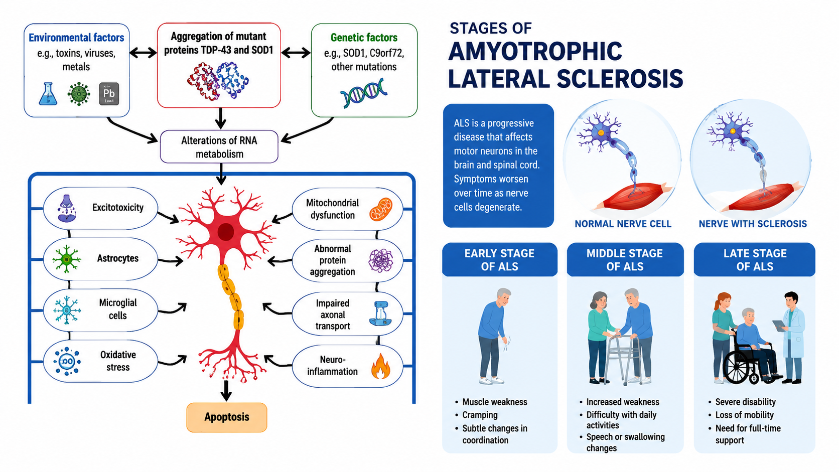
An ALS mimic is a medical condition that presents with symptoms nearly identical to the early stages of motor neuron disease (MND). These symptoms typically include muscle fasciculations (twitching), progressive weakness, muscle atrophy, and sometimes speech or swallowing difficulties. However, the distinction lies in the pathophysiology. While ALS involves the death of motor neurons in the brain and spinal cord, mimics might involve peripheral nerve compression, autoimmune responses, or metabolic deficiencies.
The stakes of differentiation are exceptionally high. An ALS diagnosis carries a significant emotional and physical weight, whereas many mimics—such as Multifocal Motor Neuropathy (MMN) or certain vitamin deficiencies—are highly treatable or even reversible. For healthcare providers and patients alike, understanding the subtle clinical "red flags" that point away from ALS is the first step toward accurate management.
Distinctive Features of Amyotrophic Lateral Sclerosis (ALS)
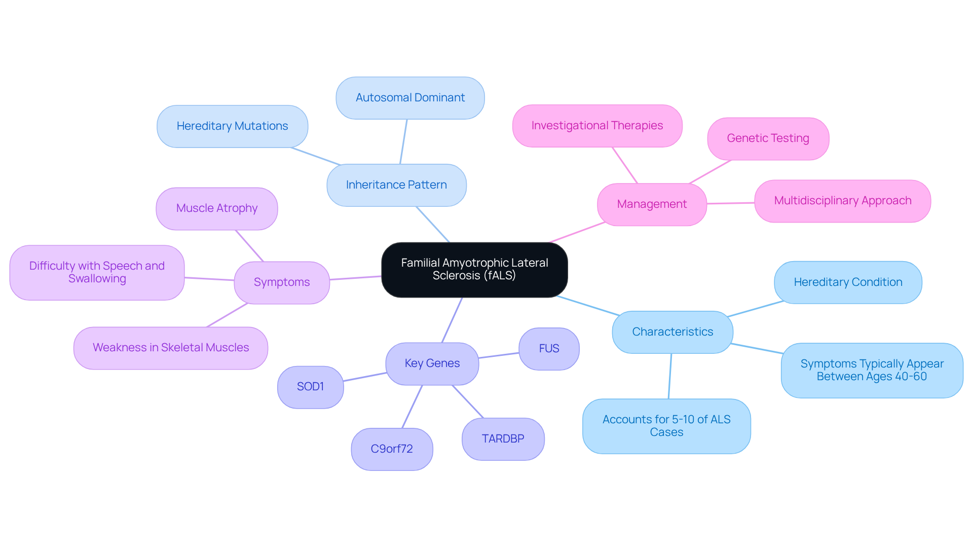
To understand the mimics, one must first master the clinical signature of ALS. The hallmark of ALS is the simultaneous presence of Upper Motor Neuron (UMN) and Lower Motor Neuron (LMN) signs across multiple body regions (bulbar, cervical, thoracic, and lumbosacral).
- Upper Motor Neuron Signs: These include spasticity (muscle stiffness), hyperreflexia (overactive reflexes), and the presence of a Babinski sign (the big toe moves upward when the sole of the foot is stimulated). These signs indicate damage to the neurons traveling from the brain down the spinal cord.
- Lower Motor Neuron Signs: These include muscle atrophy (wasting), fasciculations (twitching), and weakness. These indicate damage to the neurons traveling from the spinal cord to the muscles.
In ALS, these symptoms typically begin in one limb (limb-onset) or with speech/swallowing issues (bulbar-onset) and spread progressively to adjacent regions. Crucially, ALS is traditionally considered a "pure" motor disease, meaning it rarely involves significant sensory loss, such as numbness or tingling, in its early stages.
Common ALS Mimics: The Treatable Alternatives
Several conditions can trick even experienced clinicians. Below, we explore the most common mimics and the specific features that distinguish them from ALS.
1. Multifocal Motor Neuropathy (MMN)
Multifocal Motor Neuropathy is perhaps the most significant mimic because it is treatable with Intravenous Immunoglobulin (IVIg). MMN presents with slowly progressive, asymmetric muscle weakness, often starting in the hands.
- Distinctive Feature: MMN strictly affects the lower motor neurons. Unlike ALS, you will not see spasticity or overactive reflexes. Furthermore, MMN often shows a "conduction block" on an Electromyography (EMG) study—a sign that the nerve signal is being interrupted by an autoimmune attack on the myelin sheath, rather than the death of the neuron itself.
- The Difference: MMN is often associated with high titers of anti-GM1 antibodies, which are absent in ALS.
2. Cervical Spondylotic Myelopathy (CSM)
Cervical Spondylosis refers to the wear and tear of the spinal disks in the neck. When this leads to compression of the spinal cord (myelopathy), it can cause weakness in the hands and stiffness in the legs, mimicking the UMN/LMN split of ALS.
- Distinctive Feature: Patients with CSM frequently report sensory symptoms, such as "pins and needles" in the fingers or a loss of balance. Since ALS does not typically involve sensory pathways, the presence of numbness is a strong indicator that the issue may be structural (spinal) rather than neurodegenerative.
- The Difference: MRI imaging of the cervical spine can clearly show the physical compression of the cord, which is not present in ALS patients.
3. Inclusion Body Myositis (IBM)
IBM is an inflammatory muscle disease that occurs most often in older adults. It causes progressive weakness in the quadriceps (thighs) and the finger flexors (the muscles that allow you to grip).
- Distinctive Feature: The pattern of weakness in IBM is very specific. While ALS weakness is usually more generalized within a limb, IBM specifically targets the ability to make a fist or stand up from a chair.
- The Difference: A muscle biopsy in IBM patients will show "rimmed vacuoles" and inflammatory infiltrates, features entirely absent in the neurogenic wasting of ALS. Additionally, blood tests may show elevated levels of Creatine Kinase (CK), a marker of muscle damage.
4. Kennedy’s Disease (Spinal and Bulbar Muscular Atrophy)
Kennedy’s disease is a rare, X-linked genetic disorder that primarily affects men. It presents with muscle cramps, weakness, and speech/swallowing difficulties, making it look strikingly like bulbar-onset ALS.
- Distinctive Feature: Kennedy’s disease progresses much more slowly than ALS. Patients may also display signs of androgen insensitivity, such as gynecomastia (enlarged male breast tissue).
- The Difference: A genetic test for the CAG repeat expansion in the androgen receptor gene can definitively diagnose Kennedy's Disease.
Diagnostic Tools and Differentiation Strategies
In 2026, the diagnostic process remains rigorous. Neurologists use several key tools to distinguish between ALS and its mimics.
Electromyography (EMG) and Nerve Conduction Studies (NCS)
The EMG is the "gold standard" for evaluating motor neuron function. In ALS, the EMG reveals widespread "denervation" (loss of nerve supply to the muscle) and "reinnervation" (the body's failed attempt to fix it). In mimics like MMN, the NCS will show that the nerve itself is healthy except for specific points of blockage.
Magnetic Resonance Imaging (MRI)
MRI is primarily used to rule out "structural" mimics. It can identify brain tumors, strokes, or spinal cord compression that might be causing motor symptoms. In some advanced ALS cases, an MRI might show changes in the motor cortex (the "motor band" sign), but its primary role remains exclusionary.
Laboratory Testing
Extensive blood work is necessary to rule out metabolic and infectious mimics. These tests usually include:
- Vitamin B12 levels: Severe deficiency can cause subacute combined degeneration of the spinal cord.
- Thyroid function: Hyperthyroidism can cause muscle twitching and weakness.
- Lyme Serology: In endemic areas, Lyme disease can occasionally cause a motor neuropathy.
- Serum Protein Electrophoresis (SPEP): To look for plasma cell dyscrasias that might cause neuropathy.
Clinical Comparison Table: ALS vs. Key Mimics
| Feature | Amyotrophic Lateral Sclerosis (ALS) | Multifocal Motor Neuropathy (MMN) | Cervical Myelopathy (CSM) | Inclusion Body Myositis (IBM) |
|---|---|---|---|---|
| Neuron Involvement | Both UMN and LMN | LMN only | UMN (legs) and LMN (arms) | Muscle (Myopathic) |
| Sensory Loss | Extremely rare | No | Common (numbness) | No |
| Progression Speed | Rapid (months to years) | Slow (years) | Variable | Very slow (decades) |
| Key EMG Finding | Widespread denervation | Conduction block | Focal nerve root damage | Myopathic potentials |
| Treatment Response | Disease-modifying only | High response to IVIg | Surgical decompression | Limited (Immunosuppression) |
Identifying "Red Flags" for a Non-ALS Diagnosis
Patients and clinicians should be alert for symptoms that are "atypical" for ALS. These "red flags" often lead to the discovery of a mimic:
- Sensory Symptoms: Numbness, tingling, or "electric shock" sensations in the limbs.
- Lack of Progression: Symptoms that remain localized to one small area for several years without spreading.
- Early Bowel or Bladder Dysfunction: While this can happen in late-stage ALS, early onset suggests a primary spinal cord issue.
- Purely Lower Motor Neuron Signs: If there is no evidence of spasticity or brisk reflexes after long-term weakness, MMN or Kennedy’s Disease becomes more likely.
- Extraocular Muscle Involvement: ALS typically spares the muscles that move the eyes until the very final stages. If a patient has double vision or drooping eyelids (ptosis), conditions like Myasthenia Gravis should be considered.
The Role of the Second Opinion
Given the complexity of motor neuron disorders, seeking a second opinion at a multidisciplinary ALS center is a standard recommendation in 2026. These centers have access to specialized neuromuscular neurologists who see hundreds of cases annually and are more adept at spotting the nuances of mimic syndromes.
A second opinion often involves a "re-baselining" of the patient. This means repeating the clinical exam and sometimes the EMG to see if the symptoms have spread according to the known patterns of ALS. If the symptoms have remained static or if new sensory findings have emerged, the diagnosis is often revised.
FAQs: Understanding ALS Mimics
Can Benign Fasciculation Syndrome (BFS) be mistaken for ALS?
Yes, BFS is a common source of anxiety. It involves persistent muscle twitching (fasciculations) without muscle weakness or atrophy. The distinctive feature of BFS is the absence of "clinical weakness"—the muscle still functions at full strength despite the twitching. BFS does not progress to ALS.
Is it possible to have both ALS and a mimic?
While rare, it is possible. For example, an elderly patient might have Cervical Spondylosis (a mimic) and then develop ALS. This makes the diagnostic process even more challenging, as the neurologist must determine which symptoms are caused by the structural issue and which are caused by the neurodegenerative disease.
How long does the diagnostic process typically take?
On average, it takes 9 to 12 months from the first symptom to a confirmed ALS diagnosis. This timeframe is largely due to the "wait and see" approach required to prove that the weakness is progressive and to allow time for the exclusion of mimics through various tests.
Are there new tests in 2026 for ALS differentiation?
Research into "Neurofilament Light Chain" (NfL) levels in the blood and cerebrospinal fluid has advanced significantly. While not a standalone diagnostic test for ALS, high levels of NfL indicate active neuronal damage, which can help clinicians distinguish ALS from slower-moving mimics or non-neurodegenerative conditions.
Can Lyme Disease truly mimic ALS?
In very rare cases, a condition called Lyme Neuroborreliosis can cause motor nerve inflammation that looks like ALS. However, Lyme disease usually presents with other symptoms like joint pain, rash, or meningitis, and it responds to antibiotics, whereas ALS does not.
Final Recommendation for Patients and Caregivers
If you or a loved one are undergoing an evaluation for motor neuron symptoms, the most important action is to ensure a comprehensive "mimic screen" has been performed. This includes high-quality neuroimaging (MRI) and a detailed EMG/NCS performed by a neuromuscular specialist. Do not hesitate to ask your neurologist, "What else could this be, and how have we ruled it out?" Accurate differentiation is the only path to appropriate treatment, and for many patients, the "mimic" is a diagnosis that offers much more hope than the alternative.