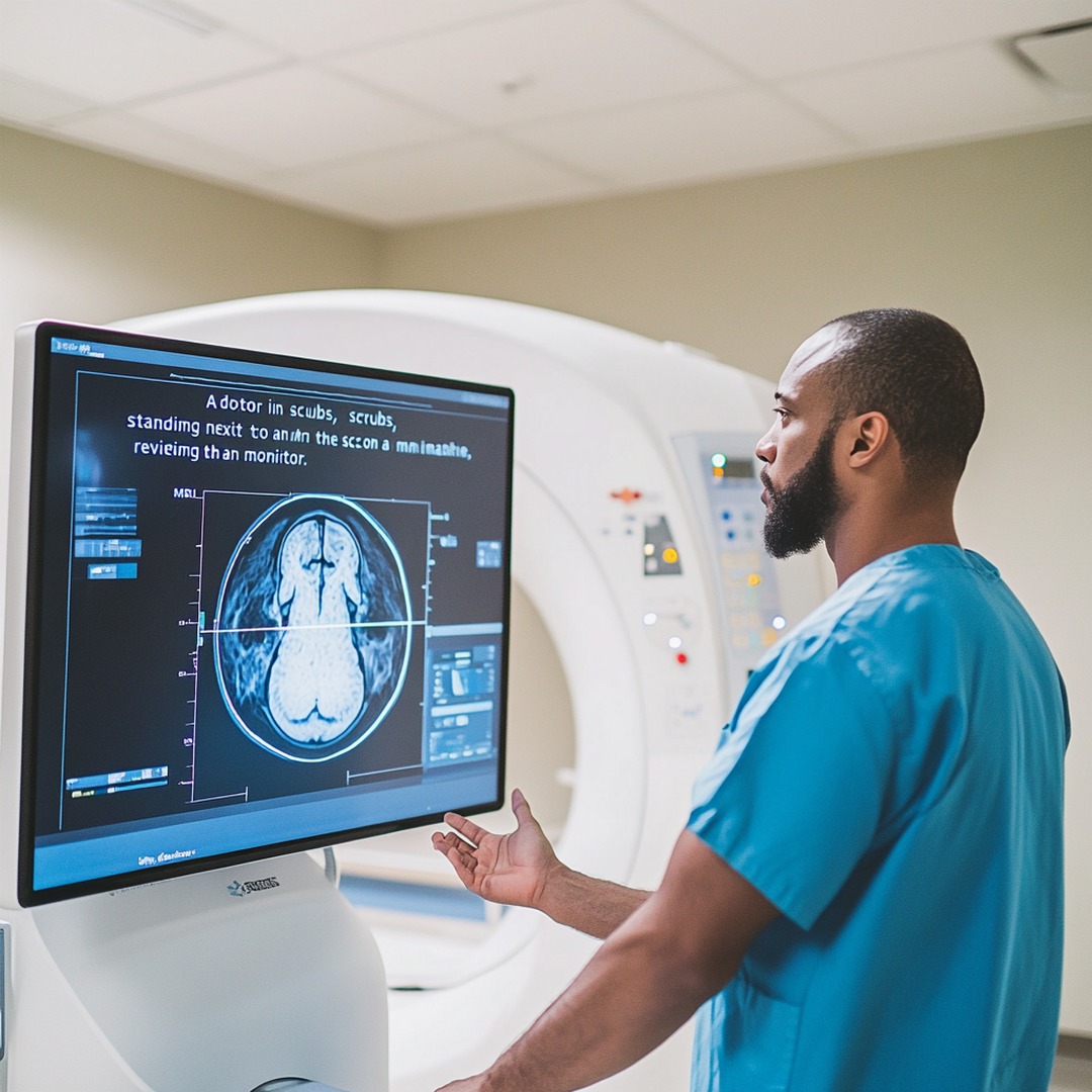
Radiology links gross anatomy with clinical pathology by utilizing electromagnetic radiation, magnetic fields, and sound waves to visualize internal anatomical structures. Selecting the appropriate imaging modality—such as projectional radiography, computed tomography, magnetic resonance imaging, ultrasound, or nuclear medicine—depends on the targeted anatomical tissue, diagnostic urgency, clinical context, patient safety considerations, and specific diagnostic goals.
Fundamentals of Anatomical Correlation in Diagnostic Radiology
Modern diagnostic radiology converts three-dimensional human anatomy into two-dimensional and three-dimensional digital representations. Interpreting these images accurately requires a firm grasp of standard anatomical orientation, cross-sectional planes, and the technical mechanisms behind tissue contrast.
Anatomical images are traditionally viewed in three orthogonal planes:
- Axial (Transverse) Plane: Divides the body into superior (head) and inferior (feet) sections. Slice-based imaging displays axial views as if standing at the patient's feet looking toward the head, placing the patient's right side on the viewer's left.
- Coronal (Frontal) Plane: Divides the body into anterior (ventral/front) and posterior (dorsal/back) sections, essential for evaluating bilateral symmetry and craniocaudal anatomical extent.
- Sagittal Plane: Divides the body into right and left portions. The mid-sagittal plane divides the body into equal halves, providing optimal visualization of midline neural structures, spine alignment, and profile anatomy.
- 3D Human Anatomy
- Axial Plane (Cross-Section)
- Coronal Plane (Front / Back)
- Sagittal Plane (Right / Left)
Diagnostic modalities categorize physical tissues using specific terminologies derived from how energy interacts with tissue density and composition:
- Radiodensity and Attenuation (X-ray and CT): Describes how effectively tissue absorbs or scatters X-ray photons. Denser tissues like cortical bone display high attenuation (hyperdense or radiopaque, appearing white), whereas low-density structures like air-filled lungs display low attenuation (hypodense or radiolucent, appearing dark).
- Echogenicity (Ultrasound): Refers to a tissue's ability to reflect sound waves back to the transducer. Tissues generating high-amplitude reflections are hyperechoic (bright white), fluid-filled structures that transmit sound without reflection are anechoic (black), and intermediate tissues are hypoechoic or isoechoic (shades of gray).
- Signal Intensity (MRI): Reflects the magnetic properties and proton density of tissues following radiofrequency excitation. Tissues with high signal intensity appear hyperintense (bright), while those with low signal intensity appear hypointense (dark).
- Radiotracer Avidity (Nuclear Medicine / PET): Measures cellular metabolic activity or receptor binding density. Areas of intense radiopharmaceutical accumulation are termed hypermetabolic or "hot spots," while non-accumulating areas are termed photopenic or "cold spots."
Primary Diagnostic Imaging Modalities: Technical Mechanisms and Indications
Radiography (X-Ray) and Fluoroscopy
Projectional radiography remains the initial imaging evaluation for many clinical presentations. High-energy ionizing photons pass through the body, casting a shadow image onto a digital detector plate. Radiography excels at evaluating osseous structures, detecting radiopaque foreign bodies, and providing baseline assessments of the chest and abdomen.
Its primary limitation is superposition—three-dimensional structures are flattened into a single two-dimensional plane, which can obscure subtle lesions behind overlapping anatomy.
Fluoroscopy uses continuous X-ray beams to evaluate real-time motion and functional processes. Combined with contrast media (such as barium or non-ionic iodinated contrast), fluoroscopy visualizes dynamic gastrointestinal motility, swallowing mechanics, arterial lumen patency during angiograms, and real-time needle placement during interventional procedures.
Computed Tomography (CT) & Hounsfield Unit Quantification
Computed Tomography overcomes superposition by rotating a localized X-ray tube and detector array around the patient, acquiring cross-sectional spatial data. Advanced Multi-Detector CT (MDCT) scanners capture volumetric data sets that can be reconstructed in any anatomical plane (Multiplanar Reconstruction or MPR) or as 3D surface-rendered models.
Tissue attenuation on CT scans is quantified using the Hounsfield Unit (HU) scale, calibrated to water at 0 HU and room air at -1000 HU:
$$\text{HU} = 1000 \times \frac{\mu_{\text{tissue}} - \mu_{\text{water}}}{\mu_{\text{water}} - \mu_{\text{air}}}$$
Where $\mu$ represents the linear attenuation coefficient of the material.
- Air: -1000 HU (black)
- Subcutaneous / Visceral Fat: -120 to -50 HU
- Pure Water / Fluid: 0 HU
- Soft Tissue (Muscle, Liver, Kidney): +30 to +80 HU
- Acute Intracranial Blood: +60 to +90 HU
- Cortical Bone: +400 to +2000+ HU (bright white)
Intravenous contrast agents (iodinated compounds) are timed precisely to highlight specific vascular or parenchymal phases, such as the early arterial phase (20–30 seconds post-injection) for angiography, the portal venous phase (60–70 seconds) for abdominal parenchymal evaluation, or delayed nephrogenic phases for renal collecting system analysis.
- Hounsfield Unit (HU) Spectrum
- -1000 HU: Air (Black)
- 0 HU: Water (Gray)
- +1000+ HU: Dense Bone (White)
Magnetic Resonance Imaging (MRI) & Tissue Relaxation Dynamics
MRI relies on non-ionizing radiofrequency pulses applied within high magnetic fields (typically 1.5 Tesla or 3.0 Tesla). Hydrogen nuclei (protons) in human tissue align with the main magnetic field ($B_0$). Radiofrequency pulses temporarily tilt these protons out of alignment. As the protons relax back to equilibrium, they emit radiofrequency signals detected by specialized receiver coils.
Soft-tissue contrast in MRI is manipulated by altering pulse sequences and timing parameters, predominantly Time to Echo (TE) and Time to Repetition (TR):
- T1-Weighted Sequences (Short TR, Short TE): Highlight anatomy. Fat yields high signal intensity (bright), while water-based fluids (such as cerebrospinal fluid or urine) appear hypointense (dark). Excellent for delineating structural borders and evaluating bone marrow infiltration.
- T2-Weighted Sequences (Long TR, Long TE): Highlight pathology and edema. Fluid displays hyperintense signal (bright), while solid organ tissue appears intermediate gray. Pathological conditions—including inflammation, ischemia, infection, and tumors—typically increase tissue water content, making them conspicuous on T2 images.
- Fluid-Attenuated Inversion Recovery (FLAIR): Suppresses the signal from free-flowing cerebrospinal fluid (CSF) while maintaining T2 weighting, allowing subtle periventricular brain lesions or subarachnoid hemorrhages to stand out clearly.
- Diffusion-Weighted Imaging (DWI): Measures microscopic Brownian motion of water molecules. Cytotoxic edema caused by acute cellular ischemia (such as acute ischemic stroke) restricts water movement, producing hyperintense signal on DWI within minutes of symptom onset.
Diagnostic Ultrasound (Ultrasonography) & Doppler Analysis
Ultrasound uses piezoelectric crystals inside a transducer probe to generate high-frequency sound waves ($2\text{ to }18\text{ MHz}$) that travel into tissue. As sound waves pass through anatomical boundaries with differing acoustic impedances, a portion of the wave reflects back to the transducer to produce a real-time image.
- High-Frequency Linear Transducers (7–18 MHz): Provide high spatial resolution with limited depth penetration, ideal for superficial structures like thyroid nodules, testicular pathology, peripheral nerves, and musculoskeletal tendons.
- Low-Frequency Curvilinear Transducers (2–5 MHz): Provide deep tissue penetration with lower spatial resolution, suitable for abdominal, transabdominal pelvic, and obstetric evaluations.
Doppler Ultrasound applies the Doppler shift principle—evaluating changes in sound wave frequency reflected off moving red blood cells—to assess vascular direction and velocity. Color Doppler maps velocity and direction onto a 2D gray-scale image (conventionally, red indicates flow toward the transducer and blue away), while Spectral Doppler plots precise quantitative flow velocities over time to evaluate vascular stenoses or organ perfusion dynamics.
Nuclear Medicine and Hybrid Molecular Imaging (PET/CT, SPECT/CT)
Unlike anatomical modalities that emit external energy through the patient, nuclear medicine introduces radioisotopes into the patient via intravenous injection, inhalation, or ingestion. These targeted radiopharmaceuticals selectively accumulate in specific tissue types or metabolic pathways.
- Positron Emission Tomography (PET): Uses positron-emitting radiotracers, most commonly $^{18}\text{F-Fluorodeoxyglucose}$ ($^{18}\text{F-FDG}$), a glucose analogue. Tissues with high metabolic rates—such as malignant tumors, active inflammatory sites, or hyperactive myocardium—rapidly take up $^{18}\text{F-FDG}$. Positrons annihilate upon encountering electrons, emitting twin gamma rays at $180^\circ$ angles that are captured by a ring detector array.
- Single-Photon Emission Computed Tomography (SPECT): Emits single gamma photons (using isotopes like Technetium-99m) to analyze organ function, such as myocardial perfusion stress testing or bone turnover scans.
- Hybrid Imaging (PET/CT and PET/MRI): Fuses functional PET/SPECT datasets with anatomical CT or MRI maps within a single scanning session. This dual approach pinpoints metabolic abnormalities to precise anatomical locations, improving diagnostic confidence in oncology, cardiology, and neurology.
Comparative Modality Matrix
The table below summarizes the key technical features, spatial advantages, and operational limitations across primary diagnostic imaging modalities:
| Imaging Modality | Underlying Energy / Physics | Primary Tissue Contrast Basis | Ionizing Radiation | Key Clinical Strengths | Operational Limitations |
|---|---|---|---|---|---|
| Radiography (X-Ray) | Transmission of high-energy photons | Differential tissue attenuation / radiodensity | Yes (Low exposure: ~0.001–0.1 mSv) | Rapid acquisition, low cost, widely accessible, excellent for bone fractures | 2D superposition, low soft-tissue resolution |
| Computed Tomography (CT) | Multi-angle X-ray photon attenuation | Quantitative tissue attenuation (Hounsfield Units) | Yes (Moderate to High: ~2–10+ mSv) | High spatial resolution, rapid cross-sectional mapping, ideal for acute trauma | Higher radiation dose, potential iodinated contrast nephrotoxicity |
| Magnetic Resonance Imaging (MRI) | Magnetic fields ($B_0$) & radiofrequency pulses | Hydrogen proton density, T1/T2 relaxation dynamics | No | Unmatched soft-tissue resolution, multiplanar capability, no radiation | Long acquisition times, motion artifact sensitivity, magnetic field safety risks |
| Ultrasonography (US) | Acoustic sound wave reflection ($2-18\text{ MHz}$) | Differences in tissue acoustic impedance | No | Real-time dynamic evaluation, portable, safe for pregnancy and pediatric care | Operator dependent, blocked by bone structures and intraluminal bowel gas |
| Nuclear Medicine (PET / SPECT) | Gamma ray emission from internal radiopharmaceutical | Tissue metabolic rate, cell surface receptor avidity | Yes (Depends on radiotracer half-life & dosage) | High metabolic sensitivity, early systemic disease detection | Low structural detail, requires hybrid CT/MRI for precise anatomical localization |
Selecting the Appropriate Imaging Modality by Clinical Context
- Clinical Presentation
- Acute Trauma
- Non-Contrast CT
- Soft Tissue/Neuro
- MRI Assessment
- Visceral/Fluid
- Ultrasound / CT
- Acute Trauma
Choosing the most effective diagnostic imaging modality depends on the patient's clinical presentation, anatomical region, target pathology, and underlying physical contraindications.
Acute Trauma and Emergency Medicine Protocols
Emergency protocols demand rapid, accurate visualization of life-threatening internal injuries.
- Pan-Scan CT (Head, Neck, Chest, Abdomen, Pelvis): Represents the diagnostic gold standard for high-energy polytrauma. Non-contrast head CT quickly identifies intracranial hemorrhages, mass effect, and skull fractures. Contrast-enhanced CT of the chest, abdomen, and pelvis reveals active arterial extravasation, solid organ lacerations (liver, spleen, kidney), aortic dissections, and bowel perforations.
- FAST Exam (Focused Assessment with Sonography for Trauma): A rapid point-of-care ultrasound protocol performed at the bedside. It screens four key anatomical regions—pericardial sac, right upper quadrant (Morison's pouch), left upper quadrant (splenorenal recess), and pelvis (rectovesical/pouch of Douglas)—for free fluid (hemoperitoneum or pericardial effusion).
Neurological Evaluation: Stroke, Seizures, and Tumors
- Acute Ischemic Stroke: Non-contrast Head CT is performed immediately to exclude hemorrhage before administering thrombolytic therapy. If non-contrast CT is negative for blood, CT Angiography (CTA) identifies large vessel occlusions amenable to endovascular thrombectomy. Diffusion-Weighted MRI (DWI) confirms early cytotoxic edema within minutes of cerebral arterial occlusion.
- Seizures and Intracranial Neoplasms: Contrast-enhanced Brain MRI provides high spatial contrast between gray and white matter, making it superior for detecting subtle cortical dysplasias, mesial temporal sclerosis, acoustic neuromas, high-grade gliomas, or cerebral metastases.
Musculoskeletal and Soft Tissue Injury Assessment
- Bone Fractures and Joint Alignment: Plain film radiography remains the baseline first-line examination. Complex intra-articular fractures (such as calcaneal, tibial plateau, or acetabular disruptions) require volumetric CT to guide surgical planning.
- Ligaments, Tendons, and Cartilage: MRI excels at resolving uncalcified soft tissues due to its soft-tissue contrast mechanisms. It is the primary modality for evaluating internal derangements of joints, such as anterior cruciate ligament (ACL) tears, meniscal disruptions, and rotator cuff pathology.
Abdominal and Pelvic Visceral Pathology
- Right Upper Quadrant Pain: Abdominal ultrasound is the first-line examination for suspected acute cholecystitis, gallstones (cholelithiasis), and biliary tree dilatation.
- Acute Appendicitis and Diverticulitis: Contrast-enhanced abdominal/pelvic CT offers high diagnostic accuracy for appendiceal inflammation, retrocecal appendicitis, pericolic fat stranding, abscess formation, or bowel wall thickening. Ultrasound or non-contrast pelvic MRI is substituted in pediatric and pregnant populations to avoid ionizing radiation.
- Gynecological & Pelvic Pathology: Transvaginal ultrasound visualizes ectopic pregnancies, ovarian cysts, adnexal torsion, and uterine fibroids. Multiparametric Pelvic MRI is reserved for staging cervical, endometrial, or rectal malignancies, as well as mapping deep infiltrating endometriosis.
Cardiothoracic Imaging and Pulmonary Evaluation
- Acute Chest Pain & Dyspnea: Standard two-view Chest Radiography (PA and Lateral) evaluates for pneumothorax, pulmonary consolidation, cardiomegaly, or pleural effusion.
- Pulmonary Embolism (PE): CT Pulmonary Angiography (CTPA) is the definitive diagnostic standard, displaying filling defects within the pulmonary arterial tree. Ventilation-Perfusion (V/Q) nuclear scintigraphy serves as an alternative for patients with severe renal failure or contrast allergies.
- Interstitial Lung Disease (ILD): High-Resolution CT (HRCT) captures thin axial slices ($\le 1\text{ mm}$) with specialized high-spatial-frequency reconstruction algorithms to characterize fine parenchymal patterns, such as honeycombing, ground-glass opacities, and traction bronchiectasis.
Safety Considerations, Contrast Media, and Radiation Stewardship
Radiological procedures require balancing diagnostic benefits against potential patient risks.
Ionizing Radiation Risk and ALARA Principles
Modalities utilizing ionizing radiation (X-ray, CT, SPECT, PET) break chemical bonds within biological tissue, creating potential DNA strand breaks. These effects fall into two broad categories:
- Deterministic Effects (Tissue Reactions): Occur above a clear dose threshold, with severity increasing proportional to dose (e.g., radiation-induced erythema, cataracts, or acute radiation sickness).
- Stochastic Effects: Radiation-induced cancer or genetic mutations where probability increases with dose, but severity is independent of dose. There is no established absolute safe threshold.
Radiological practice adheres strictly to the ALARA principle (As Low As Reasonably Achievable). Radiation exposure is minimized by adjusting kilovoltage (kV) and milliamperage-seconds (mAs), utilizing dose-modulation software, limiting scan limits, and employing alternative non-ionizing modalities (Ultrasound or MRI) whenever clinically appropriate—particularly for pediatric and pregnant patients.
Iodinated Contrast Media and Renal Safety
Intravenous iodinated contrast media improve blood vessel and organ visibility on CT scans, but carry risks of hypersensitivity reactions and renal strain:
- Anaphylactoid / Hypersensitivity Reactions: Mild reactions (hives, localized pruritus) are managed with antihistamines. Severe, life-threatening reactions (bronchospasm, laryngeal edema, anaphylactic shock) require immediate administration of intramuscular epinephrine, high-flow oxygen, and intravenous fluid resuscitation.
- Post-Contrast Acute Kidney Injury (PC-AKI): An abrupt decline in renal function following iodinated contrast administration. Serum creatinine levels or estimated Glomerular Filtration Rates (eGFR) are screened prior to contrast injection in high-risk patients (those with pre-existing renal insufficiency, chronic kidney disease, or solitary kidneys). For patients with an $\text{eGFR} < 30\text{ mL/min/1.73m}^2$, non-emergent iodinated contrast should be carefully weighed against diagnostic benefit, with volume expansion (intravenous $0.9%$ normal saline) used for prophylaxis when contrast is necessary.
Gadolinium-Based Contrast Agents (GBCAs) and MRI Safety
MRI avoids ionizing radiation, but poses distinct mechanical, thermal, and chemical safety risks:
- Ferromagnetic Projectile Hazards: The powerful static magnetic field ($B_0$) attracts unbound ferromagnetic items (such as oxygen tanks, scissors, or steel chairs), turning them into dangerous projectiles. Strict screening ensures patients do not carry compatible cardiac pacemakers, ferromagnetic cerebral aneurysm clips, or metallic intraocular foreign bodies into the MRI scanner room.
- Nephrogenic Systemic Fibrosis (NSF): A rare, debilitating systemic fibrosing disorder linked to intravenous administration of older linear Gadolinium-Based Contrast Agents (GBCAs) in patients with severe renal failure ($\text{eGFR} < 30\text{ mL/min/1.73m}^2$). Modern practice uses stable macrocyclic GBCAs, which carry an extremely low risk of NSF.
Diagnostic Workflow and Integrated Clinical Decision-Making
- Clinical Presentation
- ACR Appropriateness Criteria Selection
- Targeted Imaging Acquisition
- Systematic Image Interpretation
- Structured Reporting (BI-RADS / PI-RADS / LI-RADS)
- Multidisciplinary Clinical Action
Translating diagnostic images into actionable clinical care relies on a structured, multi-step process:
- Clinical Indication & Order Placement: The referring clinician evaluates the patient's history, physical examination, and laboratory findings to construct a differential diagnosis.
- Evidence-Based Modality Selection: Clinicians reference standardized decision support tools, such as the American College of Radiology (ACR) Appropriateness Criteria®, to choose the most suitable diagnostic exam for specific clinical scenarios.
- Protocoling and Acquisition: The radiologist tailors the scanning parameters—selecting slice thickness, spatial boundaries, dynamic timing phases, contrast volumes, or pulse sequences—to target the patient's specific presentation.
- Systematic Image Interpretation: Radiologists use structured search patterns to evaluate images. They inspect primary organ systems, systematically review high-risk anatomical transition zones, analyze incidental findings, and compare new scans with historic imaging studies to assess anatomical changes over time.
- Structured Diagnostic Reporting: Findings are recorded using standardized diagnostic classification systems to minimize ambiguity and align with established clinical management pathways:
- BI-RADS (Breast Imaging Reporting and Data System): Categorizes mammographic and breast ultrasound/MRI findings from 0 (incomplete) to 6 (biopsy-proven malignancy).
- PI-RADS (Prostate Imaging Reporting and Data System): Uses multiparametric MRI to score the probability of clinically significant prostate cancer on a 1-to-5 scale.
- LI-RADS (Liver Reporting and Data System): Standardizes the classification of focal liver lesions in patients at risk for hepatocellular carcinoma.
- TI-RADS (Thyroid Imaging Reporting and Data System): Risk-stratifies thyroid nodules on ultrasound to guide fine-needle aspiration (FNA) decision-making.
Frequently Asked Questions
How do clinicians decide between CT and MRI for acute neurological symptoms?
Non-contrast CT is typically performed first in acute settings due to its rapid scan time (often under two minutes), widespread availability, and exceptional sensitivity for acute intracranial hemorrhage. This rapidly clarifies whether a patient with stroke symptoms can safely receive thrombolytic therapy. MRI is selected when high-definition soft-tissue resolution is required—such as diagnosing early ischemic tissue changes (via Diffusion-Weighted Imaging), detecting posterior fossa and brainstem lesions, identifying subtle demyelinating plaques, or characterizing complex brain tumors.
What is the functional difference between T1-weighted and T2-weighted MRI sequences?
T1-weighted sequences feature short TR and TE times, highlighting fat signals as bright (hyperintense) and fluid signals as dark (hypointense). This provides detailed anatomical definition and tissue contrast. T2-weighted sequences feature longer TR and TE times, causing water and fluid-rich tissues to appear bright (hyperintense). Because inflammation, localized edema, and pathological lesions typically increase tissue fluid content, T2-weighted sequences excel at uncovering pathology.
Why is ultrasound preferred over CT for pediatric and pregnant patients?
Ultrasound uses non-ionizing, high-frequency sound waves, eliminating the DNA-damaging radiation risks associated with X-rays and CT scans. Developing fetuses and pediatric tissues are particularly sensitive to ionizing radiation. Ultrasound offers rapid, real-time, high-resolution diagnostic evaluation of pelvic, abdominal, and soft-tissue pathology without radiation exposure.
How do contrast media improve radiological visualization, and what screening is required?
Contrast agents change how target tissues interact with energy sources. Iodinated CT contrast absorbs X-ray photons to make blood vessels, tissue parenchyma, and inflammatory borders appear dense and bright. Gadolinium MRI contrast alters nearby hydrogen proton relaxation times, increasing T1 signal intensity. Before contrast administration, clinical teams evaluate the patient's history for past allergic reactions and review serum eGFR levels to reduce the risk of contrast-induced nephrotoxicity or Nephrogenic Systemic Fibrosis.
What are Hounsfield Units (HU) in CT scans, and how do they aid diagnosis?
Hounsfield Units quantify tissue attenuation relative to water (0 HU) and air (-1000 HU). Measuring HU values within a specific region of interest helps clinicians identify tissue composition: negative values (-120 to -50 HU) signal fat, near-zero values (0 to 15 HU) indicate simple fluid or cysts, values above 60 HU suggest acute blood accumulation or hypercellular soft tissue, and values over 400 HU indicate dense calcification or bone.
Diagnostic Recommendations and Next Actions
To select and interpret diagnostic imaging safely and effectively, medical practitioners and trainees should apply the following guidelines:
- Follow Evidence-Based Guidelines: Use established decision frameworks—such as the ACR Appropriateness Criteria or Ottawa Ankle/Knee Rules—to select the right initial imaging modality based on validated clinical indicators.
- Formulate Precise Clinical Questions: Include comprehensive clinical details and differential diagnostic considerations on imaging requisitions. Providing focused context helps radiologists select optimal scanning protocols and contrast timing phases.
- Apply Radiation Stewardship: Adhere to ALARA guidelines. When evaluating pediatric patients, pregnant women, or individuals requiring repeat exams, select non-ionizing alternatives (Ultrasound or MRI) whenever clinically appropriate.
- Perform Baseline Renal Screening: Verify serum eGFR levels before ordering exams requiring intravenous iodinated or gadolinium-based contrast agents in patients with known renal disease, diabetes, or advanced age.
- Integrate Diagnostic Findings with Clinical Data: Never interpret radiological reports in isolation. Correlate radiological findings with physical examination results, laboratory markers, and historical imaging studies to make informed clinical management decisions.
Professional Advice Disclaimer: This document is intended solely for educational and informational purposes for healthcare trainees and professionals. It does not constitute formal medical advice, clinical diagnosis, or treatment protocols. Patient care decisions must be made by qualified healthcare providers based on individual clinical evaluations and institutional guidelines.