Alexa Fluorophores are a comprehensive family of synthetic fluorescent dyes used extensively in biological research to label antibodies, proteins, and nucleic acids. Renowned for their exceptional brightness, photostability, and pH insensitivity, these dyes have largely superseded traditional fluorophores like FITC and TRITC, enabling high-resolution imaging and sensitive detection across various microscopy and flow cytometry platforms.
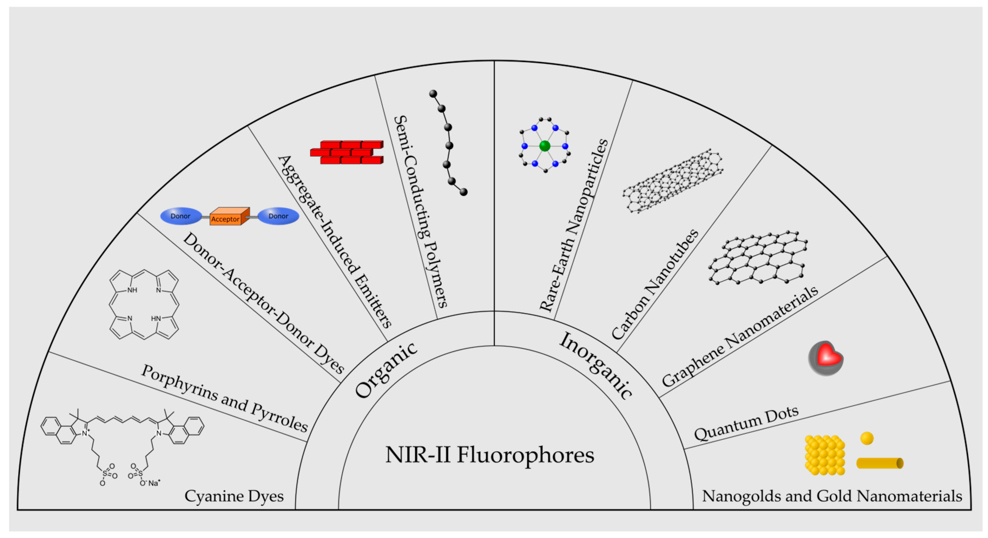
In the landscape of 2026 biological imaging, the Alexa Fluor series remains the gold standard for researchers requiring reproducible and high-contrast data. Developed originally by Molecular Probes and now offered through Thermo Fisher Scientific, these dyes are designed to span the entire visible and near-infrared spectrum. Their chemical structure—often based on sulfonated derivatives of rhodamine, coumarin, cyanine, or xanthene—provides superior water solubility and reduces the tendency of labeled molecules to aggregate, which is critical for maintaining biological function during live-cell or fixed-tissue analysis.
The Chemical Evolution of Fluorescence: Why Alexa Fluor Dyes Dominate
Before the advent of the Alexa Fluor series, biological imaging relied heavily on organic dyes such as Fluorescein Isothiocyanate (FITC) and Tetramethylrhodamine (TRITC). While functional, these older dyes suffered from significant limitations: they photobleached rapidly under intense laser illumination, their fluorescence intensity was highly dependent on the pH of the surrounding medium, and they often exhibited low water solubility, leading to self-quenching when multiple dye molecules were conjugated to a single protein.
The introduction of Alexa Fluor dyes addressed these challenges through precise chemical engineering. By incorporating sulfonic acid groups into the dye scaffolds, chemists increased the hydrophilicity of the molecules. This modification not only ensures that the dyes remain soluble in aqueous buffers but also adds a negative charge that helps prevent the "quenching" effect that occurs when dye molecules cluster together. In 2026, these characteristics are more relevant than ever as researchers push toward single-molecule detection and deep-tissue imaging where signal-to-noise ratios are paramount.
Furthermore, the spectral profiles of Alexa Fluor dyes are notably narrow. This narrow emission peak is a vital feature for multiplexing, where multiple targets are labeled simultaneously in the same sample. By reducing "bleed-through" (the leakage of signal from one color channel into another), Alexa Fluorophores allow for the clear visualization of complex cellular architectures and protein-protein interactions without the need for extensive mathematical linear unmixing in every experiment.
Core Spectral Characteristics and Naming Conventions
The naming convention of Alexa Fluor dyes is highly intuitive, designed to assist researchers in hardware configuration. Each dye is named "Alexa Fluor" followed by a number that corresponds to the approximate excitation maximum in nanometers (nm). For example, Alexa Fluor 488 has an excitation peak near 488 nm, making it a perfect match for the standard 488 nm argon-ion or solid-state lasers found in most confocal microscopes and flow cytometers.
The Visible Spectrum Range
The most commonly used dyes in molecular biology fall within the visible spectrum. Alexa Fluor 488 (green), Alexa Fluor 555 (orange), and Alexa Fluor 594 (red) are staples in almost every imaging core facility. Alexa Fluor 488, in particular, is frequently cited as the most robust replacement for FITC. It provides significantly higher brightness and does not quench as easily, even at high degrees of labeling (DOL).
The Near-Infrared (NIR) and Far-Red Range
For applications involving thick tissue sections or whole-organism imaging, NIR dyes like Alexa Fluor 647, 750, and 790 are indispensable. Light at these longer wavelengths penetrates deeper into biological tissues with less scattering and encounters lower levels of autofluorescence from endogenous molecules like collagen or NADH. In 2026, the use of Alexa Fluor 647 has also become standard in super-resolution techniques such as dSTORM (direct Stochastic Optical Reconstruction Microscopy) due to its excellent "blinking" properties and high photon yield.
Performance Metrics: Brightness, Photostability, and pH Resistance
To understand the utility of these fluorophores, one must evaluate three critical physical parameters: quantum yield, extinction coefficient, and photostability.
- Brightness: The brightness of a dye is the product of its molar extinction coefficient (how well it absorbs light) and its fluorescence quantum yield (how efficiently it converts that absorbed light into emitted photons). Alexa Fluor dyes typically exhibit high values for both, resulting in signals that are visible even at low protein expression levels.
- Photostability: This is perhaps the most famous attribute of the series. Photobleaching occurs when a dye molecule is chemically destroyed by the very light used to excite it. Alexa Fluorophores are engineered to withstand thousands of excitation cycles, which is essential for Z-stacking (3D imaging) and time-lapse cinematography where the same sample is illuminated repeatedly over hours or days.
- pH Insensitivity: Many traditional dyes lose fluorescence in acidic environments (like endosomes or lysosomes). Alexa Fluor dyes remain stable and bright across a wide pH range (typically pH 4 to 10). This stability ensures that the intensity of the signal reflects the quantity of the target rather than the local chemical environment of the cell.
Primary Applications in Modern Biotechnology
The versatility of Alexa Fluor dyes allows them to be used in nearly every facet of light-based biological inquiry. Their application is usually determined by the method of conjugation—how the dye is attached to the molecule of interest.
Immunofluorescence (IF) and Immunohistochemistry (IHC)
The most frequent use of Alexa Fluorophores is in secondary antibody labeling. In these experiments, a primary antibody binds to a specific cellular protein, and a "secondary" antibody, conjugated to an Alexa Fluor dye, binds to the primary. This provides a layered amplification of the signal. The "Alexa Fluor Plus" series, popularized in recent years, offers even higher sensitivity by utilizing proprietary signal-amplification technologies that further reduce background noise.
Flow Cytometry
In flow cytometry, cells in suspension are passed through a laser beam. Alexa Fluor dyes are used to label surface markers (like CD4 or CD8) to identify cell populations. Because flow cytometry often involves 10 or more colors simultaneously, the photostability and narrow emission peaks of Alexa Fluor 488, 532, 647, and 700 are critical for accurate data compensation and population gating.
Super-Resolution Microscopy
Techniques like Structured Illumination Microscopy (SIM), Stimulated Emission Depletion (STED), and STORM have pushed the limits of light microscopy beyond the diffraction limit (~200 nm). Alexa Fluor 647 and Alexa Fluor 594 are frequently used in these high-end applications. Their ability to produce a high number of photons before bleaching allows researchers to resolve structures as small as 20 nanometers, such as individual microtubules or synaptic vesicles.
Comparison Table: Common Alexa Fluor Dyes and Optical Properties
| Dye Name | Ex Max (nm) | Em Max (nm) | Laser Line (Common) | Common Filter Set | Primary Color |
|---|---|---|---|---|---|
| Alexa Fluor 350 | 346 | 442 | 355 (UV) | DAPI / Hoechst | Blue |
| Alexa Fluor 488 | 495 | 519 | 488 (Blue) | FITC / GFP | Green |
| Alexa Fluor 555 | 555 | 565 | 532 / 561 (Green/Yellow) | Cy3 / TRITC | Orange |
| Alexa Fluor 568 | 578 | 603 | 561 / 568 (Yellow) | Rhodamine Red | Red-Orange |
| Alexa Fluor 594 | 590 | 617 | 561 / 594 (Yellow/Red) | Texas Red | Red |
| Alexa Fluor 647 | 650 | 665 | 633 / 640 (Red) | Cy5 | Far-Red |
| Alexa Fluor 750 | 749 | 775 | 750 (NIR) | Cy7 | Near-IR |
Selection Strategies: Choosing the Right Fluorophore
Selecting the correct Alexa Fluor for an experiment requires more than just picking a favorite color. Researchers must balance the hardware capabilities of their microscope with the biological requirements of the sample.
Matching the Microscope Configuration
Before selecting a dye, the user must verify the available laser lines and emission filters on their specific instrument. Using Alexa Fluor 568 on a microscope that only has a 488 nm laser will result in negligible signal, as the excitation efficiency will be extremely low. Always consult the spectral viewing tools provided by manufacturers to ensure maximum overlap between the laser and the dye's excitation curve.
Minimizing Autofluorescence
Biological samples, especially fixed tissues or those containing heme-rich blood, often fluoresce naturally in the green spectrum (autofluorescence). If high background noise is observed in the green channel, switching the primary target of interest to a red or far-red dye like Alexa Fluor 594 or 647 can dramatically improve the signal-to-background ratio.
Considering Multiplexing and Spectral Overlap
When imaging three or more targets, spectral overlap (crosstalk) becomes a major concern. A classic four-color combination that minimizes overlap is Alexa Fluor 350 (Blue), Alexa Fluor 488 (Green), Alexa Fluor 555 (Orange/Red), and Alexa Fluor 647 (Far-Red). This combination utilizes distinct areas of the light spectrum, ensuring that the signal from the green channel does not contaminate the red channel.
Troubleshooting and Best Practices
Even with high-performance dyes like the Alexa Fluor series, experimental success depends on proper handling and optimization.
- Degree of Labeling (DOL): When conjugating dyes to antibodies in-house, the DOL—the average number of dye molecules per protein—must be optimized. A DOL that is too low results in weak signals, while a DOL that is too high can lead to "self-quenching" or changes in the antibody's binding affinity. For most IgG antibodies, a DOL of 2 to 5 is ideal.
- Storage and Handling: Alexa Fluorophores are light-sensitive. Stock solutions and labeled samples should be stored in the dark (wrapped in foil or in amber tubes) and kept at 4°C for short-term use or -20°C for long-term storage. Avoid repeated freeze-thaw cycles, which can degrade the protein-dye bond.
- Mounting Media: To further preserve fluorescence during imaging, use an anti-fade mounting medium. These reagents contain scavengers that neutralize reactive oxygen species (ROS) generated during illumination, extending the "life" of the fluorophore during long imaging sessions.
Frequently Asked Questions
Can Alexa Fluor 488 be used with standard FITC filter sets?
Yes, Alexa Fluor 488 is a direct spectral match for FITC. It can be used with any standard "Green" or "FITC" filter set. However, users will notice that Alexa Fluor 488 is significantly more photostable and produces a more consistent signal, especially in buffers that are slightly acidic.
Is Alexa Fluor 647 visible to the human eye through the eyepiece?
Alexa Fluor 647 emits in the far-red spectrum (665 nm). While the human eye can technically see light at this wavelength, it is at the very edge of our visual range where sensitivity is extremely low. Consequently, Alexa Fluor 647 usually appears very dim or invisible through the microscope eyepiece. It is designed to be detected by high-sensitivity digital cameras or photomultiplier tubes (PMTs).
What is the difference between Alexa Fluor and Alexa Fluor Plus?
Alexa Fluor Plus dyes are an evolution of the standard series designed for higher sensitivity. They typically offer a 2- to 5-fold increase in signal-to-noise ratio compared to the original Alexa Fluor secondaries. This is achieved through a combination of higher-quality antibodies and proprietary conjugation methods that minimize background binding.
Can these dyes be used for live-cell imaging?
Yes, many Alexa Fluorophores are available as "cell-permeant" versions or can be introduced into live cells via microinjection, electroporation, or specialized labeling kits (like SNAP-tag or HaloTag ligands). However, because many Alexa Fluor dyes are highly charged (to ensure water solubility), the standard sulfonated versions are generally membrane-impermeant and are most commonly used for labeling surface receptors or in fixed, permeabilized cells.
Why does my Alexa Fluor signal disappear so quickly?
While Alexa Fluor dyes are photostable, they are not invincible. Rapid signal loss is usually caused by using excessive laser power or failing to use an anti-fade mounting medium. Reducing the laser intensity to the minimum required for a clear image and using "shuttering" to only illuminate the sample during camera exposure will significantly prolong the signal.
Final Recommendation for Researchers
For researchers entering 2026, the choice of Alexa Fluorophores remains a foundational decision for successful biological imaging. For routine immunofluorescence, starting with the Alexa Fluor 488, 555, and 647 trio provides the most robust and reliable results with standard microscope configurations. If working with low-abundance targets, investing in the Alexa Fluor Plus secondary antibodies is a cost-effective way to boost signal without changing the experimental workflow. Always verify the spectral compatibility of your chosen dyes using an online spectra viewer before purchasing reagents to ensure your hardware can effectively excite and detect the chosen fluorophores.