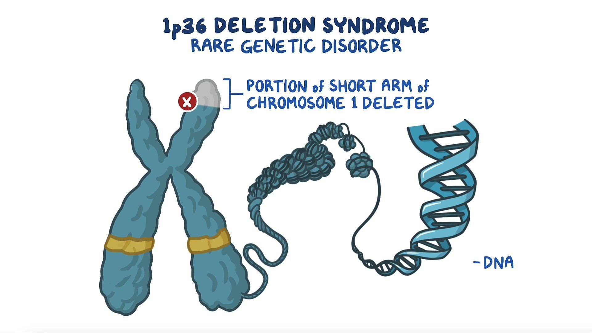
3p deletion syndrome is a rare genetic disorder caused by the microdeletion of a specific segment on the short arm (p) of chromosome 3, resulting in developmental delays, intellectual disability, distinct craniofacial features, and variable congenital malformations. The clinical presentation varies widely depending on the exact size and location of the deleted genetic material, requiring a multidisciplinary approach for comprehensive patient management.
Introduction to 3p Deletion Syndrome
Chromosome abnormalities represent a significant category of congenital disorders, among which structural deletions account for complex and diverse clinical phenotypes. 3p deletion syndrome, sometimes referred to as monosomy 3p partial, is a rare chromosomal anomaly characterized by the loss of genetic material from the short arm of chromosome 3. Because chromosomes contain thousands of individual genes, the deletion of a specific region can disrupt normal embryonic development, organogenesis, and neurological maturation.
The clinical spectrum of 3p deletion syndrome is notably heterogeneous. While some individuals experience profound developmental delays and multiple congenital anomalies, others exhibit milder cognitive impairment and subtle physical differences. This variability depends significantly on the breakpoints of the deletion—the exact locations where the chromosome broke and rejoined during genetic rearrangement—and whether the deletion is terminal (at the very tip of the chromosome) or interstitial (an internal segment missing). Understanding the nuances of this condition requires examining its genetic foundations, diagnostic procedures, phenotypic manifestations, and the long-term supportive care strategies essential for affected individuals and their families.
Medical and legal disclaimer: The following information is for educational and informational purposes only and should not be used as a substitute for professional medical advice, diagnosis, or treatment. Always seek the advice of a qualified healthcare provider regarding any medical condition.
The Genetic Basis of 3p Deletion
At the molecular level, human genetic information is organized into 23 pairs of chromosomes, each possessing a long arm (designated "q") and a short arm (designated "p"). Chromosome 3 spans approximately 198 million base pairs and contains hundreds of protein-coding genes. In 3p deletion syndrome, a portion of this short arm is missing, leading to a state of hemizygosity for the genes located within the deleted interval. Hemizygosity means that instead of having two functional copies of each gene (one inherited from each parent), the individual possesses only a single copy.
Terminal Versus Interstitial Deletions
The genomic alterations underlying 3p deletion syndrome generally fall into two structural categories:
- Terminal Deletions: These occur when a break happens near the end of the chromosome arm, and the distal segment is lost. Terminal deletions often involve the telomeric region and tend to present with a more consistent and recognizable syndrome constellation, particularly when they involve the 3p25-3p26 regions.
- Interstitial Deletions: These occur when two breaks happen within the chromosome arm, and the intervening segment is lost while the ends rejoin. Interstitial deletions can occur anywhere along the 3p arm and produce symptoms that directly correlate with the specific genes contained within the missing interval.
Dosage Sensitivity and Critical Regions
The severity of 3p deletion syndrome is largely governed by the concept of gene dosage sensitivity. Many genes require a precise double dose to function normally; when one copy is deleted, the remaining copy cannot produce sufficient protein to maintain normal cellular pathways—a phenomenon known as haploinsufficiency. Researchers have identified specific critical regions on chromosome 3p, such as the 3p25 and 3p26 bands, that house candidate genes heavily implicated in neurological development, craniofacial patterning, and ocular formation. For instance, disruptions in genes like FOXP1 or CNTN6, located on chromosome 3p, are frequently associated with speech delays, cognitive deficits, and neurodevelopmental differences.
Diagnostic Procedures and Cytogenetic Testing
Diagnosing 3p deletion syndrome requires specialized genetic testing, as the physical characteristics alone overlap with numerous other chromosomal microdeletion disorders. Because the deleted segments can be extremely small—sometimes microscopic or sub-microscopic—standard light-microscope karyotyping may fail to detect the abnormality unless it is a large deletion.
Advanced Genomic Technologies
Modern cytogenetic diagnostics employ high-resolution techniques to identify and map chromosomal deletions accurately:
- Chromosomal Microarray Analysis (CMA): Microarray technology, including array comparative genomic hybridization (aCGH) and single nucleotide polymorphism (SNP) arrays, is the gold standard for detecting copy number variations. CMA can precisely determine the size of the deletion, identify the exact breakpoints, and reveal which specific genes are missing.
- Fluorescence In Situ Hybridization (FISH): FISH utilizes fluorescently labeled DNA probes that bind to specific sequences on chromosome 3p. This technique is highly effective for confirming suspected deletions and for testing parents to determine whether the deletion arose de novo (spontaneously during gametogenesis) or was inherited from a parent carrying a balanced translocation.
- G-Banded Karyotyping: While lower-resolution karyotypes may miss small microdeletions, they remain valuable for identifying larger chromosomal rearrangements, such as translocations or inversions involving chromosome 3, which may have led to the unbalanced deletion in the child.
| Diagnostic Method | Primary Purpose | Resolution Level | Limitation |
|---|---|---|---|
| G-Banded Karyotype | Detect large structural rearrangements and major chromosomal anomalies. | Moderate (visible under light microscope, ~5–10 megabases) | Cannot detect small microdeletions. |
| FISH Analysis | Confirm specific targeted deletions or rearrangements using fluorescent probes. | High for targeted regions | Only tests pre-selected loci; does not scan the whole genome. |
| Chromosomal Microarray (CMA) | Detect sub-microscopic gains and losses across the entire genome. | Very High (can detect deletions of kilobase scale) | May not detect balanced structural rearrangements (e.g., balanced translocations). |
Clinical Characteristics and Phenotype
The phenotypic expression of 3p deletion syndrome encompasses a wide array of physical, developmental, and medical findings. Because the syndrome is rare—with only a few hundred cases documented in medical literature—the full clinical spectrum continues to be defined as diagnostic testing identifies individuals with milder or atypical deletions.
Craniofacial and Physical Features
Many individuals with 3p deletion syndrome exhibit subtle or distinctive facial dysmorphism. While these features rarely cause medical complications on their own, they serve as important diagnostic clues for clinical geneticists. Common observations include:
- Microcephaly (an abnormally small head size) or trigonocephaly (a triangular-shaped forehead caused by premature fusion of the metopic suture).
- Hypertelorism (increased distance between the eyes) or epicanthal folds.
- A depressed nasal bridge, broad nasal tip, or anteverted nares (upturned nostrils).
- Micrognathia (a small lower jaw) or retrognathia (receding chin).
- Low-set or abnormally shaped ears.
Neurological and Developmental Manifestations
Neurological involvement is a core component of 3p deletion syndrome. The impact on the central nervous system typically manifests across multiple developmental domains:
- Global Developmental Delay: Milestones such as sitting, crawling, and walking are frequently delayed. Gross and fine motor skill acquisition often requires physical and occupational therapy intervention.
- Intellectual Disability: The degree of cognitive impairment ranges from mild to severe, heavily influenced by the extent of the genetic deletion and the specific neural genes involved.
- Speech and Language Impairment: Expressive language is often significantly more affected than receptive language. Many individuals experience motor speech disorders, such as childhood apraxia of speech, or structural oral challenges that hinder articulation.
- Behavioral Differences: Some children exhibit features of autism spectrum disorder, attention-deficit/hyperactivity disorder (ADHD), anxiety, or sensory processing sensitivities.
Congenital Malformations and Systemic Health Issues
Depending on the exact genetic material lost, individuals with 3p deletion syndrome may present with structural anomalies affecting various internal organ systems:
- Cardiac Defects: Congenital heart anomalies, such as ventricular septal defects (VSD), atrial septal defects (ASD), or patent ductus arteriosus (PDA), occur in a subset of patients and require pediatric cardiology evaluation.
- Ophthalmologic Findings: Structural eye anomalies, strabismus (crossed eyes), ptosis (drooping eyelids), or refractive errors are frequently reported.
- Gastrointestinal and Feeding Difficulties: Infants may experience hypotonia (low muscle tone) affecting suck-and-swallow reflexes, gastroesophageal reflux disease (GERD), and failure to thrive in early infancy.
- Skeletal and Limb Differences: Clinodactyly (curved fingers), syndactyly (webbed fingers or toes), or delayed bone age are occasionally observed.
Multidisciplinary Care and Management Pathways
Because 3p deletion syndrome is a multisystem condition, effective management requires a coordinated, multidisciplinary team of medical specialists, therapists, and educators. There is no cure for the underlying chromosomal deletion; therefore, treatment focuses on supportive care, early intervention, and symptom management to maximize the individual's functional independence and quality of life.
Pediatric and Specialty Medical Monitoring
Upon diagnosis, a comprehensive baseline evaluation is essential to identify congenital anomalies and establish ongoing surveillance protocols:
- Clinical Genetics: To discuss the specific deletion, recurrence risks for future pregnancies, and interpretation of microarray results.
- Pediatric Cardiology: To perform an echocardiogram and monitor for structural heart defects.
- Ophthalmology and Audiology: To screen for vision impairments, strabismus, and hearing loss, ensuring sensory deficits do not compound developmental delays.
- Neurology: To evaluate hypotonia, monitor for seizure disorders (which occur in a minority of cases), and manage neurodevelopmental concerns.
Early Intervention and Therapy Services
Therapeutic interventions should begin as early as infancy to capitalize on neuroplasticity and support developmental progress:
- Physical Therapy (PT): Focuses on improving gross motor skills, core strength, balance, and coordination to help children achieve milestones like walking.
- Occupational Therapy (OT): Addresses fine motor skills, sensory processing, and activities of daily living, such as self-feeding and dressing.
- Speech-Language Pathology (SLP): Essential for addressing feeding difficulties in infancy and supporting verbal communication, alternative augmentative communication (AAC) systems, and oral-motor coordination as the child grows.
- Special Education and Individualized Education Programs (IEPs): Tailored educational plans within school systems ensure appropriate accommodations, specialized instruction, and cognitive support.
Prognosis and Long-Term Outlook
The prognosis for individuals with 3p deletion syndrome varies widely and depends primarily on the severity of associated medical complications, particularly congenital heart defects and the degree of intellectual disability. With modern medical care, early surgical correction of congenital anomalies, and proactive developmental therapies, many individuals with 3p deletion syndrome live into adulthood, achieving varying degrees of independence.
Support groups and advocacy organizations play a vital role for families navigating the challenges of rare genetic disorders. Connecting with communities of other families affected by chromosome 3 deletions provides practical guidance, emotional support, and shared resources for long-term care planning, transition to adult medical care, and community integration.
Frequently Asked Questions
What causes 3p deletion syndrome?
3p deletion syndrome is caused by the accidental loss of a small segment of DNA from the short arm of chromosome 3. This deletion typically occurs spontaneously during the formation of reproductive cells (eggs or sperm) or early embryonic development, though it can occasionally be inherited from a parent with a balanced chromosomal translocation.
Is 3p deletion syndrome inherited from parents?
In the vast majority of cases, the deletion occurs de novo, meaning neither parent carries the genetic change, and the recurrence risk for future pregnancies is very low. However, in a small percentage of cases, a parent may carry a balanced structural rearrangement that predisposes them to producing gametes with unbalanced deletions. Parental chromosomal testing via karyotype or microarray is recommended following a child's diagnosis.
How is 3p deletion syndrome diagnosed?
The condition is diagnosed using advanced cytogenetic testing, most commonly a chromosomal microarray analysis (CMA). CMA can precisely detect the loss of genetic material, measure the size of the deleted region, and identify which specific genes are involved. Fluorescence in situ hybridization (FISH) is also used to confirm specific deletions.
What are the most common developmental challenges associated with this condition?
Most individuals experience global developmental delays, intellectual disability of varying severity, and significant speech and language impairments. Motor delays stemming from infant hypotonia are also common, requiring structured physical and occupational therapy.
Can individuals with 3p deletion syndrome live independently?
The level of independence an affected individual can achieve depends heavily on the size of the deletion and the severity of intellectual and physical impairments. Some individuals with smaller interstitial deletions or milder phenotypes may acquire skills for semi-independent living with appropriate support, while those with larger deletions and significant medical complexities require lifelong care and assistance.