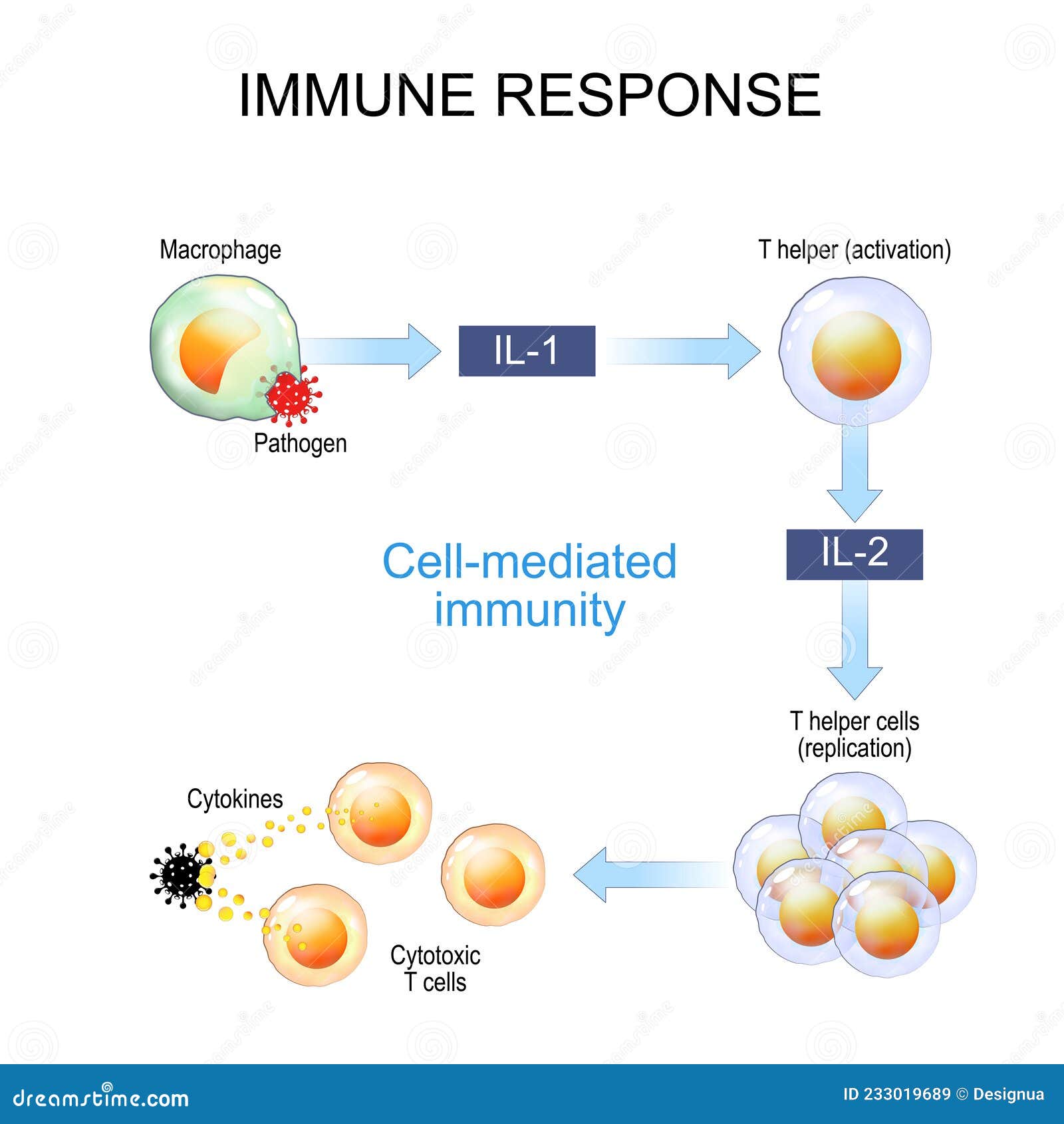
Cell-mediated immunity is a specialized immune response that identifies and destroys intracellular pathogens, such as viruses and certain bacteria, as well as cancerous cells. This process is driven primarily by T-lymphocytes and does not rely on antibodies, instead using a precise sequence of antigen recognition, T-cell activation, proliferation, and direct cellular attack.
Cell-mediated immunity (CMI) represents one of the two primary branches of the adaptive immune system, the other being humoral immunity. While humoral immunity focuses on pathogens circulating in the "humors" (fluids like blood and lymph) through B-cells and antibodies, cell-mediated immunity is the body’s primary defense against "hidden" threats—those that have already invaded host cells or those that have become malignant.
As of 2026, our understanding of the nuances within T-cell signaling has reached a point where we can precisely map the journey of a T-lymphocyte from a naive state to an effector powerhouse. This journey is traditionally categorized into seven distinct steps that ensure the immune system responds with enough force to clear an infection while maintaining enough control to prevent damage to healthy tissue.
1. Antigen Entry and Processing by Phagocytes
The process begins when a pathogen, such as a virus, intracellular bacterium (like Mycobacterium tuberculosis), or parasite, enters the body. Unlike B-cells, which can recognize antigens in their native, three-dimensional form, T-cells are "blind" to whole pathogens. They can only "see" short peptide fragments of an antigen that are displayed on the surface of other cells.
The first step involves specialized cells known as Antigen-Presenting Cells (APCs), most notably dendritic cells and macrophages. These cells act as sentinels. When they encounter a foreign invader, they engulf it through a process called phagocytosis or endocytosis. Once inside the APC, the pathogen is trapped in a vesicle (a phagosome), which then fuses with a lysosome containing digestive enzymes.
These enzymes break down the complex proteins of the pathogen into smaller peptide chains, typically 8 to 20 amino acids long. This "processing" is critical; without it, the molecular "fingerprint" of the pathogen would remain hidden inside the cell, and the rest of the immune system would never be alerted to its presence.
2. Antigen Presentation via MHC Molecules
Once the antigen has been processed into manageable fragments, the APC must "present" these fragments to the T-cells. This is achieved using specialized transport proteins called Major Histocompatibility Complex (MHC) molecules.
There are two primary types of MHC molecules involved in this step:
- MHC Class I: These are found on nearly all nucleated cells in the body. They typically present "endogenous" antigens—proteins produced inside the cell, such as viral proteins or mutated cancer proteins.
- MHC Class II: These are found almost exclusively on professional APCs (dendritic cells, macrophages, and B-cells). They present "exogenous" antigens that have been brought into the cell from the outside.
The processed peptide fragment is loaded onto the MHC molecule within the endoplasmic reticulum or endocytic vesicles and then transported to the cell surface. The resulting MHC-peptide complex acts as a biological "billboard," signaling to any passing T-cell that an intruder has been detected. For a comprehensive look at how these structures interact, resources such as the National Center for Biotechnology Information (NCBI) provide detailed molecular maps of MHC loading pathways.
3. T-Cell Recognition and Binding
In the third step, a naive T-cell—one that has not yet encountered its specific antigen—interacts with the APC. This is the moment of high specificity. Each T-cell possesses thousands of identical T-cell Receptors (TCRs) on its surface, and each TCR is unique to a specific antigen.
The T-cell "scans" the MHC-peptide complexes on the APC. For an immune response to move forward, the TCR must recognize and bind to both the MHC molecule and the specific peptide fragment nestled within it. This is often referred to as "MHC restriction."
To stabilize this connection, co-receptors are required:
- CD4+ T-cells (Helper T-cells): Bind to MHC Class II molecules.
- CD8+ T-cells (Cytotoxic T-cells): Bind to MHC Class I molecules.
This binding event forms what is known as the immunological synapse, a specialized junction where signals are exchanged between the two cells. However, binding alone is not enough to activate the T-cell; a "safety catch" is in place to prevent accidental activation against the body’s own tissues.
4. Co-stimulation: The Second Signal
The fourth step is the critical "fail-safe" of the immune system. For a T-cell to become fully activated, it requires a second signal in addition to the TCR-MHC binding. This is known as co-stimulation.
The most well-studied co-stimulatory interaction involves the B7 protein on the surface of the APC and the CD28 receptor on the T-cell. APCs only express high levels of B7 when they have been "alarmed" by signals from the innate immune system, such as Pathogen-Associated Molecular Patterns (PAMPs).
If a T-cell binds to an antigen but does not receive this second signal, it does not activate. Instead, it enters a state of anergy (a form of cellular "sleep") or undergoes apoptosis (programmed cell death). This mechanism is vital for preventing autoimmunity; it ensures that T-cells only attack when there is a genuine, confirmed threat accompanied by inflammatory "danger" signals.
5. T-Cell Activation and Clonal Expansion
Once both signals are received, the T-cell undergoes a dramatic transformation. It enters the cell cycle and begins to produce a potent growth factor called Interleukin-2 (IL-2), while simultaneously expressing high-affinity IL-2 receptors on its surface.
This creates an autocrine feedback loop: the T-cell stimulates itself to divide. Over the next several days, the single T-cell that recognized the antigen undergoes rapid, repeated rounds of mitosis. This process, called clonal expansion, turns one specific T-cell into an "army" of thousands of identical cells, all programmed to recognize the exact same antigen.
During this phase, the metabolic demands on the T-cells increase exponentially. They shift from oxidative phosphorylation to aerobic glycolysis (the Warburg effect) to support the rapid synthesis of biomass needed for such massive proliferation. This expansion ensures that there are enough effector cells to track down and eliminate the pathogen throughout the entire body.
6. Differentiation into Effector and Memory Cells
As the T-cell population expands, the cells begin to differentiate into different functional types based on the cytokine environment provided by the APCs and the surrounding tissue.
- Effector T-cells: These are the "soldiers" ready for immediate action. CD4+ cells differentiate into various subtypes (Th1, Th2, Th17) that secrete cytokines to coordinate the broader immune response. CD8+ cells differentiate into Cytotoxic T-Lymphocytes (CTLs), which are the primary executioners of the cell-mediated response.
- Memory T-cells: A subset of the expanded clones becomes long-lived memory cells. These cells do not participate in the current fight but instead remain in the lymph nodes, blood, or peripheral tissues for years—sometimes a lifetime. If the same pathogen ever returns, these memory cells can bypass several of the initial activation steps, mounting a faster and more aggressive response.
The 2026 guidelines for vaccine development emphasize the importance of generating "Tissue-Resident Memory T-cells" (Trm), which stay at the site of initial entry (like the lungs or skin) to provide immediate protection against reinfection.
7. Elimination: The Effector Phase and Target Cell Death
The final step is the execution of the immune mission. Effector T-cells leave the lymphatic system and migrate to the site of infection, guided by chemical signals called chemokines.
When a Cytotoxic T-cell (CTL) encounters an infected cell presenting the specific foreign antigen on its MHC Class I molecules, it docks with the target cell and delivers a "lethal hit" through two primary mechanisms:
- Perforin and Granzymes: The CTL releases perforin, a protein that creates pores in the target cell's membrane. It then injects granzymes (proteases) through these pores. Granzymes trigger a cascade within the target cell that leads to apoptosis (programmed suicide).
- Fas-FasL Interaction: The CTL expresses a protein called Fas Ligand (FasL) that binds to the "death receptor" (Fas) on the target cell, further signaling it to undergo apoptosis.
Because the target cell dies via apoptosis rather than bursting (necrosis), the pathogens inside are not simply spilled out. Instead, they are contained within "apoptotic bodies" that are quickly and cleanly cleaned up by macrophages. Once the threat is neutralized, most effector cells die off to prevent chronic inflammation, leaving only the memory cells behind.
Comparison of Key T-Cell Subsets in Cell-Mediated Immunity
| Feature | CD4+ Helper T-Cells | CD8+ Cytotoxic T-Cells |
|---|---|---|
| MHC Interaction | MHC Class II | MHC Class I |
| Primary Goal | Orchestration and signaling | Direct destruction of infected cells |
| Target Cells | Professional APCs | Any nucleated cell (infected/malignant) |
| Effector Molecules | Cytokines (IL-2, IFN-γ, TNF-α) | Perforin, Granzymes, Fas Ligand |
| Key Function | Activates B-cells and Macrophages | Induces apoptosis in target cells |
| Clonal Expansion | High | Very High |
The Role of Cytokines in Coordinating the Response
While the seven steps describe the lifecycle of a T-cell, the entire process is "oiled" by cytokines—small signaling proteins. Interferon-gamma (IFN-γ), for example, is produced by Th1 cells and is essential for activating macrophages, making them much more efficient at killing the pathogens they have ingested.
Without these chemical messengers, cell-mediated immunity would be a series of isolated cellular events rather than a coordinated system-wide defense. Research published by the Nature Portfolio indicates that the specific "flavor" of the cytokine cocktail during Step 6 determines whether the body focuses on fighting a virus, a parasite, or an extracellular fungus.
Clinical Relevance and Therapeutic Applications
Understanding the 7 steps of cell-mediated immunity has revolutionized modern medicine. By the mid-2020s, several therapeutic fields have matured by leveraging these pathways:
- Cancer Immunotherapy: Treatments like CAR-T cell therapy involve taking a patient’s T-cells and genetically engineering them to recognize cancer antigens, essentially skipping Steps 1 through 4 and jumping straight to clonal expansion and elimination.
- mRNA Vaccines: While early vaccines focused on antibodies, modern mRNA platforms are designed to robustly stimulate the cell-mediated branch, ensuring long-term T-cell memory which is often more effective against viral variants than antibodies alone.
- Autoimmune Management: By understanding the "second signal" in Step 4, researchers have developed drugs (co-stimulation blockers) that prevent the immune system from attacking the body’s own organs, such as in rheumatoid arthritis or organ transplant rejection.
Common Obstacles to Effective Cell-Mediated Immunity
The cell-mediated response is not infallible. Certain pathogens and conditions have evolved to interfere with specific steps in the process:
- MHC Downregulation: Viruses like Herpes Simplex Virus (HSV) and many cancer cells can stop the production of MHC Class I molecules. If there is no MHC "billboard" (Step 2), the T-cell cannot "see" the antigen (Step 3).
- T-cell Exhaustion: In chronic infections like HIV or Hepatitis C, constant antigen exposure leads to "exhaustion," where T-cells lose their ability to proliferate or kill (Steps 5 and 7).
- Checkpoint Inhibition: Some tumors express proteins (like PD-L1) that send a "stop" signal to T-cells, effectively hijacking the co-stimulation/inhibitory pathways of Step 4 to turn the T-cell off.
FAQs about Cell-Mediated Immunity
Does cell-mediated immunity involve antibodies?
No, cell-mediated immunity does not utilize antibodies. Antibodies are the primary tools of the humoral immune system, produced by B-cells. However, the two systems often work together; for example, Helper T-cells (CD4+) are required to signal B-cells to produce high-quality antibodies.
How long does it take for the 7 steps to complete?
A primary response—the first time the body sees a pathogen—usually takes between 5 to 10 days to reach its peak. This is because it takes time for the single naive T-cell to find its matching antigen and undergo the massive clonal expansion required in Step 5.
What is the difference between MHC I and MHC II?
MHC Class I is present on almost all cells and tells the immune system "I am infected; kill me." MHC Class II is only on professional immune cells and tells the immune system "I found this intruder elsewhere; here is what it looks like so you can go find others."
Can you have cell-mediated immunity without a thymus?
The thymus is where T-cells "mature" and learn to distinguish self from non-self. In conditions like DiGeorge Syndrome, where the thymus is absent or underdeveloped, cell-mediated immunity is severely compromised, leading to frequent and life-threatening infections.
Is cell-mediated immunity permanent?
The effector cells die off once an infection is cleared, but the memory T-cells created in Step 6 can last for decades. This is the biological basis for the "long-term immunity" provided by many vaccines.
Practical Recommendations for Supporting Immune Health
Maintaining a robust cell-mediated immune system involves supporting the health of your T-cells and the APCs that prime them.
- Nutritional Support: Ensure adequate intake of Vitamin D and Zinc. Vitamin D is a known modulator of T-cell activation, and deficiency has been linked to impaired T-cell responses.
- Vaccination: Stay current with vaccinations, particularly those designed to elicit a T-cell response. As of 2026, many annual boosters for respiratory viruses are optimized to refresh the memory T-cell pool.
- Chronic Stress Management: High levels of cortisol are known to suppress T-cell proliferation (Step 5) and cytokine production, making the body more susceptible to intracellular pathogens.
- Clinical Screening: If you experience recurrent viral or fungal infections, consult an immunologist. They can perform tests like a "Delayed-Type Hypersensitivity" (DTH) skin test or T-cell count panels to verify if your cell-mediated pathways are functioning correctly.