A pseudocyst is a circumscribed, fluid-filled collection that lacks an epithelial lining, distinguishing it from a true cyst which is encased by a distinct cellular membrane. In clinical practice, the term most frequently refers to a pancreatic pseudocyst, a common complication of pancreatitis characterized by an accumulation of enzyme-rich fluid, tissue debris, and blood sequestered by inflammatory fibrous walls.
Distinguishing Between True Cysts and Pseudocysts
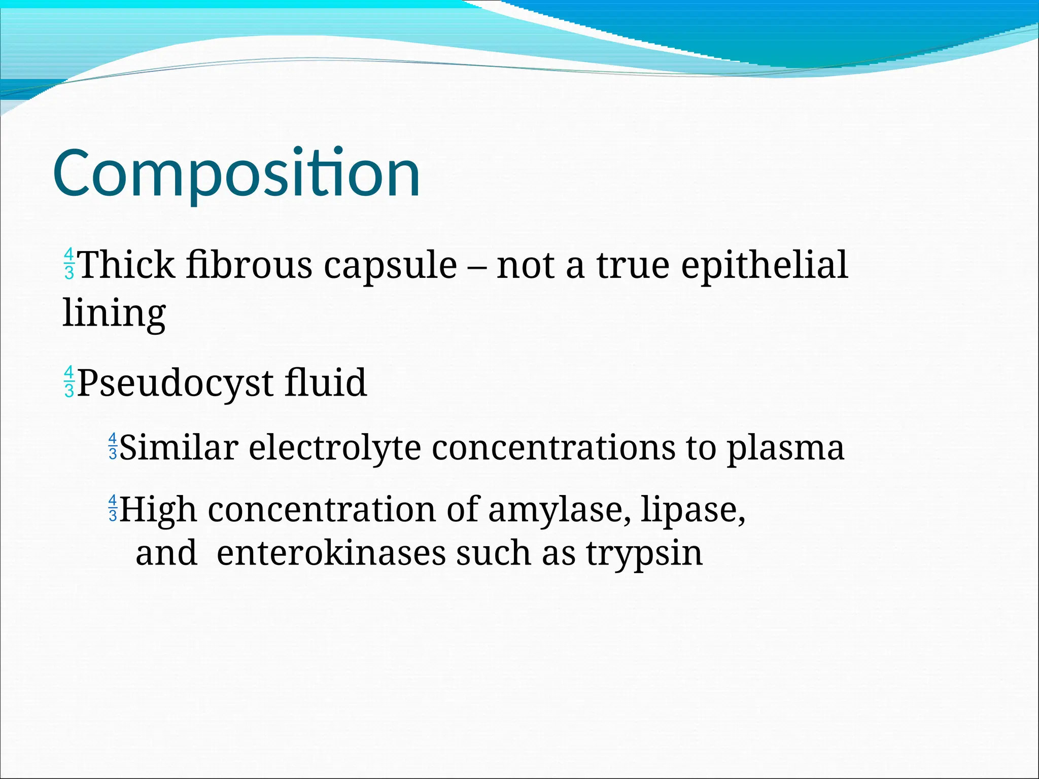
The fundamental difference between a true cyst and a pseudocyst lies in the biological construction of the "wall" that contains the fluid. A true cyst is a pathological cavity lined by an epithelium—a layer of specialized cells that actively secrete or regulate the fluid content within the sac. Because these cells belong to the body's natural lining, they provide a stable, distinct barrier between the cystic contents and the surrounding healthy tissue.
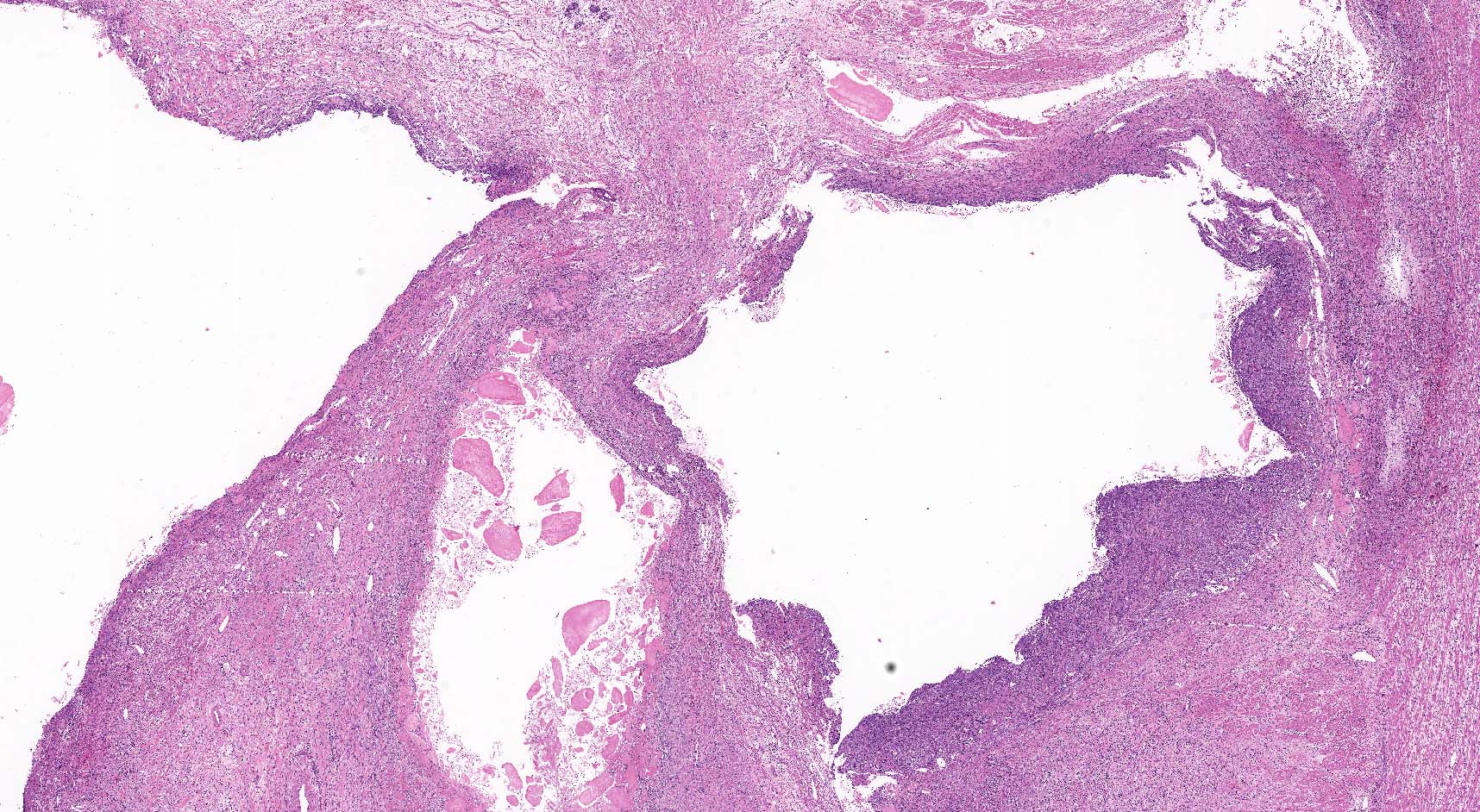
In contrast, a pseudocyst is defined by the absence of this epithelial lining. Instead, the boundary is formed by reactive granulation tissue and fibrous connective tissue. This inflammatory wall forms when fluid—often originating from a localized injury, trauma, or organ inflammation—leaks into the surrounding anatomical spaces. The body attempts to "wall off" this irritating fluid to prevent it from damaging nearby organs, creating a sac out of hardened, scar-like tissue rather than functional, protective epithelium.
The Most Common Context: Pancreatic Pseudocysts
While the medical literature describes various fluid collections throughout the body (such as post-traumatic seromas or specific types of renal collections), the most clinically significant usage of the term is in relation to the pancreas. A pancreatic pseudocyst typically develops several weeks after an episode of acute or chronic pancreatitis.
When the pancreatic ducts become damaged due to inflammation or blockage, pancreatic juices—which contain potent digestive enzymes—leak out of the ductal system into the lesser sac of the abdomen or the retroperitoneal space. The body’s immune response initiates a localized inflammatory process, laying down a fibrous capsule around the leaked enzymes. Because these enzymes are designed to break down organic matter, the resulting collection inside the pseudocyst remains highly caustic. If the wall of the pseudocyst becomes compromised, these enzymes can cause severe damage to adjacent structures, including blood vessels (leading to pseudoaneurysms) or the stomach and duodenum.
Diagnostic Criteria and Imaging Techniques
Diagnosing a pseudocyst requires advanced imaging, as clinical symptoms alone—such as abdominal pain, nausea, or a palpable mass—are often non-specific. Medical professionals rely on several modalities to confirm the definition and physical characteristics of the collection:
- Computed Tomography (CT) Scans: This is the gold standard for identifying the location, size, and relationship of the pseudocyst to surrounding organs. A CT with intravenous contrast allows radiologists to visualize the thick, fibrous rim of the pseudocyst and assess whether it is exerting mass effect on the bile ducts or the gastrointestinal tract.
- Magnetic Resonance Imaging (MRI) and MRCP: Magnetic Resonance Cholangiopancreatography (MRCP) is particularly useful for visualizing the pancreatic ductal system. It helps doctors determine if the pseudocyst communicates with the main pancreatic duct, which is a critical factor in determining whether the condition will resolve spontaneously or require intervention.
- Endoscopic Ultrasound (EUS): This procedure involves passing an endoscope with an integrated ultrasound probe into the stomach or duodenum. It provides the highest resolution images of the pseudocyst wall and its contents. EUS also allows for fine-needle aspiration (FNA) if there is clinical doubt regarding whether the mass is a pseudocyst or a cystic neoplasm (a potential cancer that masquerades as a benign cyst).
Clinical Implications of Fluid Composition
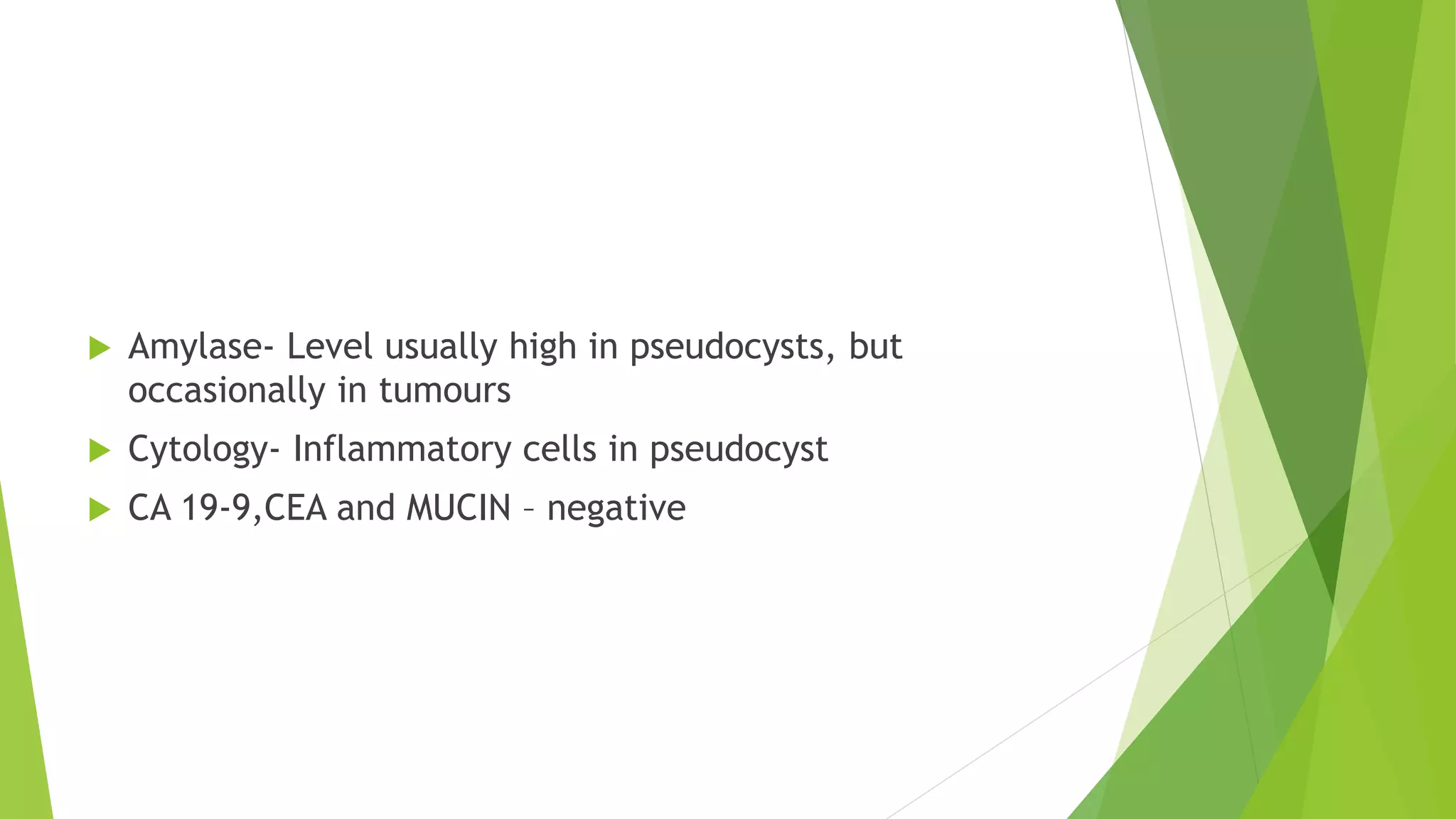
The contents of a pseudocyst are heterogeneous. In the context of the pancreas, the fluid typically contains high levels of amylase and lipase. Because the wall is not an epithelium but a fibrous reaction, the stability of the pseudocyst depends entirely on the thickness and maturity of this wall.
A significant danger associated with these collections is infection. If bacteria enter the pseudocyst, it can evolve into an infected pseudocyst or a pancreatic abscess. This is a life-threatening complication that changes the treatment priority from observation to urgent drainage and aggressive antibiotic therapy. Furthermore, the pressure exerted by a large pseudocyst can cause early satiety (feeling full quickly), gastric outlet obstruction, or biliary obstruction, which manifests as jaundice.
Comparison of Diagnostic and Therapeutic Features
The following table summarizes the key distinctions between various fluid collections often confused with a pseudocyst.
| Feature | Pancreatic Pseudocyst | True Cyst | Abscess |
|---|---|---|---|
| Lining | Fibrous tissue (no epithelium) | Epithelial lining | Inflammatory pus/debris |
| Origin | Post-pancreatitis or trauma | Developmental or neoplastic | Infection/Bacterial |
| Fluid Type | Enzyme-rich pancreatic juice | Clear or mucinous fluid | Purulent (pus) |
| Communication | Often with pancreatic duct | Usually isolated | N/A |
| Management | Observation or drainage | Surgery or surveillance | Drainage and antibiotics |
Management and Treatment Strategies
Management of a pseudocyst depends heavily on its size, the presence of symptoms, and evidence of complications. Historically, a "wait and see" approach was common, as many pseudocysts resolve on their own as the underlying inflammation subsides. However, modern 2026 protocols favor a more proactive approach if the patient is symptomatic.
- Observation: Small, asymptomatic pseudocysts are often monitored with serial imaging to ensure they do not expand or cause secondary complications.
- Endoscopic Drainage: This has become the preferred intervention for stable, symptomatic pseudocysts. Using EUS guidance, a stent is placed between the stomach or duodenum and the pseudocyst, allowing it to drain internally. This avoids the need for major abdominal surgery.
- Percutaneous Drainage: In cases where endoscopic access is difficult or the patient is not a candidate for sedation, a catheter may be placed through the skin under ultrasound or CT guidance to drain the fluid into an external bag.
- Surgical Intervention: Open or laparoscopic surgery (such as cystogastrostomy) is generally reserved for complex cases where there is suspicion of malignancy or if endoscopic techniques have failed.
Important Limitations and Professional Advice
It is essential to recognize that a "pseudocyst" is a broad descriptive term. While this article focuses on the clinical definition in the context of general pathology and gastroenterology, the appearance of any unexplained abdominal mass necessitates professional medical evaluation.
Medical Disclaimer: This information is for educational purposes only and does not constitute medical advice, diagnosis, or treatment. Always seek the advice of a physician or other qualified health provider with any questions regarding a medical condition. Never disregard professional medical advice or delay in seeking it because of something you have read here.
Frequently Asked Questions
1. Is a pseudocyst considered a type of cancer? No, a pseudocyst is a benign, reactive fluid collection. However, some cystic tumors of the pancreas can appear similar to pseudocysts on imaging. It is crucial for a specialist to perform diagnostics, such as an endoscopic ultrasound, to rule out cystic neoplasms.
2. Can a pseudocyst disappear on its own? Yes. Many pseudocysts, particularly those that are small and not associated with active pancreatic duct leakage, will undergo spontaneous regression as the body reabsorbs the fluid during the healing phase of pancreatitis.
3. Why is it called a "pseudo" cyst? It is called a "pseudo" (false) cyst because it lacks the specific biological epithelial lining that defines a "true" cyst. It is essentially a collection of fluid held together by a scar-tissue barrier, rather than a self-contained biological sac.
4. What happens if a pseudocyst bursts? A ruptured pseudocyst is a medical emergency. The leakage of concentrated digestive enzymes into the peritoneal cavity causes chemical peritonitis, leading to severe, acute abdominal pain, systemic inflammation, and potential organ failure. Immediate surgical or intensive care intervention is required.
5. How long does it take for a pseudocyst to form? A pseudocyst typically takes several weeks to develop following an acute inflammatory event. It is rarely present immediately at the onset of symptoms and usually appears as the patient is recovering from the acute phase of pancreatitis.
Recommended Next Steps for Patients
If you have been diagnosed with a pancreatic fluid collection, the most important step is to be evaluated by a gastroenterologist specializing in pancreatic diseases.
- Request a Detailed Imaging Report: Ensure your physician has completed a cross-sectional imaging study (CT or MRI) to establish the baseline size and anatomical location of the collection.
- Monitor for "Red Flag" Symptoms: Seek immediate emergency care if you experience high fever, uncontrollable vomiting, severe jaundice (yellowing of the skin/eyes), or sharp, stabbing abdominal pain that is not relieved by standard medications.
- Review the Underlying Cause: Since pseudocysts are usually a consequence of pancreatitis, focus your long-term management on the cause of that inflammation (e.g., alcohol cessation, gallstone management, or dietary changes) to prevent recurrent pseudocyst formation.