T2c prostate cancer is a specific clinical classification within the TNM staging system indicating that a tumor is palpable, visible, or clinically identifiable, involving more than half of one lobe of the prostate gland without extending outside the capsule. Understanding this diagnosis requires looking closely at how staging defines tumor volume, the clinical implications for treatment selection, and the role of ongoing active surveillance versus intervention.
1. What Is T2c Prostate Cancer? Defining the Clinical Stage
The journey toward understanding any cancer diagnosis begins with accurate staging. In oncology, staging describes the severity of an individual's cancer based on the size and extent of the primary tumor and whether it has spread to lymph nodes or distant sites. For prostate cancer, physicians universally rely on the TNM staging system, maintained by the American Joint Committee on Cancer (AJCC) and the Union for International Cancer Control (UICC). Within this framework, the "T" category specifically describes the extent of the primary tumor within the prostate itself.
Clinically localized prostate cancer stages range from T1 to T4. Stages T1 and T2 describe tumors that are confined entirely within the prostate gland. While T1 tumors are typically undetectable by digital rectal examination (DRE) or imaging and are discovered incidentally—often during surgery for benign prostatic hyperplasia (BPH) or through elevated prostate-specific antigen (PSA) levels leading to a biopsy—T2 tumors are large enough to be felt during a physical examination or visualized clearly on advanced imaging modalities such as multiparametric magnetic resonance imaging (mpMRI).
The T2 category is further subdivided into a, b, and c based on how much of the prostate lobe the tumor occupies:
- T2a: The tumor involves one-half of one lobe of the prostate or less.
- T2b: The tumor involves more than half of one lobe, but still only involves a single lobe.
- T2c: The tumor involves both lobes of the prostate gland.
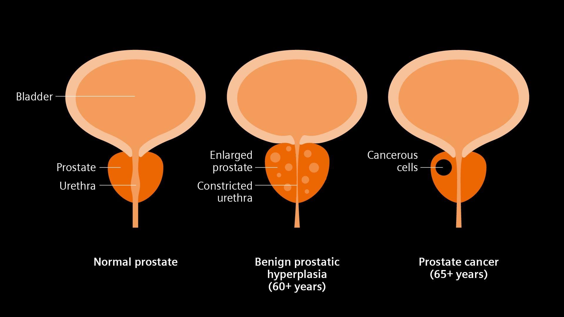
A T2c classification means that the physical examination or imaging has confirmed cancer in both the left and right halves of the prostate. Despite this bilateral involvement, the tumor remains organ-confined. There is no clinical evidence that the cancer has broken through the fibrous outer capsule of the prostate to invade surrounding tissues, such as the seminal vesicles, bladder neck, or pelvic wall, nor has it spread to regional lymph nodes (N0) or distant organs (M0).
Accurate clinical staging like T2c relies heavily on a combination of digital rectal exams, transrectal ultrasound (TRUS), mpMRI, and systematic or targeted prostate biopsies. Pathological findings from biopsy cores—such as the Gleason score and the Grade Group—work hand in hand with the clinical T2c stage to give urologists and oncologists a complete picture of the cancer's aggressiveness and biological potential.
2. Clinical Implications: Tumor Volume and Risk Stratification
A diagnosis of T2c prostate cancer signals a higher tumor burden within the organ compared to T2a or T2b classifications because the disease is bilateral. In medical oncology, tumor volume and extent are critical variables that influence how physicians calculate a patient's overall risk profile. While T2c remains clinically localized, the presence of disease on both sides of the prostate increases the likelihood of higher grade disease or multi-focal cancer growth patterns.
To determine the most appropriate management path, physicians do not rely on staging alone. They combine clinical T-stage with the serum PSA level and the Gleason score (or Grade Group derived from biopsy pathology) to stratify patients into risk groups, as outlined by organizations like the National Comprehensive Cancer Network (NCCN).
| Risk Category | Clinical T-Stage | Gleason Score / Grade Group | Serum PSA Level (ng/mL) |
|---|---|---|---|
| Low Risk | T1 to T2a | Gleason $\le$ 6 (Grade Group 1) | Less than 10 |
| Intermediate Risk | T2b to T2c (or) | Gleason 7 (Grade Group 2 or 3) (or) | 10 to 20 |
| High Risk | T3a or higher (or) | Gleason 8–10 (Grade Group 4 or 5) (or) | Greater than 20 |
Because a T2c classification places the tumor at the upper boundary of the intermediate-risk threshold (even in the absence of adverse pathological features), patients with T2c disease are generally categorized as having unfavorable intermediate-risk or high-risk prostate cancer, depending on their specific Gleason score and PSA velocity.
Higher tumor volume in both lobes means there is a greater statistical probability of microscopic extracapsular extension—cancer cells breaching the prostate capsule invisibly—or microscopic involvement of the seminal vesicles that imaging might miss. Consequently, treatment planning for T2c prostate cancer must account for this increased biological risk. Physicians evaluate whether the cancer is likely to remain locally controlled by prostate-directed therapies or whether it requires more aggressive multi-modality treatment regimens to prevent biochemical recurrence.
3. Treatment and Management Pathways for T2c Disease
Because T2c prostate cancer is localized to the gland but involves both lobes and carries an intermediate-to-high risk profile, management decisions require a nuanced, personalized discussion between the patient and their multidisciplinary care team, typically comprising a urologist, a radiation oncologist, and sometimes a medical oncologist.
While active surveillance—closely monitoring the cancer with periodic PSA tests, DREs, and repeat biopsies—is frequently recommended for low-risk, low-volume localized prostate cancer, it is less commonly chosen for higher-volume T2c disease unless the patient has significant competing medical comorbidities or a limited life expectancy. Most patients with T2c tumors pursue active, curative-intent treatments.
Radical Prostatectomy
Surgical removal of the entire prostate gland and seminal vesicles, known as radical prostatectomy, is a primary option for healthy patients with T2c disease.
- Procedure: Performed via open, laparoscopic, or robot-assisted techniques, the surgeon removes the prostate and meticulously reconstructs the urinary tract.
- Lymph Node Dissection: For T2c tumors, surgeons frequently perform a pelvic lymph node dissection during the procedure to accurately stage the regional nodes and remove any microscopic metastatic disease.
- Considerations: While surgery offers definitive pathological staging, potential side effects include temporary or permanent urinary incontinence and erectile dysfunction, depending on whether nerve-sparing techniques can be safely utilized without compromising cancer control.
Radiation Therapy
External beam radiation therapy (EBRT) combined with advanced delivery techniques, such as intensity-modulated radiation therapy (IMRT) or stereotactic body radiation therapy (SBRT), offers a non-surgical alternative with comparable long-term cancer control rates.
- Combination Therapy: For intermediate-to-high risk presentations like T2c, radiation is frequently combined with a short or long course of androgen deprivation therapy (ADT)—hormone therapy that lowers testosterone levels—to shrink the tumor and eradicate microscopic out-of-field cancer cells.
- Brachytherapy: Permanent seed implantation (low-dose-rate brachytherapy) or high-dose-rate brachytherapy boosts may also be integrated into the treatment plan, either alone or supplementing EBRT.
Multidisciplinary Guidance
Selecting between surgery and radiation depends on patient age, baseline urinary and sexual function preferences, anatomical findings on imaging, and detailed pathology reports. Patients should consult specialized cancer centers, such as those designated by the National Cancer Institute (NCI), to explore clinical trials and individualized treatment roadmaps.
Frequently Asked Questions
Does a T2c staging mean my prostate cancer has spread outside the gland?
No. By definition, clinical stage T2c indicates that the cancer is entirely confined within the prostate capsule. The "T2" designation specifies organ-confined disease, while the "c" subpart simply denotes that the tumor is large enough or multifocal enough to be detected in both lobes of the prostate during examination or imaging. However, because T2c represents a higher tumor volume, physicians remain vigilant about the potential for microscopic spread that cannot be seen on standard scans.
Can active surveillance be an option for T2c prostate cancer?
Active surveillance is primarily reserved for low-risk, low-volume localized prostate cancer (typically T1c or T2a with a low Gleason score and low PSA). Because T2c involves both lobes and carries an intermediate-to-high risk profile, active surveillance is rarely the standard recommendation. However, it may be considered for older adults or individuals with significant underlying health conditions where the risks of curative treatment outweigh the risks posed by the slow-growing cancer.
How is T2c prostate cancer diagnosed?
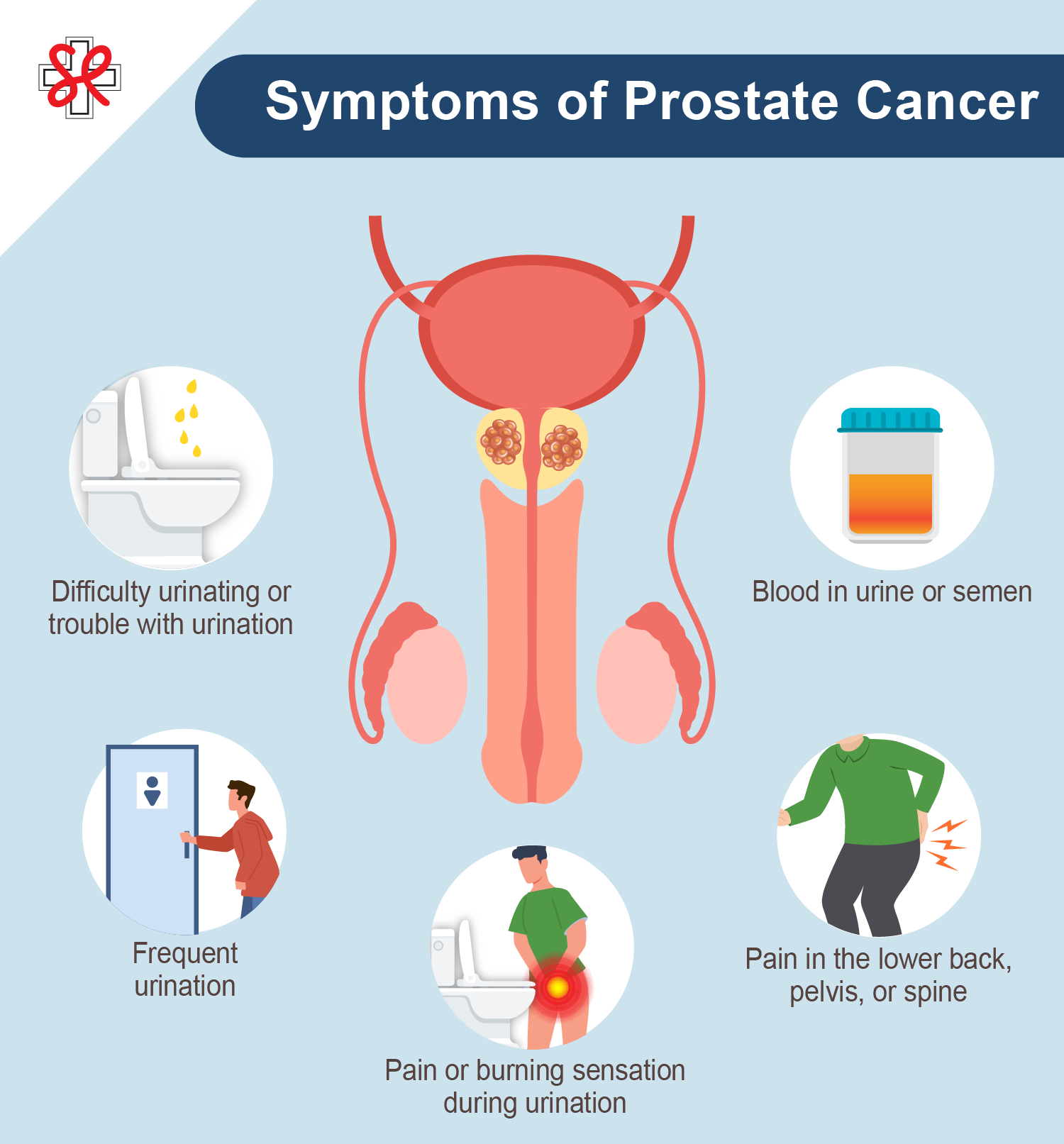
Diagnosing T2c involves a multi-step process. It typically begins with an abnormal digital rectal exam where a physician feels nodules or firmness in both lobes of the prostate, or an elevated PSA test that prompts further imaging. A multiparametric MRI is then often performed to visualize suspicious lesions within both lobes, followed by an ultrasound-guided or MRI-targeted needle biopsy. The pathology from the biopsy confirms the cancer diagnosis and provides the Gleason score necessary for complete risk stratification.
What is the difference between T2b and T2c prostate cancer?
The primary difference lies in the anatomical distribution of the tumor within the prostate gland. The prostate is divided into a right lobe and a left lobe. A T2b tumor involves more than half of a single lobe, but the cancer remains confined to that one side. In contrast, a T2c tumor involves both the right and left lobes of the prostate. This bilateral involvement increases the overall tumor volume and typically places the patient in a higher risk category for treatment planning.
What role does hormone therapy play in treating T2c prostate cancer?
Androgen deprivation therapy (ADT) is frequently used in combination with external beam radiation therapy for intermediate- and high-risk prostate cancers, including T2c disease. Hormone therapy works by reducing testosterone levels, which starves prostate cancer cells of the hormone they need to grow and multiplies the effectiveness of radiation therapy. ADT is rarely used as a standalone treatment for localized T2c disease, as it is not curative on its own in this setting.
Next Steps and Professional Guidance
Receiving a T2c prostate cancer diagnosis requires careful navigation and timely decision-making. Because treatment choices—ranging from robotic-assisted radical prostatectomy to advanced radiation therapy combined with hormone therapy—carry distinct long-term impacts on quality of life, gathering comprehensive information is essential. Patients should request a complete copy of their pathology report, discuss all available options with both a urologist and a radiation oncologist, and consider seeking a second opinion at a comprehensive cancer center to ensure an optimized, individualized treatment plan.