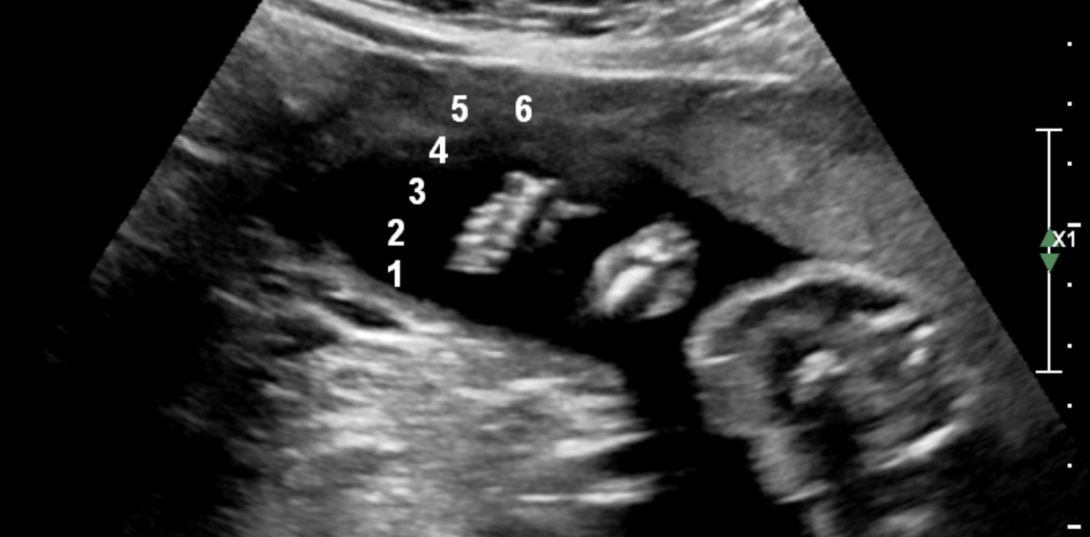
The 20-week scan, also known as the mid-pregnancy anatomy ultrasound, is a detailed screening conducted between 18 and 22 weeks of gestation. It evaluates fetal growth through specific biometrics—Head Circumference (HC), Biparietal Diameter (BPD), Abdominal Circumference (AC), and Femur Length (FL)—while systematically examining internal organs and placental positioning to monitor healthy development.
Medical Disclaimer: The information provided in this article is for educational purposes only and does not constitute medical advice. Always consult with a qualified healthcare provider or sonographer regarding specific ultrasound results or clinical concerns.
The Purpose and Timing of the Mid-Pregnancy Anatomy Scan
In the clinical landscape of 2026, the 20-week scan remains a cornerstone of prenatal care. While earlier screenings focus on viability and genetic markers, the mid-pregnancy scan is primarily an anatomical survey. By 20 weeks, the fetus has reached a size where major organs—such as the heart, kidneys, and brain—are sufficiently developed to be visualized in detail, yet the fetus still has enough room to move, allowing the sonographer to capture various angles.
The primary goals of this scan are threefold:
- Biometric Assessment: Measuring the physical dimensions of the fetus to ensure growth is tracking along expected percentiles.
- Anatomical Survey: A "head-to-toe" inspection of the internal and external structures to rule out structural anomalies.
- Environmental Evaluation: Checking the volume of amniotic fluid, the position of the placenta, and the integrity of the umbilical cord.
As of 2026, many diagnostic centers have integrated AI-assisted biometry software. This technology helps sonographers identify the correct planes for measurement more rapidly, though the final verification and interpretation remain the responsibility of the human clinician.
Core Biometric Measurements Defined
When parents receive their ultrasound report, they are often met with a series of acronyms. These represent the four standard biometric measurements used to calculate the Estimated Fetal Weight (EFW) and confirm gestational age.
Biparietal Diameter (BPD)
The BPD is the diameter of the fetal head measured from one parietal bone to the other. In technical terms, it is a transverse section of the head, typically measured from the "leading edge" (outer surface) of the proximal skull bone to the "leading edge" (inner surface) of the distal skull bone.
The BPD is a reliable indicator of gestational age in the second trimester. However, its accuracy can be influenced by the shape of the head. For instance, if the head is flattened (dolichocephaly) or rounded (brachycephaly), the BPD may appear smaller or larger than expected, necessitating the use of the Cephalic Index to normalize the data.
Head Circumference (HC)
Because head shape can vary, the HC is often considered more reliable than the BPD. The HC measures the total distance around the outside of the fetal skull. The sonographer captures this measurement in the same plane as the BPD, ensuring that specific brain structures—namely the thalami and the cavum septum pellucidum—are visible. This measurement is crucial for monitoring neurological development and ensuring there is no evidence of microcephaly or hydrocephalus.
Abdominal Circumference (AC)
The AC is perhaps the most critical measurement for assessing fetal nutrition and growth. It is taken at the level of the fetal liver and stomach. Specifically, the sonographer looks for the "J-shape" where the umbilical vein meets the left portal vein.
The AC reflects the size of the liver and the amount of subcutaneous fat. If the AC is significantly below the 10th percentile, it may trigger a diagnosis of Fetal Growth Restriction (FGR). Conversely, an elevated AC can sometimes indicate gestational diabetes or macrosomia.
Femur Length (FL)
The FL measures the length of the thigh bone, the longest bone in the human body. The sonographer measures only the ossified (hardened) portion of the diaphysis, excluding the cartilaginous ends. This measurement is a proxy for longitudinal skeletal growth. Significant deviations in FL can occasionally prompt further investigation into skeletal dysplasias or genetic markers, though slight variations are often just a reflection of the parents' stature.
The Anatomical Checklist: What the Sonographer Sees
Beyond the numbers, the 20-week scan involves a rigorous qualitative assessment of fetal anatomy. In many jurisdictions, this follows a standardized checklist often referred to as the "20-point check."
Neurological Structures
The sonographer examines the brain's symmetry and the presence of the cerebellum (responsible for motor control) and the cisterna magna. They also measure the lateral ventricles; if these are enlarged, it may indicate a backup of cerebrospinal fluid. The spine is also viewed in both longitudinal and transverse planes to ensure the vertebrae are properly aligned and the skin covering the spine is intact, ruling out conditions like spina bifida.
The Fetal Heart
The heart is one of the most complex organs to evaluate. The sonographer looks for the "four-chamber view" to ensure the atria and ventricles are proportional. They also check the "outflow tracts"—the pathways where the aorta and pulmonary artery exit the heart—to ensure blood is flowing in the correct directions and that the vessels are not transposed.
The Face and Profile
The scan looks for the presence of the nasal bone and checks the upper lip for any signs of a cleft. In 2026, 3D and 4D imaging are frequently used as adjuncts to 2D imaging to provide a clearer view of facial features if a potential issue is detected on the standard 2D view.
The Abdominal Organs
The sonographer confirms the presence and proper location of the stomach, kidneys, and bladder. Observing fluid in the stomach and bladder is a positive sign, as it indicates the fetus is swallowing amniotic fluid and the kidneys are functioning well enough to produce urine.
Environmental and Placental Considerations
The health of the pregnancy depends heavily on the "support system" inside the uterus.
Placental Location
The placenta's position is noted relative to the cervix. If the placenta is low-lying or covering the cervix (Placenta Previa), it can lead to bleeding later in pregnancy. If a low-lying placenta is noted at 20 weeks, a follow-up scan is usually scheduled for 32–36 weeks, as most placentas "migrate" upward as the uterus expands.
Amniotic Fluid Volume
Amniotic fluid protects the fetus and allows for lung development. The sonographer may estimate the volume subjectively or use the Amniotic Fluid Index (AFI) or Maximum Vertical Pocket (MVP) measurement. Too little fluid (oligohydramnios) or too much (polyhydramnios) requires clinical follow-up.
Umbilical Cord
The cord is checked to ensure it contains three vessels: two arteries and one vein. A "Single Umbilical Artery" (SUA) is a common finding that, while often isolated and harmless, may prompt a more detailed look at the fetal heart and kidneys.
Interpreting the Data: Percentiles and Growth Curves
It is vital for parents to understand that ultrasound measurements are estimates, not absolute values. Results are typically expressed as percentiles.
| Measurement | Definition | Clinical Significance |
|---|---|---|
| BPD | Diameter of the head (ear-to-ear). | Used for dating and detecting head shape anomalies. |
| HC | Total circumference of the skull. | Primary marker for brain volume and growth. |
| AC | Circumference of the mid-abdomen. | Best indicator of fetal weight and nutritional status. |
| FL | Length of the femur (thigh bone). | Reflects skeletal growth and overall height. |
| EFW | Estimated Fetal Weight. | Calculated using a formula (e.g., Hadlock) combining HC, AC, and FL. |
If a measurement is in the 50th percentile, it means 50% of fetuses at that gestational age are smaller and 50% are larger. Anything between the 10th and 90th percentile is generally considered "normal." Even measurements outside this range are not necessarily cause for alarm; they often represent normal biological variation or the genetic influence of the parents.
Practical Considerations for the Day of the Scan
To ensure the best possible images, there are several practical factors for expectant parents to keep in mind.
1. Preparation and Hydration
In 2026, clinical guidelines generally suggest a moderately full bladder for the 20-week scan, especially if the patient has a retroverted (tilted) uterus. A full bladder helps lift the uterus out of the pelvis, providing a better window for the ultrasound waves. However, an overfull bladder can be uncomfortable and may actually distort the view of the cervix. Drinking 16–20 ounces of water an hour before the appointment is usually sufficient.
2. The Impact of BMI and Fetal Position
Ultrasound uses sound waves, which must travel through maternal tissue to reach the fetus. A higher Body Mass Index (BMI) can sometimes result in "suboptimal visualization" because the sound waves attenuate as they pass through adipose tissue. Similarly, if the fetus is in a "breech" or "transverse" position, or has its back to the probe, the sonographer may struggle to see certain structures like the heart or face.
3. Soft Markers
Occasionally, the scan reveals "soft markers." These are not birth defects but are minor variations in anatomy that can be associated with an increased risk of chromosomal conditions. Examples include an Echogenic Intracardiac Focus (EIF) (a bright spot in the heart) or Choroid Plexus Cysts (CPC) (small fluid-filled spaces in the brain). In the absence of other findings, these markers are frequently "normal variants" that resolve on their own.
4. Managing Anxiety
The 20-week scan is often an emotional experience. While it is a chance to "see" the baby, it is also a diagnostic medical procedure. It is helpful to discuss with the sonographer beforehand whether they will talk through the scan or wait until the end to share their findings. Many clinics allow a partner or support person to attend, which can help manage the nerves associated with the anatomy check.
What Happens if an Issue is Identified?
If the sonographer cannot see everything they need to, or if they identify a measurement that falls outside the expected range, several steps may follow:
- Rescan: If the fetus was in a difficult position, you may be asked to walk around, drink cold water, or return in 1–2 weeks for a repeat scan.
- MFM Referral: You may be referred to a Maternal-Fetal Medicine (MFM) specialist (perinatologist) for a "Level II" or "Targeted" ultrasound. These specialists have high-resolution equipment and advanced training in fetal anomalies.
- Genetic Counseling: If structural issues are found, a genetic counselor can explain the options for further testing, such as NIPT (Non-Invasive Prenatal Testing) or amniocentesis.
Frequently Asked Questions
Can the 20-week scan accurately predict the baby's weight at birth?
No. The 20-week scan measures growth at that moment. While it can identify if a fetus is currently smaller or larger than average, it cannot account for the significant growth spurts that occur in the third trimester. It is a snapshot of current health, not a final prediction of birth weight.
Why is my baby's head measurement different from the abdomen measurement?
It is very common for different parts of the body to track on different percentiles. For example, a baby might have a head in the 70th percentile and an abdomen in the 40th. As long as the measurements are within a healthy range and the growth is proportional over time, these differences are usually just a matter of individual genetics.
Is the 20-week scan 100% accurate in detecting anomalies?
While ultrasound technology in 2026 is highly advanced, it is not 100% accurate. Some conditions, such as small heart defects or certain types of brain dysplasia, may not be visible at 20 weeks or may develop later in pregnancy. The scan is a screening tool designed to catch the majority of major structural issues, but it cannot guarantee a "perfect" baby.
Will I definitely find out the sex of the baby at this scan?
In most cases, yes, provided the baby is in a position that allows the sonographer to see the genital area. However, the primary purpose of the scan is medical. If the baby's legs are crossed or the umbilical cord is in the way, the sonographer may not be able to confirm the sex with 100% certainty.
Does a short femur length always mean there is a problem?
Not necessarily. While a very short femur (below the 3rd or 5th percentile) can be a marker for certain conditions, it is often just a sign that the baby will be shorter in stature, especially if the parents are not tall. Clinical context, such as the results of earlier genetic screenings, is essential for interpreting this measurement.
Practical Recommendation for Expectant Parents
As you prepare for your 20-week scan, the best approach is to stay informed but remains patient. Review your previous screening results (like the first-trimester NIPT or NT scan) so you can discuss them with your provider if "soft markers" appear. Wear comfortable, two-piece clothing to allow easy access to your abdomen, and don't hesitate to ask the sonographer to explain what you are seeing on the screen. If the report shows measurements that worry you, schedule a follow-up with your OB-GYN or midwife to discuss the results in the context of your overall health and family history.