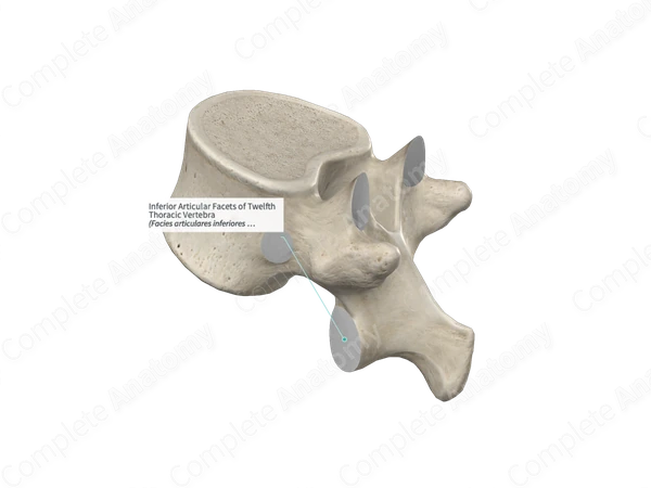
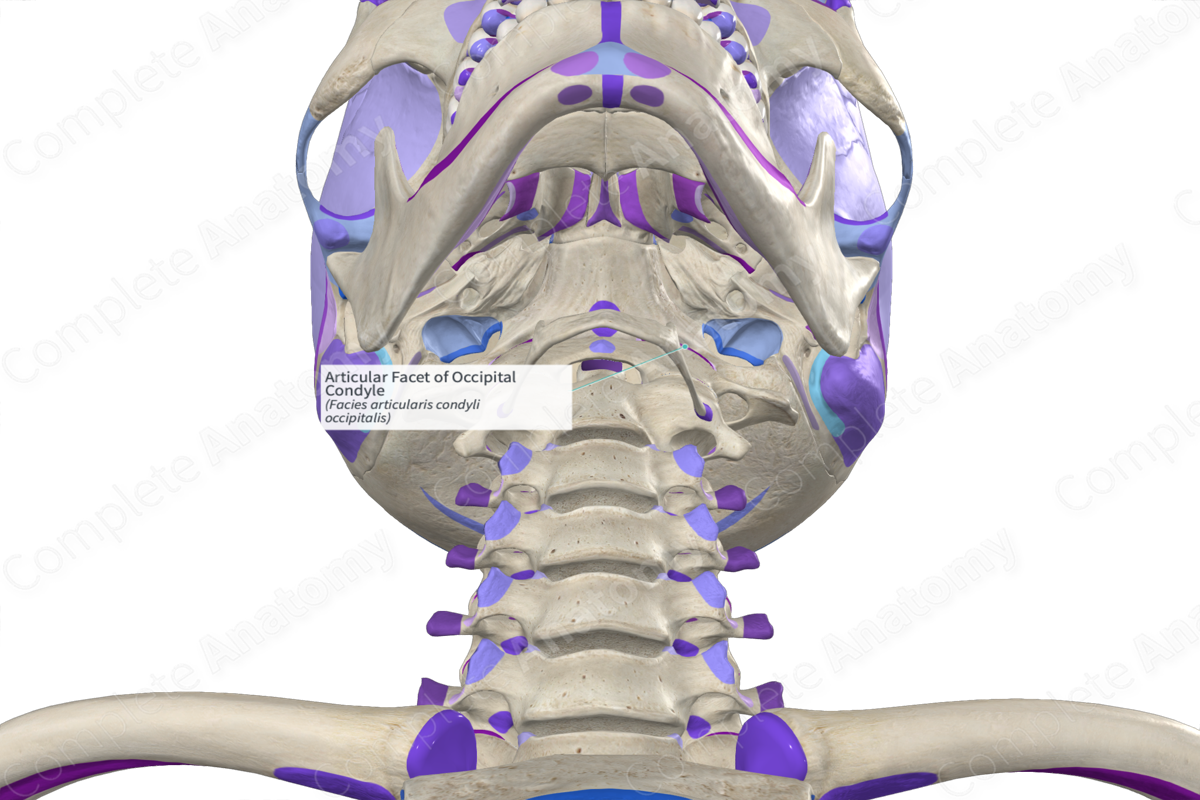
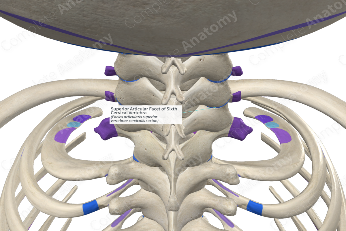
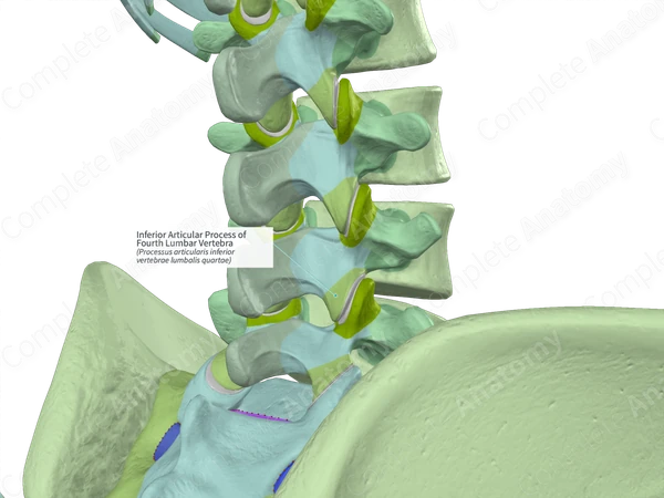
An articular process is a structural bony projection that extends from the vertebral arch, while an articular facet is the specific, smooth, cartilage-covered surface located on that process. In the human spine, the processes act as the physical levers and supports, whereas the facets serve as the functional interface for joint movement and articulation.
The human vertebral column is one of the most complex mechanical structures in the biological world. To understand how it maintains stability while allowing for a wide range of motion—including bending, twisting, and tilting—one must distinguish between the various bony landmarks of a vertebra. Among the most frequently confused terms in musculoskeletal anatomy are "process" and "facet." While they are intimately related and physically connected, they represent two distinct anatomical concepts: the macro-structure (the process) and the micro-functional surface (the facet).
The Articular Process: The Bone's Structural Extension
In anatomical terminology, a process (from the Latin processus) refers to a projection or outgrowth of tissue from a larger body. In the context of the skeletal system, processes serve as attachment points for muscles and ligaments or as structural components that help form joints.
Each typical vertebra in the human body possesses seven processes. These include the single spinous process (which points posteriorly), two transverse processes (which point laterally), and four articular processes. The articular processes are divided into two superior articular processes (pointing upward) and two inferior articular processes (pointing downward).
The Anatomy of the Process
The articular process is a dense, cortical bone structure that emerges from the junction of the pedicle and the lamina, an area often referred to as the "pars interarticularis." As of 2026, clinical biomechanics continues to emphasize the role of these processes in weight-bearing. While the vertebral bodies and intervertebral discs handle the majority of the spine's axial load (approximately 80%), the articular processes and their associated joints manage the remaining 20%.
These processes act as physical barriers that prevent the vertebrae from sliding too far forward or backward (spondylolisthesis). Because they are extensions of the vertebral arch, they provide the necessary leverage for the deep muscles of the back to stabilize the spine. If you were to think of a vertebra as a specialized tool, the process would be the handle or the arm that reaches out to connect with the next tool in the sequence.
The Articular Facet: The Functional Interface
While the process is the "arm," the articular facet is the "palm." A facet is a small, smooth, flat, or slightly specialized surface on a bone. In the spine, the facets are located on the posterior aspect of the superior articular processes and the anterior aspect of the inferior articular processes.
The Composition of the Facet
The articular facet is not merely bare bone. For a joint to function without debilitating friction, the facet is covered in a layer of hyaline cartilage. This cartilage is incredibly smooth and serves to distribute loads across the joint surface while providing a low-friction environment for movement.
When the superior articular facet of one vertebra meets the inferior articular facet of the vertebra above it, they form a zygapophysial joint, more commonly known as a facet joint. These are synovial joints, meaning they are encased in a fibrous capsule and lubricated by synovial fluid. The facet itself is the actual "mating surface" where the two bones communicate.
Orientation and Kinematics
The orientation of the articular facets varies significantly depending on the region of the spine, and this orientation dictates the type of movement permitted:
- Cervical Spine: Facets are oriented at approximately a 45-degree angle to the transverse plane. This "shingle-like" arrangement allows for extensive rotation, flexion, and extension.
- Thoracic Spine: Facets are oriented primarily in the frontal plane. This setup favors lateral bending and some rotation but significantly limits flexion and extension to protect the thoracic cavity and the heart/lungs.
- Lumbar Spine: Facets are oriented in the sagittal plane (curving into a "J" or "C" shape). This orientation is designed to allow significant flexion and extension while strictly limiting rotation, which could otherwise damage the large lumbar intervertebral discs.
Key Differences: Process vs. Facet
To differentiate these two terms effectively, one must look at their scale, composition, and primary role within the kinetic chain.
| Feature | Articular Process | Articular Facet |
|---|---|---|
| Definition | A bony projection or outgrowth from the vertebral arch. | The smooth surface on the process that forms a joint. |
| Physical Nature | Large, three-dimensional structural component. | A specialized, two-dimensional (or slightly curved) area. |
| Tissue Type | Primarily cortical and cancellous bone. | Covered in hyaline (articular) cartilage. |
| Primary Function | Provides leverage and structural "stops" for motion. | Provides a low-friction gliding surface for articulation. |
| Quantity | Four per vertebra (2 superior, 2 inferior). | Four per vertebra (located on each process). |
| Clinical Focus | Fractures (e.g., pars defects), hypertrophy. | Arthritis, cartilage degradation, synovial cysts. |
The Role of Facets and Processes in Spinal Motion
The relationship between the process and the facet is the foundation of spinal biomechanics. When you lean forward (flexion), the inferior articular processes of the upper vertebra slide upward and forward across the superior articular facets of the lower vertebra.
Because the processes are rigid, they determine the "end-feel" of a movement. For example, in the lumbar spine, when you attempt to twist your torso, the bony articular processes physically bump into each other almost immediately. This "bony block" is an essential safety mechanism that prevents the twisting of the spinal cord and the shearing of the intervertebral discs.
The facets, meanwhile, ensure that this movement is fluid. If the hyaline cartilage on the facet wears down—a condition known as facet arthropathy—the smooth gliding motion is replaced by bone-on-bone grinding. This triggers the growth of bone spurs (osteophytes) on the process. In 2026, advanced diagnostic imaging increasingly focuses on the "joint space" between facets to determine the health of the motion segment.
Clinical Implications and Pathologies
Understanding the distinction is vital for interpreting medical reports or studying pathology. Disorders can affect the process, the facet, or both.
Facet Syndrome and Arthropathy
Facet syndrome is a degenerative condition where the cartilage on the articular facet deteriorates. Because the facet joints are highly innervated by the medial branch of the dorsal primary rami (nerves), this wear and tear causes significant localized back pain. Patients often feel "stiff" in the morning because the synovial fluid has not yet lubricated the facet surfaces.
Process Hypertrophy
When facets are stressed due to disc height loss, the body attempts to stabilize the area by thickening the bone. This is known as hypertrophy of the articular processes. While the body intends to help, the enlarged processes can grow into the spinal canal or the neural foramina (the holes where nerves exit), leading to spinal stenosis or radiculopathy (pinched nerves).
Fractures and Spondylolysis
A "stress fracture" typically occurs not on the facet itself, but on the part of the process or the bridge of bone leading to it (the pars interarticularis). This is common in athletes who perform repetitive hyperextension, such as gymnasts or football linemen. If the process or the bridge fails, the structural integrity of the "bony stop" is lost, allowing the vertebra to slip.
Diagnostic Imaging and Identification
In a clinical setting in 2026, radiologists use specific views to visualize these structures:
- Oblique X-rays: Often used to see the "Scotty Dog" appearance of the lumbar spine. The "ear" of the dog is the superior articular process, and the "front leg" is the inferior articular process.
- MRI: The best tool for evaluating the health of the facet, as it can show cartilage thinning, synovial fluid buildup (effusion), and inflammation.
- CT Scan: The gold standard for evaluating the process, as it provides high-definition views of bone density, fractures, and osteophyte (bone spur) formation.
Practical Application for Healthcare Professionals and Students
For students of anatomy or healthcare professionals, a helpful mnemonic is that the Process is the Part (the whole structural piece), while the Facet is the Face (the part that looks at and touches the other bone).
When palpating a patient or reviewing a model:
- You cannot directly feel a facet through the skin; it is deep within the joint capsule.
- You can, however, palpate the effects of a thickened process or the general area of the processes if there is significant paraspinal muscle guarding.
- Treatment for facet-related pain often involves "facet injections" (anesthetic/steroids delivered directly to the facet surface) or "medial branch blocks" (nerving the sensation coming from the joint).
FAQ
Can you have a process without a facet?
In the spine, the transverse and spinous processes do not have articular facets for vertebral joining, though transverse processes in the thoracic spine have costal facets for rib attachment. However, every articular process must have an articular facet to fulfill its purpose as a joint component.
Is "facet joint" the same as "zygapophysial joint"?
Yes. "Facet joint" is the common clinical term, while "zygapophysial joint" (often abbreviated as Z-joint) is the formal anatomical name. The name comes from the Greek zygos (yoke/bridge) and apophysis (process).
What causes facet joint pain?
Facet pain is typically caused by the inflammation of the synovial membrane or the degradation of the hyaline cartilage on the facet surface. This is often secondary to degenerative disc disease, as the loss of disc height places more pressure on the articular processes and their facets.
How does facet orientation affect my flexibility?
Your flexibility is largely determined by the "shape" of your processes and the "angle" of your facets. For example, if your lumbar facets are more coronally oriented than average, you might have slightly more rotation but less stability in flexion/extension.
Can a broken process heal?
Yes, a fractured articular process or a pars interarticularis can heal with bracing and rest, especially in younger patients. However, if the fracture is complete and the vertebra has slipped significantly, surgical stabilization may be required to replace the mechanical "stop" that the process once provided.
Next Steps for Improving Spinal Health
If you are experiencing localized back pain that worsens when leaning backward (extension) or twisting, the issue may lie with the articular facets or the processes. As of 2026, the recommended course of action involves:
- Core Stabilization: Strengthening the multifidus and transverse abdominis muscles to reduce the load on the articular processes.
- Posture Correction: Avoiding chronic hyperextension, which "jams" the superior and inferior facets together.
- Professional Consultation: Seeking a physical therapist or orthopedic specialist who can perform a manual "facet loading" test to confirm if these specific structures are the source of your discomfort.
Understanding your anatomy is the first step toward effective rehabilitation and long-term spinal health. By distinguishing between the structural process and the functional facet, you can better communicate with healthcare providers and tailor your recovery exercises to the specific needs of your spine.