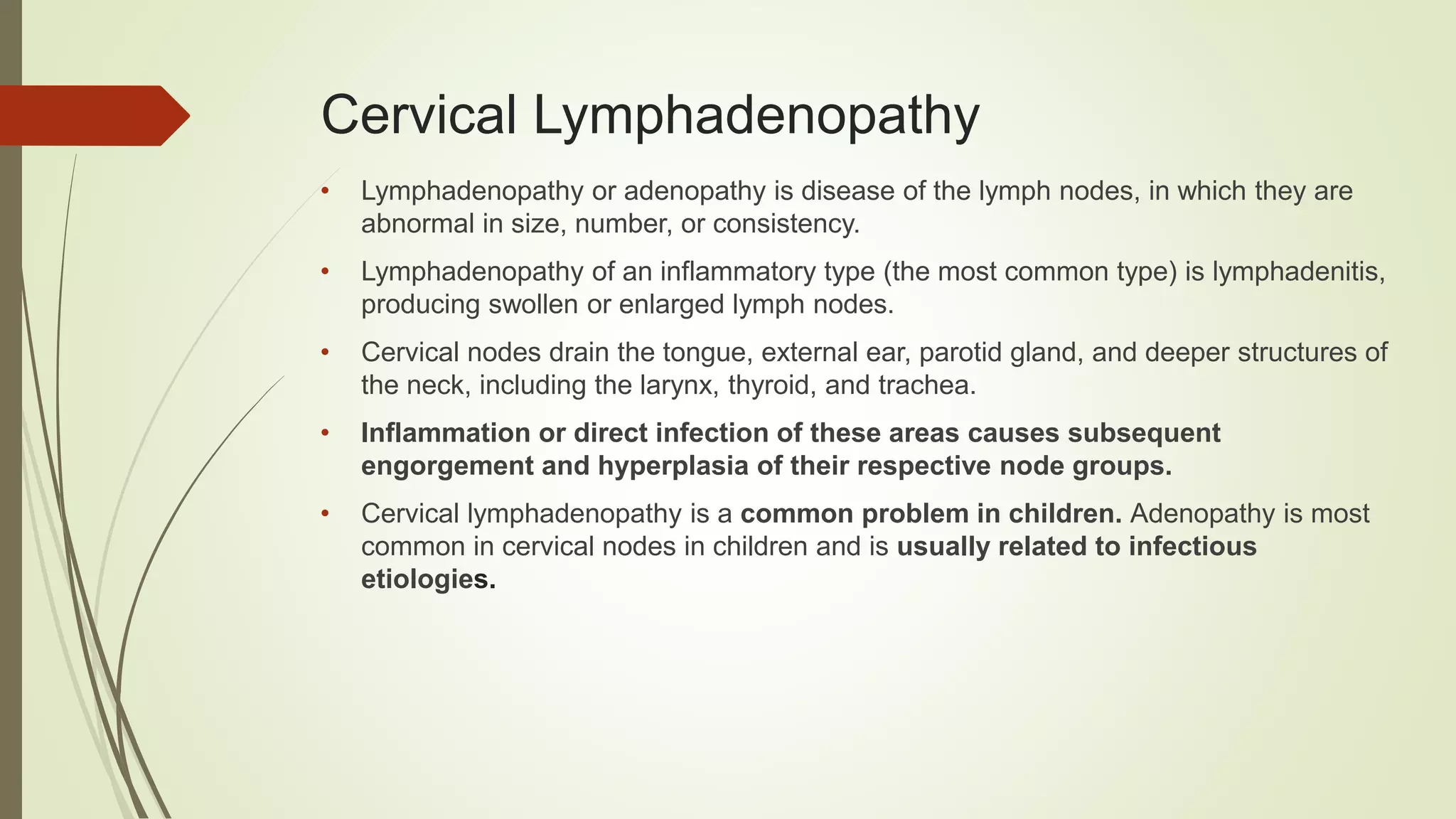
Abnormal neck lymph nodes are swellings or enlargements of the bean-shaped structures located throughout the neck that usually indicate an underlying immune response, infection, or, less commonly, malignancy. When these nodes increase in size, change in consistency, or remain persistent, they require medical evaluation to determine the precise underlying cause and guide appropriate clinical management.
Introduction to the Cervical Lymphatic System
The human body contains hundreds of lymph nodes, with a significant concentration located in the neck, known medically as the cervical region. These nodes act as crucial filters for the lymphatic system, trapping foreign particles, bacteria, viruses, and abnormal cells before they can circulate further into the body. When a person encounters an infection, injury, or systemic illness, the immune cells inside these nodes multiply rapidly to fight off the threat, causing the nodes to swell and become noticeable.
While most instances of enlarged or abnormal neck lymph nodes are benign and resolve spontaneously as an underlying infection clears, persistent changes require careful clinical analysis. Differentiating between a normal reactive response and a pathological condition involves assessing the duration, physical characteristics, and associated symptoms of the swelling. Because the neck houses complex anatomical structures—including the thyroid gland, salivary glands, major blood vessels, and nerves—evaluating abnormal neck lymph nodes demands a structured medical approach that balances diagnostic thoroughness with patient reassurance.
Normal Anatomy versus Abnormal Presentation
To understand what constitutes an abnormal neck lymph node, one must first recognize the baseline state of the cervical lymphatic system. Under normal conditions, many superficial lymph nodes in the neck are either completely impalpable or feel like small, soft, mobile, pea-sized grains beneath the skin. They are typically painless and soft to the touch.
An abnormal presentation generally involves lymphadenopathy, which is the clinical term for lymph nodes that are abnormal in size, consistency, or number. Healthcare providers typically categorize a lymph node as enlarged if it exceeds one centimeter in diameter, though certain regions, such as the epitrochlear or inguinal areas, have different thresholds. In the neck, nodes are divided into specific anatomical levels (Levels I through VII) to help clinicians document their precise location, which aids significantly in differential diagnosis.
Physical attributes that transform a routine finding into an abnormal one include:
- Size: Diameters exceeding 1 to 1.5 centimeters, or nodes that continue to grow over several weeks.
- Consistency: Nodes that feel hard, woody, rubbery, or firm rather than soft or rubbery-elastic.
- Mobility: Nodes that feel fixed, matted together, or tethered to underlying deep structures rather than freely movable under the skin.
- Tenderness: While tender nodes often indicate acute inflammation or infection, painless nodes that persist for extended periods can sometimes warrant higher clinical suspicion.
- Location: Supraclavicular nodes (located just above the collarbone) carry a statistically higher probability of underlying systemic pathology compared to submandibular or upper cervical nodes.
Common Causes of Cervical Lymphadenopathy
The etiology of abnormal neck lymph nodes spans a broad spectrum of medical conditions, ranging from minor self-limiting infections to complex hematologic malignancies. Clinicians generally group these causes into categories to streamline the diagnostic workup.
Infectious Causes (Benign and Reactive)
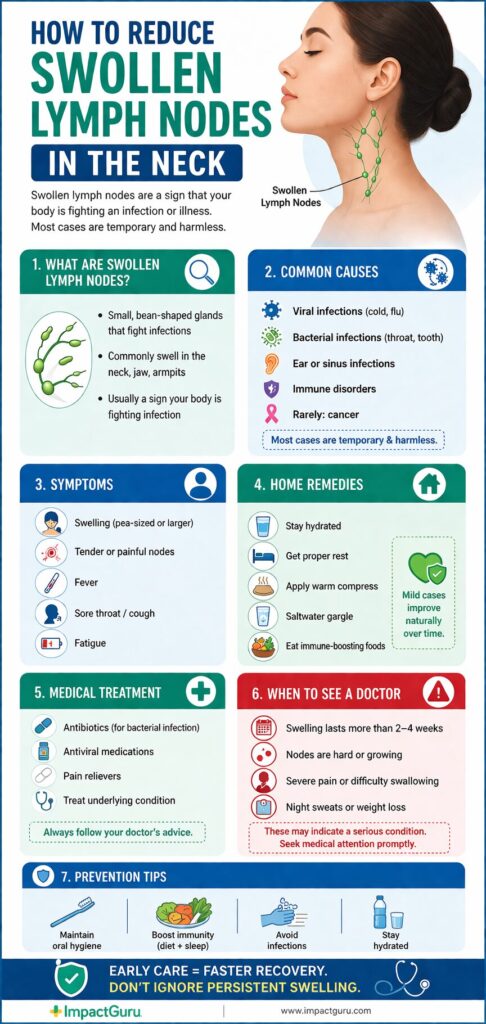
Infections represent by far the most frequent trigger for cervical lymphadenopathy. Because the head and neck drain areas exposed to environmental pathogens, upper respiratory tract infections, pharyngitis, dental abscesses, and otitis media frequently cause reactive enlargement of nearby lymph nodes.
- Viral Infections: Common pathogens include the Epstein-Barr virus (causing infectious mononucleosis), cytomegalovirus, adenovirus, and influenza. These often present with bilateral, tender lymphadenopathy accompanied by systemic symptoms like fatigue and fever.
- Bacterial Infections: Strep throat, dental infections, and skin infections of the face or scalp can lead to tender, rapidly enlarging nodes. In more severe cases, bacterial invasion can cause lymphadenitis, where the node itself becomes infected and inflamed.
- Specific Pathogens: Conditions like cat-scratch disease (Bartonella henselae), tuberculosis, and toxoplasmosis present with distinct clinical histories and characteristic patterns of lymph node involvement.
Inflammatory and Autoimmune Conditions
Non-infectious inflammatory disorders can also stimulate the immune system, resulting in chronic or recurrent lymph node enlargement. Rheumatoid arthritis, systemic lupus erythematosus, and sarcoidosis frequently feature generalized or localized lymphadenopathy. In these cases, the immune system mistakenly targets healthy tissues, leading to sustained activation and growth of lymphoid tissue.
Malignant Neoplasms
Though less common than infections, malignancy is a primary concern during the evaluation of abnormal neck lymph nodes. Malignant causes fall into two main categories:
- Metastatic Cancer: The vast majority of malignant neck masses in adults are metastases from primary squamous cell carcinomas of the head and neck (arriving from the oral cavity, larynx, pharynx, or thyroid). Cancer cells break away from the primary tumor, travel through lymphatic channels, and settle in the cervical lymph nodes.
- Primary Hematologic Malignancies: Lymphomas (both Hodgkin lymphoma and non-Hodgkin lymphomas) and leukemias originate within the lymphatic system itself. These conditions often present with firm, rubbery, painless, and progressively enlarging lymph nodes that may occur alongside systemic "B symptoms" such as unexplained weight loss, drenching night sweats, and persistent fevers.
| Category | Typical Node Characteristics | Associated Symptoms | Common Examples |
|---|---|---|---|
| Acute Infection | Soft, tender, mobile, warm | Fever, sore throat, cough, localized pain | Pharyngitis, dental abscess, viral URI |
| Chronic/Granulomatous | Firm, rubbery, slow-growing | Low-grade fever, fatigue, night sweats | Tuberculosis, cat-scratch disease |
| Autoimmune | Firm, movable, often generalized | Joint pain, skin rashes, chronic fatigue | Lupus, rheumatoid arthritis |
| Malignant (Metastatic) | Hard, fixed, painless, irregular | History of smoking/alcohol, mucosal lesions | Squamous cell carcinoma, thyroid cancer |
| Malignant (Lymphoma) | Firm, rubbery, matted, painless | Unexplained weight loss, night sweats, fevers | Hodgkin lymphoma, non-Hodgkin lymphoma |
Diagnostic Evaluation and Clinical Workup
When a patient presents with abnormal neck lymph nodes, a physician initiates a systematic diagnostic evaluation. The primary goal is to distinguish between benign, self-limiting processes and serious medical conditions that require specialized intervention.
Detailed Medical History
The clinical history provides critical clues. Key elements include the duration of the swelling, whether the node has changed in size, recent upper respiratory infections, dental pain, travel history, animal exposures, and systemic symptoms like unexplained fevers, drenching night sweats, or unintended weight loss. Social history, particularly tobacco and alcohol use, is heavily weighed because these substances are major risk factors for head and neck malignancies.
Physical Examination
A thorough physical examination assesses the size, shape, consistency, tenderness, and mobility of the abnormal neck lymph nodes. The clinician meticulously maps out which cervical levels are involved. Additionally, a complete head and neck examination is performed, inspecting the oral cavity, oropharynx, nasal passages, and thyroid gland to identify potential primary sources of infection or malignancy.
Laboratory Testing
Blood tests are often ordered to support the physical findings. A complete blood count (CBC) helps detect signs of infection, anemia, or hematologic malignancies. Inflammatory markers such as erythrocyte sedimentation rate (ESR) and C-reactive protein (CRP) can indicate the presence of acute or chronic inflammation. Specific serological tests may also be utilized if viral, bacterial, or autoimmune etiologies are suspected based on the patient's history.
Diagnostic Imaging
Imaging modalities play an essential role in visualizing nodes that cannot be adequately assessed by physical touch alone:
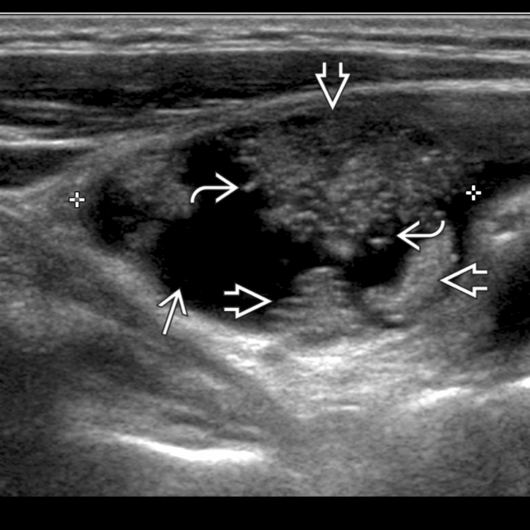
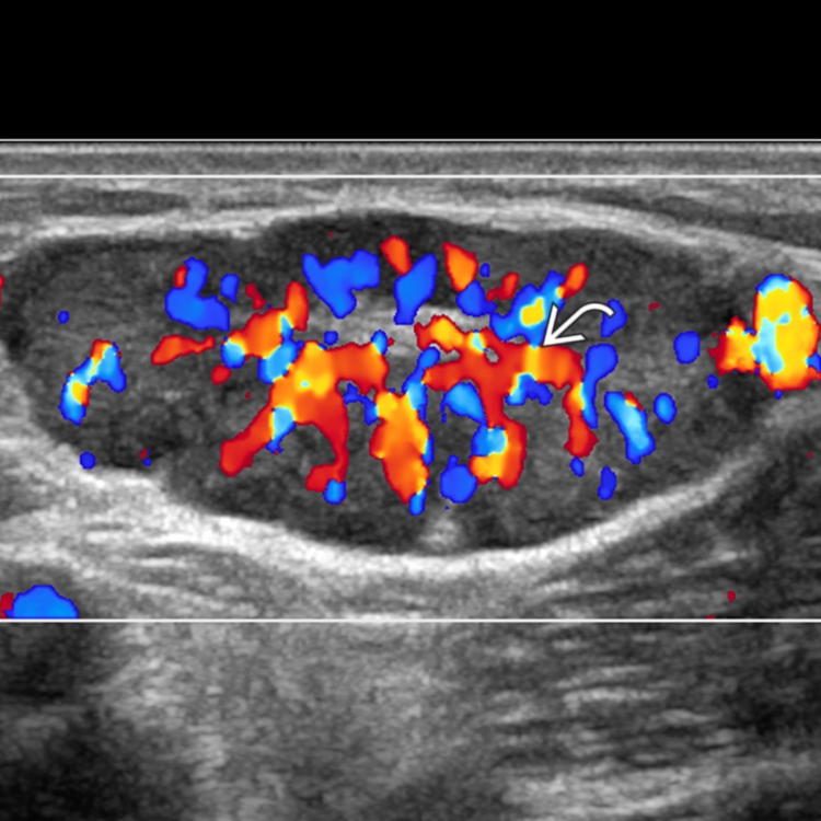
- Ultrasound: Often the first-line imaging choice, especially in younger patients or when evaluating superficial neck structures. Ultrasound assesses internal architecture, blood flow patterns (vascularity), and margins without exposing the patient to ionizing radiation.
- Computed Tomography (CT) Scan: Contrast-enhanced CT scans of the neck provide detailed cross-sectional views, helping clinicians evaluate the exact size, relationship to major blood vessels, and central necrosis of abnormal nodes, as well as checking for primary mucosal lesions.
- Magnetic Resonance Imaging (MRI): Offers superior soft-tissue contrast, making it particularly useful for evaluating the nasopharynx, skull base, and deep tissue spaces of the neck.
Biopsy Procedures
If imaging and laboratory evaluations do not establish a definitive benign diagnosis—or if high-risk features are present—a tissue diagnosis becomes necessary.
- Fine-Needle Aspiration (FNA): A thin needle is inserted into the lymph node to withdraw a sample of cells for cytological examination. FNA is minimally invasive, highly accurate in experienced hands, and capable of differentiating between infection, reactive hyperplasia, and malignancy.
- Core Needle Biopsy or Excisional Biopsy: If FNA results are inconclusive or if lymphoma is strongly suspected, a larger tissue core or the removal of the entire lymph node may be required for histopathological and flow cytometry analysis.
Red Flag Symptoms and Urgent Indicators
While the majority of abnormal neck lymph nodes are benign, recognizing clinical red flags ensures that patients with potentially serious conditions receive timely evaluation. Medical providers urge immediate or prompt assessment if a patient notices any of the following features:
- Persistence: A neck lump that remains unchanged or continues to grow for more than two to four weeks without an obvious infectious cause.
- Consistency and Mobility: Nodes that feel hard as a stone or are firmly fixed to underlying tissue structures.
- Location: Any swelling located in the supraclavicular fossa (above the collarbone).
- Size: A lymph node measuring greater than 2 centimeters in diameter with no signs of acute inflammation.
- Systemic Red Flags: Accompanying symptoms such as unexplained weight loss exceeding 10% of body weight, soaking night sweats, persistent unexplained fevers, or fatigue.
- Aerodigestive Symptoms: Difficulty swallowing (dysphagia), pain when swallowing (odynophagia), hoarseness lasting more than two weeks, or unexplained ear pain (otalgia), which can signal a primary tumor in the upper aerodigestive tract.
Management Strategies and Treatment Approaches
Management of abnormal neck lymph nodes depends entirely on the underlying diagnosis established during the clinical workup. Because the root causes vary widely, treatment plans are highly individualized.
Observational Management and Reassurance
If clinical findings strongly suggest a benign reactive process—such as a tender node following a viral upper respiratory tract infection—a period of observation (typically two to four weeks) is standard practice. Patients are advised to monitor the area and return if the swelling persists, grows larger, or develops concerning features. Antibiotics are prescribed only when there is clear clinical evidence of a bacterial infection.
Treatment of Infections
When a specific infectious agent is identified, targeted antimicrobial therapy is initiated. This may range from oral antibiotics for common bacterial lymphadenitis to specialized regimens for atypical mycobacterial infections or systemic fungal illnesses. Supportive care, including hydration, rest, and anti-inflammatory medications, helps manage associated pain and fever.
Oncologic Interventions
If a biopsy confirms malignancy, management shifts to a multidisciplinary oncology team consisting of head and neck surgeons, medical oncologists, and radiation oncologists.
- Metastatic Squamous Cell Carcinoma: Treatment often involves surgical resection of the primary tumor and neck dissection (removal of involved lymph nodes), frequently combined with adjuvant radiation therapy or concurrent chemoradiotherapy.
- Lymphomas: Management typically relies on systemic therapies such as chemotherapy, immunotherapy, or targeted biologic agents, sometimes supplemented by localized radiation therapy depending on the specific subtype and staging.
Frequently Asked Questions
What does it mean if a neck lymph node is hard and painless?
A lymph node that feels hard, firm, and painless is generally viewed with higher clinical suspicion than a soft, tender node. While infection usually causes tender, softer swelling, hard and fixed nodes can sometimes indicate metastatic cancer or lymphoma, necessitating prompt medical evaluation and potential diagnostic imaging or biopsy.
How long do reactive lymph nodes take to go away after an infection?
Lymph nodes that swell in response to a common viral or bacterial infection typically begin to decrease in size within two weeks as the immune response subsides. However, it is not uncommon for benign reactive nodes to remain palpable as small, pea-sized bumps for several weeks or even months afterward, especially in children.
Is an ultrasound sufficient to diagnose the cause of an abnormal neck node?
Ultrasound is an exceptionally useful initial imaging tool because it evaluates the size, shape, borders, and internal blood flow characteristics of a lymph node. While it can reliably distinguish many benign features from suspicious ones, it cannot provide a definitive cellular diagnosis. If high-risk ultrasound features are identified, a fine-needle aspiration or tissue biopsy is usually required to confirm the diagnosis.
Can stress cause neck lymph nodes to swell?
Stress does not directly cause lymph nodes to swell on its own. However, chronic psychological stress can temporarily suppress or alter immune function, making an individual more susceptible to viral infections or reactivation of latent viruses, which in turn can lead to reactive lymphadenopathy.
When should I see a doctor for a lump in my neck?
You should schedule an evaluation with a healthcare provider if a neck lump persists for more than two to three weeks without shrinking, if it continues to grow, if it feels hard and immobile, or if it is accompanied by systemic symptoms such as unexplained weight loss, persistent fevers, drenching night sweats, or difficulty swallowing.
Conclusion and Next Actions
Discovering abnormal neck lymph nodes can be an unsettling experience, but understanding the underlying physiological mechanisms helps replace anxiety with informed action. Because the vast majority of cervical lymphadenopathies stem from benign, treatable infections and inflammatory responses, panic is rarely warranted. However, maintaining vigilance regarding persistent or atypical features is essential for safeguarding your health. If you or someone you know has noticed a persistent neck mass or swelling that does not resolve within a reasonable timeframe, schedule an appointment with a primary care physician or an otolaryngologist for a thorough clinical examination and appropriate diagnostic verification.