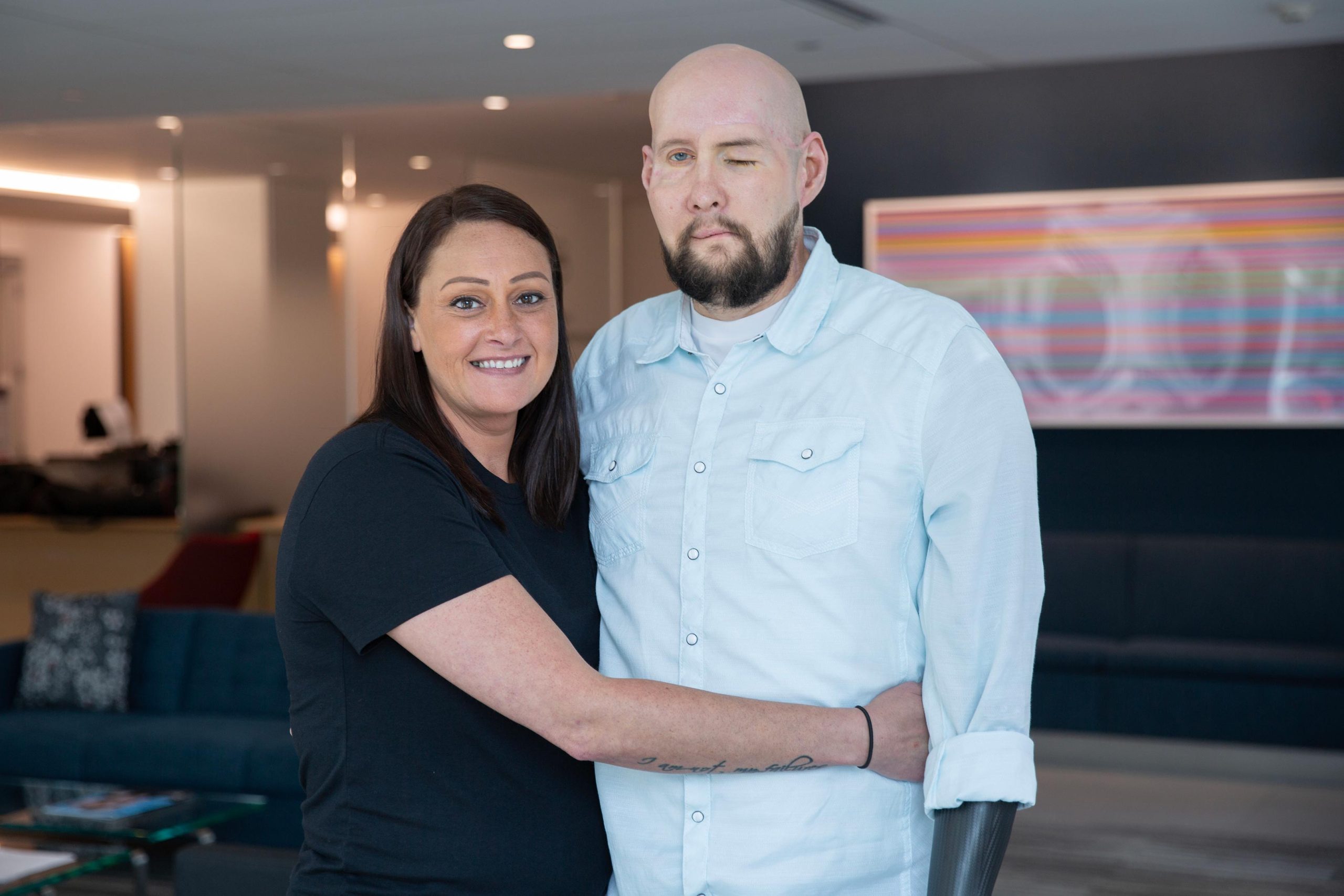
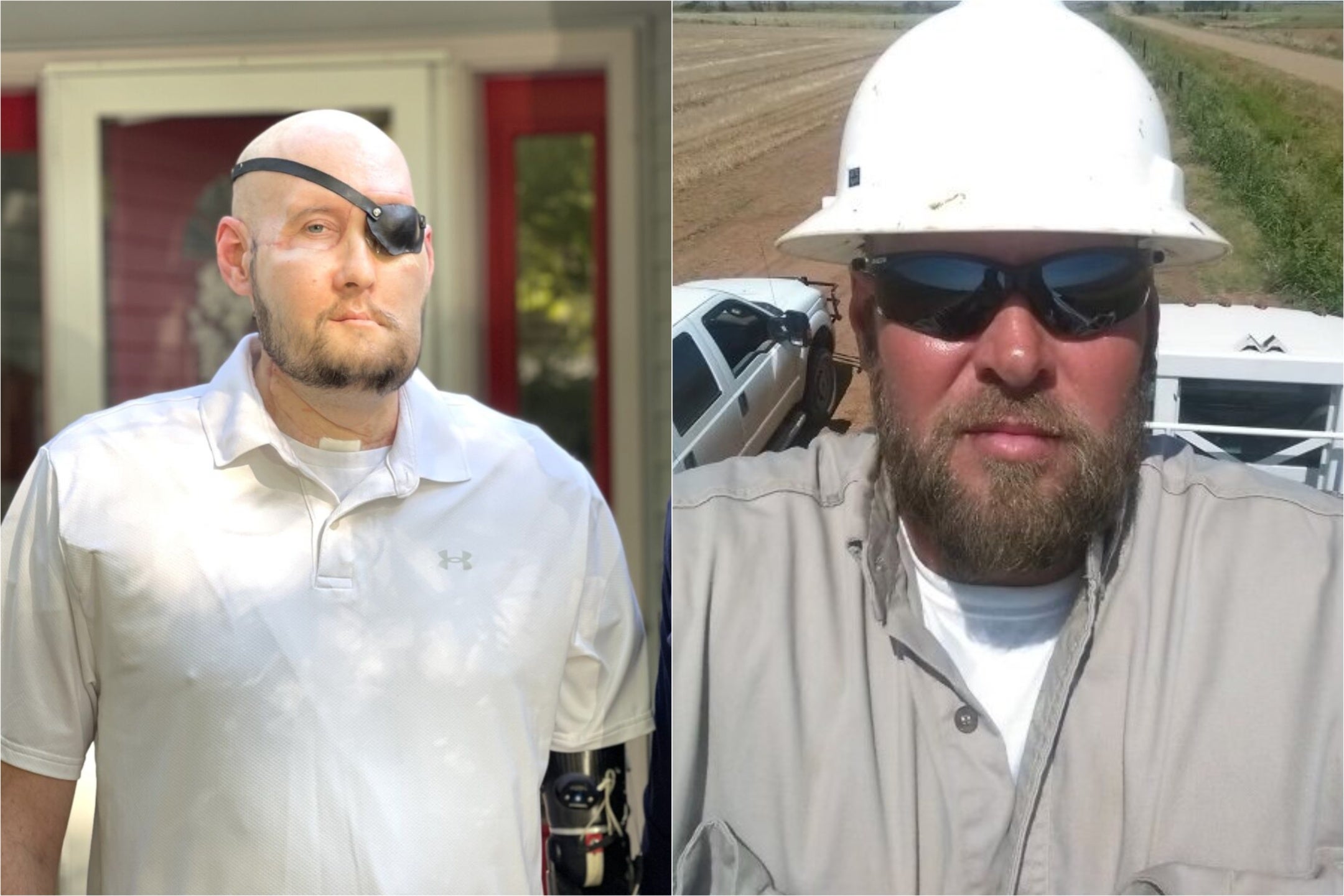
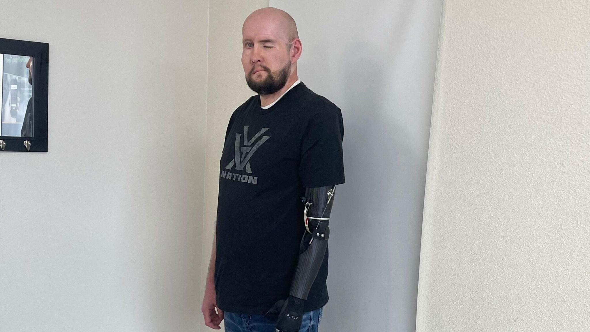
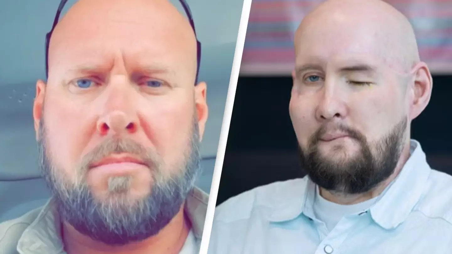
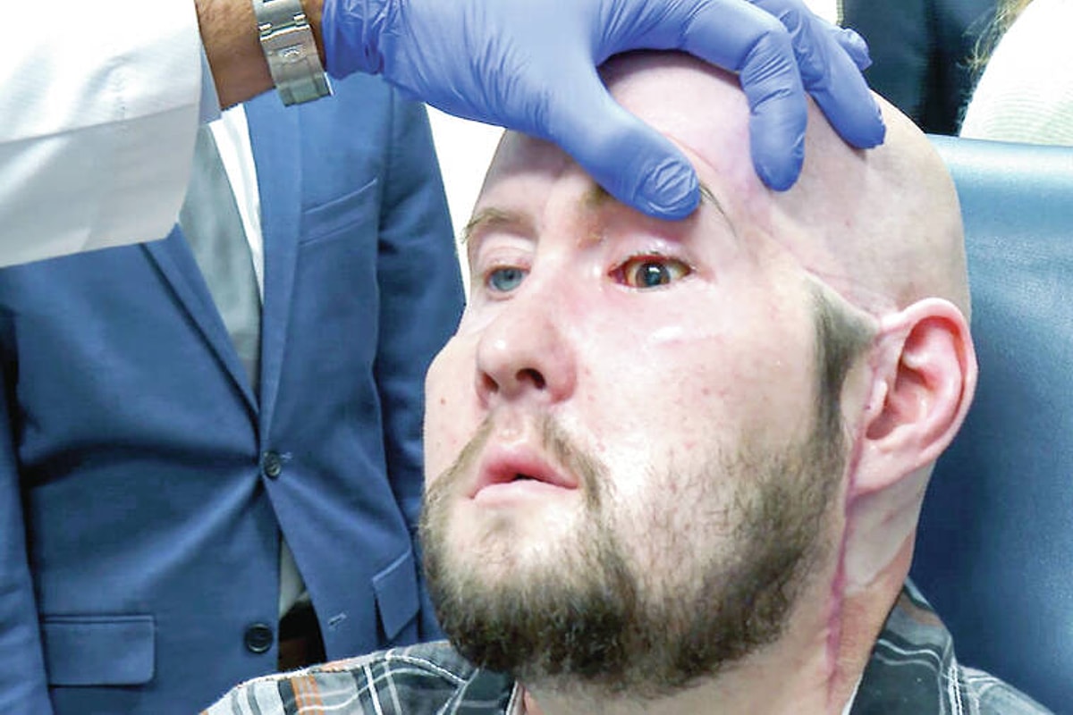
Aaron James, a utility lineman from Arkansas who survived a high-voltage electrical accident, became the recipient of the world’s first successful combined partial face and whole eye transplant at NYU Langone Health. This milestone medical procedure involved replacing his damaged facial structures and left eye with organs from a single human donor, marking a massive leap forward in reconstructive surgery, ophthalmology, and the neurology of sight restoration.
Understanding the Aaron James Case and the Nature of the Injury
In June 2021, Aaron James suffered a catastrophic occupational accident when his face accidentally brushed against a 7,200-volt live power line. The electrical current caused severe, life-threatening trauma, destroying the left side of his face, his left eye, his nose, his lips, and large portions of underlying bone and tissue. Despite enduring numerous traditional reconstructive surgeries, James faced permanent disfigurement, the loss of his left vision, and significant functional deficits regarding breathing, eating, and communication.
His case was ultimately evaluated by the multidisciplinary facial transplant team at NYU Langone Health, led by Dr. Eduardo D. Rodriguez. Because James was young, highly motivated, and maintained exceptional overall health despite his traumatic injuries, the medical team determined he was an ideal candidate for an experimental, highly complex procedure: a combined partial face and whole eye transplant (also known scientifically as an ocular globe transplant).
Prior to this landmark surgery, whole eye transplants had been successfully performed in animal models, but never in a living human being. While partial and full face transplants had grown increasingly common since the mid-2000s, transplanting an entire functioning human eye—including the delicate retina, choroid, and the optic nerve—had long been considered an insurmountable barrier in modern medicine due to the immense challenges of re-establishing microscopic blood supply and nervous system integration.
Surgical Protocol and Medical Execution
The historic operation took place on May 27, 2023, lasting approximately 21 hours and requiring a coordinated surgical team of more than 140 healthcare professionals. The procedure was meticulously planned using advanced three-dimensional virtual surgical imaging and custom-printed cutting guides to ensure exact alignment of donor and recipient bone structures.
| Surgical Phase | Primary Objective | Key Clinical Challenges |
|---|---|---|
| Preparation & Excision | Removal of scarred recipient tissue and precise harvesting of donor tissue. | Preserving fragile vascular pathways and matching bony architecture. |
| Vascular Anastomosis | Connecting microscopic blood vessels to restore immediate blood flow. | Preventing thrombosis and ensuring adequate perfusion to ocular structures. |
| Nerve Coaptation | Splicing recipient and donor motor and sensory nerves. | Promoting axonal regeneration across the optic nerve junction. |
| Skeletal Fixation | Rebuilding the midface using titanium plates and screws. | Restoring facial symmetry, structural integrity, and eye socket depth. |
During the operation, surgeons transplanted both the lower half of the donor's face and the entire left human eye, encompassing the eyeball, the surrounding orbit, extraocular muscles, and a significant segment of the donor optic nerve. A critical innovation of the procedure involved the injection of adult hematopoietic stem cells—harvested from the donor's bone marrow—directly into the recipient's optic nerve during surgery. The medical team hypothesized that these stem cells would protect nerve cells from dying and encourage nerve regeneration between the donor eye and the recipient's brain.
Post-Operative Recovery, Monitoring, and Medical Findings
Following the surgery, Aaron James was closely monitored in the intensive care unit and subsequent inpatient recovery units at NYU Langone Health. The primary immediate concerns in any vascularized composite allotransplantation (VCA) are acute tissue rejection and vascular complications. James was placed on a rigorous, lifelong regimen of immunosuppressive medications designed to prevent his immune system from attacking the foreign facial and ocular tissues.
Subsequent evaluations by the surgical and ophthalmological teams yielded several significant medical findings:
- Structural Integration: The transplanted face and eye healed remarkably well, with robust blood flow circulating through the retina and choroid weeks and months after the procedure.
- Intraocular Pressure: The transplanted eye maintained normal intraocular pressure independently, avoiding common post-surgical complications such as hypotony (low eye pressure) or severe glaucoma.
- Aesthetic and Functional Restoration: James regained significant facial symmetry, the ability to breathe more naturally through his reconstructed nose, and a dramatically improved quality of life.
- Neurological Status: While the eye successfully integrated into the socket and maintained structural viability, medical evaluations confirmed that visual signals are not yet transmitting from the transplanted retina to the visual cortex of the brain. The optic nerve shows signs of healing, but functional sight remains unachieved.
Scientific Implications and Future Outlook in Ocular Transplantation
The Aaron James eye transplant represents a foundational proof-of-concept for human whole eye transplantation. For decades, the ophthalmology community debated whether an excised human eye could survive transplantation without degenerating. This case definitively answers that question: with modern microsurgical techniques and targeted immunosuppression, an entire human eye can be successfully transplanted and maintained in a living host without immediate ischemic failure or necrosis.
However, the medical community remains cautious about classifying the procedure as a complete functional success regarding sight. The human optic nerve is part of the central nervous system, and unlike peripheral nerves, it does not readily regenerate once severed. Researchers are actively studying how to bridge the gap between the donor optic nerve and the recipient's brain using advanced neurotrophic factors, gene therapy, and artificial neural interfaces.
The insights gained from tracking James's recovery are shaping global protocols for future clinical trials involving ocular transplantation. By demonstrating that whole eye transplants are surgically feasible and immunologically manageable under controlled hospital conditions, this landmark case has opened new horizons for millions of individuals suffering from blinding ocular trauma, advanced glaucoma, and retinal diseases.
Frequently Asked Questions
Did Aaron James regain his sight from the eye transplant?
No. While the transplanted left eye remains healthy, maintains normal pressure, and has strong blood flow, it does not currently transmit visual signals to the brain. The primary barrier is the reconnection and regeneration of the optic nerve, which remains a monumental challenge in modern neuroscience.
Why was this specific transplant considered a world first?
While partial and full face transplants have been performed globally since 2005, Aaron James is the first human in medical history to receive a combined partial face and whole eye transplant from a single human donor in one continuous surgical procedure.
What kind of immunosuppression is required for a face and eye transplant?
Recipients of vascularized composite allotransplants must take systemic immunosuppressive drugs for the rest of their lives. These medications suppress the immune system to prevent organ rejection, but they also carry long-term risks such as increased susceptibility to infections and kidney toxicity.
How long did the surgical procedure take?
The complex surgery was performed on May 27, 2023, at NYU Langone Health, requiring approximately 21 hours of continuous operation by a multidisciplinary team of over 140 surgeons, nurses, and specialized clinical staff.
What role did stem cells play in the procedure?
Surgeons injected adult stem cells harvested from the donor's bone marrow into the optic nerve during the transplant. The experimental goal was to promote nerve cell survival and encourage neural regeneration between the donor eye and the recipient's nervous system.